دانلود کتاب اطلس روشهای هدایت تصویری ستون فقرات + ویدئو
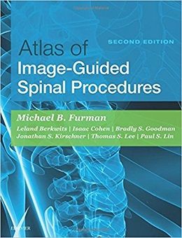
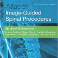
Atlas of Image-Guided Spinal Procedures, 2ed + Video
The second edition of Atlas of Image-Guided Spinal Procedures features a highly visual atlas format to illustrate exactly how to perform each technique. This medical reference walks you through each procedure, step-by-step, to safely and efficiently relieve patients’ pain. This book presents an algorithmic, image-guided approach for each technique; trajectory view (demonstrates fluoroscopic “setup”); multiplanar confirmation views (AP, lateral, oblique); and “safety view” (what should be avoided during injection), along with optimal and suboptimal contrast patterns. Each fluoroscopic and ultrasound chapter also has the same “voice” so it is easy to follow.
- Safely and efficiently relieve your patients’ pain with consistent, easy-to-follow chapters that guide you through each technique.
- Presents an algorithmic, image-guided approach for each technique: trajectory view (demonstrates fluoroscopic “setup”); multiplanarconfirmation views (AP, lateral, oblique); and “safety view” (what should be avoided during injection), along with optimal and suboptimal contrast patterns.
- Special chapters on Needle Techniques, Procedural Safety, Fluoroscopic and Ultrasound Imaging Pearls, Radiation Safety, and L5-S1 Disc Access provide additional visual instruction.
- View drawings of radiopaque landmarks and key radiolucent anatomy that cannot be viewed fluoroscopically.
- Videosincludingprocedural “set-up” and optimal and suboptimal constrast flow are available in the Expert Consult eBook version.
- Includes new unique and diagrams demonstrating cervical, thoracic and lumbar radiofrequency probe placement and treatment zones on multiplanar views, as well as newunique tables and examples differentiating between optimal and suboptimal epidural contrast flow
- Features new coverage of ultrasound techniques, as well as new presentation of procedures for “spine masqueraders” such as the hip and shoulder.
- Expert Consult eBook version included with purchase. This enhanced eBook experience allows you to access and search all of the text, figures, images, videos, and references from the book on a variety of devices
Contents
۱. Introduction: How to Use This Atlas
۲. Needle Techniques
۳. Fluoroscopic Techniques: Anatomy and Set-Up
۴. Ultrasound Techniques and Pearls
۵. Optimizing Patient Safety and Positioning
۶. Radiation Safety
۷A Caudal Epidural Steroid Injection–Shallow Angle Approach: Fluoroscopic Guidance
۷B Caudal Epidural Spinal Injection–Steep Angle Approach: Fluoroscopic Guidance
۷C Caudal Epidural Steroid Injection: Ultrasound Guidance
۸A Ganglion Impar Injection: Fluoroscopic Guidance
۸B Ganglion Impar Injection: Ultrasound Guidance
۹. Sacral Insufficiency Fracture Repair/Sacroplasty: Fluoroscopic Guidance
۱۰A Sacroiliac Intraarticular Joint Injections–Posterior Approach, Inferior Entry: Fluoroscopic Guidance
۱۰B Sacroiliac Intraarticular Joint Injection: Ultrasound Guidance
۱۱. S1 Transforaminal Epidural Steroid Injection
۱۲. Lumbar Interlaminar Epidural Steroid Injection–Paramedian Approach: Fluoroscopic Guidance
۱۳A Lumbar Transforaminal Epidural Steroid Injection–Supraneural, Traditional Single-Needle Technique: Fluoroscopic Guidance
۱۳B Lumbar Transforaminal Epidural Steroid Injection–Supraneural, Two-Needle Technique: Fluoroscopic Guidance
۱۳C Lumbar Transforaminal Epidural Steroid Injection–Infraneural: Fluoroscopic Guidance
۱۳D Lumbar Transforaminal Epidural Steroid Injection–Needle Localization Diagrams
۱۴. Lumbar Myelography: Fluoroscopic Guidance
۱۵A Lumbar Zygapophysial Joint Intraarticular Joint Injection–Posterior Approach: Fluoroscopic Guidance
۱۵B Lumbar Zygapophysial Joint Nerve (Medial Branch) Injection–Oblique Approach: Fluoroscopic Guidance
۱۵C Lumbar Zygapophysial Joint Nerve (Medial Branch) Radiofrequency Neurotomy, Posterior Approach: Fluoroscopic Guidance
۱۵D Lumbar Medial Branch Blocks–Midline: Ultrasound Guidance
۱۵E Lumbar Zygapophysial Joint Innervation–Anatomy Diagram and Lesion Zone Diagram
۱۶. Lumbar Sympathetic Block: Fluoroscopic Guidance
۱۷A Lumbar Provocation Discography/Disc Access: Standard Fluoroscopic Techniques
۱۷B L5-S1 Disc Access: Fluoroscopic Guidance
۱۸. Thoracolumbular Spinal Cord Stimulation: Fluoroscopic Guidance
۱۹. Vertebral Augmentation (Vertebroplasty/Kyphoplasty), Transpedicular Approach: Fluoroscopic Guidance
۲۰. Thoracic Interlaminar Epidural Steroid Injection–Paramedian Approach, Fluoroscopic Guidance
۲۱. Thoracic Transforaminal Epidural Steroid Injection–Infraneural Approach
۲۲A Thoracic Zygapophysial Joint Intraarticular Injection–Posterior Approach: Fluoroscopic Guidance
۲۲B Thoracic Zygapophysial Joint Nerve (Medial Branch) Injection–Posterior Approach: Fluoroscopic Guidance
۲۲C Thoracic Zygapophysial Joint Nerve (Medial Branch) Radiofrequency Neurotomy–Posterior Approach: Fluoroscopic Guidance
۲۲D Thoracic Zygapophysial Joint Innervation–Anatomy Diagram
۲۳A Intercostal Nerve Injections: Fluoroscopic Guidance
۲۳B Intercostal Nerve Injection–In-Plane Approach: Ultrasound Guidance
۲۴. Thoracic Disc Access: Fluoroscopic Guidance
۲۵. Cervical Interlaminar Epidural Steroid Injection–Paramedian Approach: Fluoroscopic Guidance
۲۶. Cervical Spinal Cord Stimulation, Fluoroscopic Guidance
۲۷. Cervical Transforaminal Epidural Steroid Injection: Fluoroscopic Guidance
۲۸A Stellate Ganglion Injection: Fluoroscopic Guidance
۲۸B Stellate Ganglion Injection: Ultrasound Guidance
۲۹. Cervical Discography/Disc Access
۳۰A Cervical Zygapophysial Joint Intraarticular Injection–Posterior Approach: Fluoroscopic Guidance
۳۰B Cervical Zygapophysial Joint Intraarticular Injection–Lateral Approach: Fluoroscopic Guidance
۳۰C Cervical Zygapophysial Nerve (Medial Branch) Injection–Lateral Approach: Ultrasound Guidance
۳۰D Cervical Zygapophysial Joint Nerve (Medial Branch) Injection–Lateral Approach: Fluoroscopic Guidance
۳۰E Cervical Zygapophysial Joint Intraarticular Injection–Posterior Approach: Ultrasound Guidance
۳۰F Cervical Zygapophysial Joint Nerve (Medial Branch) Radiofrequency Neurotomy and Nerve Injection–Posterior Approach: Fluoroscopic Guidance
۳۰G Cervical Zygapophysial Joint Nerve (Medial Branch) Radiofrequency Neurotomy and Nerve Injection–Posterior Approach: Ultrasound Guidance
۳۰H Cervical Zygapophysial Joint Innervation–Anatomy Diagram and Lesion Zone Diagrams
۳۱. Atlantoaxial Joint Intraarticular Injection: Fluoroscopic Guidance
۳۲. Atlantooccipital Joint Intraarticular Injection: Fluoroscopic Guidance
۳۳. Greater Occipital Nerve Steroid Injection–In Plane Approach: Ultrasound Guidance
۳۴A Shoulder/Glenohumeral Joint Intraarticular Injection–Anterior Approach: Fluoroscopic Guidance
۳۴B Intraarticular Shoulder/Glenohumeral Joint Injection–Posterior Approach: Ultrasound Guidance
۳۴C Shoulder/Subacromial/Subdeltoid Injection–Lateral Approach: Ultrasound Guidance
۳۴D Shoulder/Acromioclavicular Joint Injection–Posterior Approach: Ultrasound Guidance
۳۴E Shoulder/Suprascapular Nerve Injection–Posterior Approach: Ultrasound Guidance
۳۴F Shoulder/Biceps Tendon Sheath Injection–Anterior Approach: Ultrasound Guidance
۳۵A Intra-articular Hip Injection–Anterior Approach: Fluoroscopic Guidance
۳۵B Intra-articular Hip Injection–Lateral Approach: Fluoroscopic Guidance
۳۵C Intraarticular Hip Injection–Anterior Approach: Ultrasound Guidance
۳۵D Greater Trochanteric/Gluteus Medius Injection: Ultrasound Guidance
۳۵E Lateral Femoral Cutaneous Nerve Injection: Ultrasound Guidance
۳۶A Iliac Crest Biopsy/Aspiration: Fluoroscopic Guidance
۳۶B Iliac Crest Biopsy: Ultrasound Guidance
Video Contents
۱-۱ Demonstration of How to Use This Atlas
۳-۱ Digital Subtraction Demonstrating Right L5 Transforaminal ESI After Repositioning
۱۰A-1 Optimal Intraarticular Sacroiliac Joint Contrast Filling Joint
۱۳A-1A Fluoroscopic Live of an Optimal L3 Transforaminal Epidural Spinal Injection
۱۳A-1B AP Digital Subtraction Live of the Same L3 Transforaminal Epidural Spinal Injection
۱۳A-2 AP Left L5 Transforaminal Epidural Spinal Injection With Vascular Injection Observed With Standard
۱۳A-3 AP Suboptimal Filling of a Left L4 Transforaminal Epidural Spinal Injection Observed With Digital Subtraction Fluoroscopy
۱۳A-4A AP Suboptimal Filling of a Left L5 Transforaminal Epidural Spinal Injection Observed With Standard Fluoroscopy
۱۳A-4B Same Case With Suboptimal Filling of a Left L5 Transforaminal Epidural Spinal Injection Observed With Digital Subtraction Fluoroscopy
۱۵C-1 Medial Branch Neurotomy: Electrode Placement Overview — Farzad Karkvandeian, Jesse Bernstein, and
۱۵C-2 L4 Medial Branch Neurotomy: Electrode Placement Demonstration
۱۵C-3 L5 Dorsal Ramus Neurotomy: Electrode
۱۶-۱ Lateral View Sympathetic Block, Digital Subtraction Imaging
۲۲A-1 Digital Subtraction Demonstrating Optimal Contrast Filling of a Thoracic Z-Joint
۲۵-۱ Cervical Interlaminar Epidural Steroid Injection Flow (Standard Fluoroscopy in Contralateral Oblique) Demonstrating Optimal Interlaminar Flow (right of needle tip) Simultaneously Flowing Suboptimally Posteriorly (left of screen) Out of the Epidural Space Into the Soft Tissues
۲۵-۲ Cervical Interlaminar Epidural Steroid Injection Flow (Standard Fluoroscopy in Contralateral Oblique) Demonstrating Optimal Interlaminar Flow (left of needle tip) Simultaneously Flowing Suboptimally Anteriorly (left of screen) Vascular
۲۵-۳ Suboptimal Cervical Interlaminar Epidural Steroid Injection Flow (Standard Fluoroscopy in AP) Demonstrating Vascular Flow
۲۷-۱ Optimal C6 Transforaminal Epidural Spinal Injection (Digital Subtraction Fluoroscopy in AP)
۳۴B-1 Ultrasound-Guided Glenohumeral Joint, Medial to Lateral In-Plane Needle Placement
۳۴B-2 Glenohumeral Joint Injection, In-Plane, Lateral to Medial, Initial Placement
۳۴B-3 Glenohumeral Joint Injection, In-Plane, Lateral to Medial With Capsular Distension
۳۴C-1 Subacromial Subdeltoid Bursa Injection, In-Plane, in a Patient With Calcifc Tendinosis
۳۴C-2 Subacromial Subdeltoid Bursa Injection, In-Plane, Demonstrating Bursal Distension
۳۴C-3 Supraspinatous Percutaneous Needle Tenotomy and Subacromial/Subdeltoid Bursa PRP Injection, Out-ofPlane
۳۴F-1 Biceps Tendon Sheath Injection, Short Axis, Lateral to Medial Approach
۳۵C-1 Hip Injection, In-Plane — Jonathan Kirschner and
۳۵C-2 USG Femoroacetabular Intraarticular Joint, Distal to Proximal (LAX) In-Plane Injection With Capsular Distention
۳۵C-3 Ultrasound-Guided Femoroacetabular Intraarticular Joint, Distal to Proximal (LAX) In-Plane Needle Injection With Capsular Distension
۳۵D-1 Greater Trochanteric (Sub-Gluteus Maximus) Bursa Injection, In-Plane (LAX), Distal to Proximal — Jonathan
۳۵D-2 Greater Trochanteric (Sub-Gluteus Maximus) Bursa Injection, In-Plane (SAX), Posterior to Anterior
۳۵D-3 Greater Trochanteric (Sub-Gluteus Maximus) Bursa Injection, In-Plane (SAX), Posterior to Anterior
لینک کوتاه : https://bookbaz.ir/?p=83292
نویسنده : Michael Bruce Furman MD , Leland Berkwits
ناشر : Elsevier; 2 edition
سال انتشار : 2018
زبان کتاب : انگلیسی
نوع فایل : MP4 + PDF (کیفیت اصلی)
تعداد صفحات : 683
(ISBN) شابک : 0323401538
قیمت کتاب درآمازون : $133.61
حجم فایل : 400 MB


