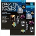
دانلود کتاب تصویربرداری تشخیصی کودکان کافی (ویرایش ۲۰۱۹) + ویدئو
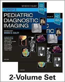
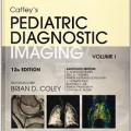
Caffey’s Pediatric Diagnostic Imaging, 2-Volume Set, 13ed + Video
For more than 70 years, Caffey’s Pediatric Diagnostic Imaging has been the comprehensive, go-to reference that radiologists have relied upon for dependable coverage of all aspects of pediatric imaging. In the 13th Edition, Dr. Brian Coley leads a team of experts to bring you up to date with today’s practice standards in radiation effects and safety, as well as in head and neck, neurologic, thoracic, cardiac, gastrointestinal, genitourinary, and musculoskeletal pediatric imaging. This two-volume bestselling reference is a must-have resource for pediatric radiologists, general radiologists, pediatric subspecialists, pediatricians, hospitals, and more – anywhere clinicians need to ensure safe, effective, and up-to-date imaging of children.
- Provides access to ۵۰ online videos, including hypertrophic pyloric stenosis, disorders of swallowing, fetal swallowing, fetal bowel obstruction, upper GI and ultrasound evaluation of malrotation and volvulus, congenital heart disease MRI evaluation, and many more.
- Includes separate chapters on radiation effects and safety, pre-natal imaging, neoplasms, trauma, techniques, embryology, genetic anomalies, and common acquired conditions.
-
- Takes an updated, contemporary approach with more focused and consistently formatted content throughout. Clinical content includes Overview; Etiologies, Pathophysiology, and Clinical Presentation; Imaging, including pros and cons, costs, evidence-based data, findings, and differential diagnostic considerations; and Treatment, including follow-up.
- Features ۸,۵۰۰ high-quality images – ۱,۰۰۰ new or updated.
-
- Provides expanded coverage of advanced imaging and diagnostics, including genetics and fetal imaging, MRI and advanced MR techniques, low-dose CT, ultrasound, nuclear medicine, and molecular imaging, as well as the latest quality standards, evidence-based data, and practice guidelines.
- Features new Key Points boxes and more tables and flowcharts that make reference faster and easier.
-
- Focuses on safety, particularly in radiation dosing, as part of the Image Gently® campaign to improve pediatric imaging while limiting radiation exposure and unneeded studies.
- Expert Consult™ eBook version included with purchase. This enhanced eBook experience allows you to search all of the text, figures, and references from the book on a variety of devices.
Contents
۱. Radiation Bioeffects, Risks, and Radiation Protection in Medical Imaging in Children
۲. Magnetic Resonance Safety
۳. Complications of Contrast Media
۴. Embryology, Anatomy, Normal Findings, and Imaging Techniques
۵. Prenatal, Congenital, and Neonatal Abnormalities
۶. Orbit Infection and Inflammation
۷. Orbital Neoplasia
۸. Nose and Sinonasal Cavities
۹. Embryology, Anatomy, Normal Findings, and Imaging Techniques
۱۰. Congenital and Neonatal Abnormalities
۱۱. Infection and Inflammation
۱۲. Neoplasia
۱۳. Embryology, Anatomy, Normal Findings, and Imaging Techniques
۱۴. Prenatal, Congenital, and Neonatal Abnormalities
۱۵. Infection and Inflammation
۱۶. Neoplasia
۱۷. Thyroid and Parathyroid
۱۸. Embryology, Anatomy, Normal Findings, and Imaging Techniques
۱۹. Prenatal Imaging
۲۰. Craniosynostosis, Selected Craniofacial Syndromes, and Other Abnormalities of the Skull
۲۱. Neoplasms, Neoplasm-like Lesions, and Infections of the Skull
۲۲. The Mandible
۲۳. Traumatic Lesions of the Skull and Face
۲۴. Embryology and Brain Development .
۲۵. Magnetic Resonance Spectroscopy and Positron Emission Tomography
۲۶. Diffusion-Weighted Magnetic Resonance Imaging: Principles and Implementation in Clinical and Research Settings
۲۷. Functional Magnetic Resonance Imaging
۲۸. Perfusion Imaging and Magnetoencephalography
۲۹. Prenatal Imaging
۳۰. Neonatal Brain Injury
۳۱. Congenital Brain Malformations
۳۲. Hydrocephalus
۳۳. Inherited Metabolic and Neurodegenerative Disorders
۳۴. Infection and Inflammation
۳۵. Pediatric Brain Neoplasms
۳۶. Cerebrovascular Disorders
۳۷. Stroke
۳۸. Neuroimaging in Pediatric Epilepsy
۳۹. Trauma
۴۰. Embryology, Anatomy, and Normal Findings
More…
۱۳۰. Prenatal Musculoskeletal Imaging
۱۳۱. Congenital Anomalies of Bone
۱۳۲. Skeletal Dysplasias and Selected Chromosomal Disorders
۱۳۳. Alignment Disorders
۱۳۴. Scoliosis
۱۳۵. Developmental Dysplasia of the Hip
۱۳۶. Arthritis and Differential Inflammatory Joint Disorders
۱۳۷. Musculoskeletal Infections
۱۳۸. Soft Tissue and Bone Tumors
۱۳۹. Metabolic Bone Disease
۱۴۰. Endocrine Disorders
۱۴۱. Skeletal Manifestations of Systemic Disease
۱۴۲. Skeletal Trauma
۱۴۳. Child Abuse
۱۴۴. Sports Medicine
Video Contents
۱. Fetal Swallowing
۲. Normal Fetal Swallowing
۳. Mucoepidermoid Carcinoma
۴. Aortic Stenosis, Sagittal and Coronal Time-Resolved
۵. Aortic Coarctation
۶. Aortic Stenosis
۷. Cardiac MRI in a Patient with Previously Repaired Tetralogy of Fallot, View 1
۸. Cardiac MRI in a Patient with Previously Repaired Tetralogy of Fallot, View 2
۹. Marginally Hypoplastic Left Ventricle to Evaluate for a Two-Ventricle Versus Single-Ventricle Repair
۱۰. Bicuspid Aortic Valve in a 25-Year-Old with History of Aortic Coarctation
۱۱. Bicuspid Aortic Valve in a 25-Year-Old with History of Aortic Coarctation, Showing Upward Ballooning of the Aortic Valve Lea?ets and Turbulent Poststenotic Jet
۱۲. Systolic Flow Void Beyond the Coarctation Consistent with Turbulent Flow Across the Coarctation
۱۳. Tetralogy of Fallot, Status Post-Repair, Status Post– Pulmonary Valve Replacement
۱۴. Tetralogy of Fallot, Status Post-Repair, Status Post– Transcatheter Pulmonary Valve Replacement
۱۵. Markedly Dilated Right Ventricle with Overall Mildly Depressed Systolic Function
۱۶. Dilated Right Ventricular Out?ow Tract with Dyskinetic Anterior Wall in the Region of the Prior Transannular Patch
۱۷. To-and-Fro Flow in the Main Pulmonary Artery
۱۸. Hypertrophy of the Right Ventricle with Bowing of the Interventricular Septum into the Lower Pressure Left Ventricle
۱۹. Anterior and Leftward Aorta Arising From the Morphologic Right Ventricle
۲۰. Dephasing Jet at the Pulmonary Valve
۲۱. Tortuous Aorta and Iliac Arteries with Multiple Stenoses: Infant with Loeys Dietz Syndrome
۲۲. Diffuse Arterial Tortuosity and Multiple Stenoses
۲۳. Thickening of the Right and Left Ventricle Myocardium with Small Ventricular Cavities: 6-Month-Old Girl with Features of LEOPARD Syndrome
۲۴. Spectrum of Anatomic Abnormalities: 2-Day-Old Female with Cantrell Syndrome (Pentalogy of Cantrell)
۲۵. Hypertrophic Cardiomyopathy with Severe Asymmetric Thickening of the Left Ventricular Septal Wall
۲۶. Cardiac Fibroma with Gorlin Syndrome
۲۷. Duodenal Obstruction with Hyperperistalsis
۲۸. Laryngeal Penetration
۲۹. Laryngeal Penetration and Tracheal Aspiration
۳۰. Sonographic Sweep to Find Antropyloric Canal: Sweep Terminating at Normal Pylorus 100-1
۳۱. Sonographic Sweep to Find Antropyloric Canal: Sweep Terminating at Abnormal, Hypertrophied Pylorus of HPS
۳۲. Transient Appearance of HPS during Normal Peristaltic Activity in a Normal Infant
۳۳. Passage of Air Through Abnormal Pyloric Canal
۳۴. Changes in the Length and Thickness of the Abnormal Antropyloric Canal in Real Time
۳۵. UGI Evaluation of the Duodenum
۳۶. Midgut Volvulus
۳۷. Midgut Volvulus
۳۸. Chronic Midgut Volvulusxxx Video Contents
۳۹. Crohn Stricture Causing Partial Small Bowel Obstruction
۴۰. Crohn Disease Ileal Stricture
۴۱. Active Peristalsis in Transient Small Bowel Intussusception
۴۲. Air Reduction of Intussusception
۴۳. Fetal Autosomal Recessive Polycystic Kidney Disease on Ultrasound
۴۴. Amniotic Band Syndrome
لینک کوتاه : https://bookbaz.ir/?p=89317
نویسنده : Brian D. Coley MD
ناشر : Elsevier; 13 edition
سال انتشار : 2019
زبان کتاب : انگلیسی
نوع فایل : MP4 + PDF (کیفیت اصلی)
تعداد صفحات : 2745
(ISBN) شابک : 0323497489
قیمت کتاب درآمازون : $346.58
حجم فایل : 350 MB


