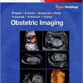
دانلود کتاب تصویربرداری زنان و زایمان: تشخیص و مراقبت از جنین + ویدئو
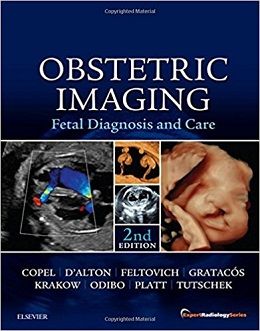
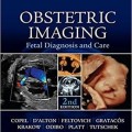
Obstetric Imaging: Fetal Diagnosis and Care, 2ed + Video
Richly illustrated and comprehensive in scope, Obstetric Imaging, 2nd Edition, provides up-to-date, authoritative guidelines for more than 200 obstetric conditions and procedures, keeping you at the forefront of this fast-changing field. This highly regarded reference covers the extensive and ongoing advances in maternal and fetal imaging in a concise, newly streamlined format for quicker access to common and uncommon findings. Detailed, expert guidance, accompanied by superb, high-quality images, helps you make the most of new technologies and advances in obstetric imaging.
- Features more than 1,350 high-quality images, including 400 in color, and over 85 videos online.
- Helps you select the best imaging approaches and effectively interpret your findings with a highly templated, bulleted, at-a-glance organization.
- Reflects all the latest developments in the field, including genetics, open fetal surgery, fetal echocardiography, Zika virus, and 3D imaging, so you can provide the safest and most responsive care to both mother and fetus.
- Includes new chapters on Limbs and Bones Overview; Open Fetal Surgery; Biophysical Profile; Ultrasound Physics; Elastography; Doppler; MRI; Echogenic Bowel; Pregnancy of Unknown Location (PUL), Failed Pregnancy and Ectopic Pregnancy, Cesarean Scar Pregnancy; Cytomegalovirus (CMG), Rubella, Toxoplasmosis, Herpes, Varicella; and Congenital Syphilis; plus a new chapter on Zika Virus written by imaging experts from the “hot zone.”
- Keeps you up to date with the latest developments in multimodality imaging and optimizing diagnostic accuracy from ultrasound, 3D ultrasound, Doppler, MRI, elastography, image-guided interventions, and much more.
- Expert Consult™ eBook version included with purchase. This enhanced eBook experience allows you to search all of the text, figures, Q&As, and references from the book on a variety of devices.
Contents
Part 1: Atlas of Selected Normal Images
Part 2: Thorax
Part 3: Retroperitoneum
Part 4: Abdomen
Part 5: Central Nervous System
Part 6: First-Trimester Complications
Part 7: Skeletal Dysplasias: An Overview
Part 8: Head and Neck
Part 9: Heart and Great Vessels
Part 10: Placenta and Cord
Part 11: Fetal Growth
Part 12: Procedures
Part 13: Miscellany
Part 14: Syndromes
Part 15: Chromosomes
Part 16: Multiple Gestation
Part 17: Multiple Gestation
Part 18: Multiple Gestation
Video Contents
۴.۱ Bilateral Fetal Pleural Effusion or Hydrothorax
۴.۲ Massive Fetal Pericardial Effusion
۲۰.۱ Color Doppler Image of the Umbilical Cord
۲۰.۲ Four-Dimensional Ultrasound of Fetal Torso With Gastroschisis
۲۱.۱ Omphalocele With Levorotated Heart
۲۴.۱ Partial Resection of Ovarian Cyst in a Neonate
۲۴.۲ Endoscopic Retrieval of a Detached Right Ovary With Hemorrhagic Cyst in a Neonate
۲۴.۳ Endoscopic Resection of Gastric Duplication in a Neonate
۲۴.۴ Normal Fetal Small Bowel Mesentery
۲۷.۱ Ultrasound of Isolated Left-Sided Congenital Diaphragmatic Hernia
۲۷.۲ Fetoscopic Removal of Tracheal Balloon in a Fetus With Isolated Congenital Diaphragmatic Hernia
۲۸.۱ Gray Scale Imaging of Hepatic Hemangioma
۲۸.۲ Power Doppler Imaging of a Hepatic Hemangioma
۳۲.۱ Double Vessel Sign in Left-Isomerism: Transverse View
۳۲.۲ Double Vessel Sign in Left-Isomerism: Sagittal View
۳۹.۱ Coronar Slices Through the Monocavity of the HPE-Brain in an 18-Week-Old Fetus
۳۹.۲ Approximately 13-Week-Old Fetus With Alobar HPE: Horizontal Slices Through the Brain
۳۹.۳ Approximately 13-Week-Old Fetus With Alobar HPE: Coronar Slices Through the Brain
۳۹.۴ Approximately 13-Week-Old Fetus With Alobar HPE: Coronar Slices Through the Brain
۶۵.۱ Three-Dimensional Multiplanar Mode of a Fetus With a Bilateral Cleft Lip and Palate at 21 Weeks’ Gestation
۶۸.۱ Agnathia
۶۸.۲ Three-Dimensional Reconstruction of a Fetus With Micrognathia
۶۸.۳ Four-Dimensional Reconstruction of a Fetus With Micrognathia
۶۹.۱ Otocephaly and Micrognathia
۷۱.۱ Unilocular, Encapsulated Complex Tumor of Fetal Neck
۷۱.۲ Primarily Solid Neck Tumor
۷۱.۳ Neck Teratoma
۷۴.۱ Normal Drainage of Superior Pulmonary Veins to the Left Atrium
۷۴.۲ Normal View of Right Ventricle, Pulmonary Valve, and Ductal Arch
۷۴.۳ Normal Ductal Arch
۷۴.۴ Normal Aortic Arch
۷۴.۵ Normal Aortic Arch
۷۴.۶ Drainage of the Superior and Inferior Vena Cava Into the Right Atrium
۷۴.۷ Presence of a Normal Ductus Venosus
۷۴.۸ Normal Heart Evaluated by Transvaginal Route
۷۴.۹ Transposition of Great Arteries
۷۴.۱۰ Examination of a Normal Heart by the Five Short-Axis Views
۷۵.۱ Small Apical Ventricular Septal Defects
۷۵.۲ Large Perimembranous Ventricular Septal Defects
۷۶.۱ Unbalanced Complete Atrioventricular Septal Defects
۷۶.۲ Unbalanced Complete Atrioventricular Septal Defects
۷۶.۳ Incomplete Atrioventricular Septal Defects
۷۶.۴ Incomplete Atrioventricular Septal Defects
۷۷.۱ Gray Scale Imaging of Tricuspid Atresia With Ventricular Septal Defect
۷۷.۲ Gray Scale and Color Doppler Imaging of Tricuspid Atresia With Ventricular Septal Defect
۷۷.۳ Tricuspid Atresia Without Ventricular Septal Defect
۷۸.۱ Gray Scale and Color Doppler Images of Ebstein Anomaly of Tricuspid Valve
۷۸.۲ Ebstein Anomaly of Tricuspid Valve
۷۹.۱ Fetus With Pulmonary Stenosis and Intact Interventricular Septum
۷۹.۲ Pulmonic Stenosis
۸۱.۱ Hypoplastic Left Heart Syndrome in a Case of Aortic Atresia
۸۲.۱ Fetal Aortic Coarctation
۸۳.۱ Fetus With Interrupted Aortic Arch
۸۴.۱ Right Aortic Arch
۸۴.۲ Aberrant Right Subclavian Artery
۸۵.۱ Tetralogy of Fallot
۸۵.۲ Tetralogy of Fallot/Absent Pulmonary Valve Syndrome
۸۶.۱ Transposition of the Great Arteries and an Intact Ventricular Septum
۸۶.۲ Corrected Transposition of the Great Vessels
۸۷.۱ Double-Outlet Right Ventricle With Subpulmonary Ventricular Septal Defect
۸۸.۱ Truncus Arteriosus
۸۹.۱ Four-Chamber and Out?ow Views of Single Ventricle With Transposition
۹۰.۱ Sweep of Abdomen and Thorax Showing Stomach on One Side and Apex of the Heart on the Opposite Side
۹۰.۲ Stomach on the Right Side and Apex of the Heart on the Opposite Side
۹۱.۱ Anomalous Pulmonary Venous Return
۹۲.۱ Extrahepatic Agenesis of the Ductus Venosus
۹۳.۱ Cardiomegaly With Dilated Myocardiopathy
۹۳.۲ Noncompacted Cardiomyopathy
۹۴.۱ Multiple Rhabdomyomata
۹۴.۲ Huge Pericardial Teratoma
۹۵.۱ Fetal Tachycardia
۹۵.۲ Fetal Tachycardia
۹۵.۳ Fetal Atrial Flutter With Variable Block
۹۵.۴ Fetal Complete Heart Block
۱۰۳.۱ Umbilical Vein Varix
۱۰۳.۲ Umbilical Vein Varix
۱۱۵.۱ Fetoscopic Selective Laser Photocoagulation
۱۱۵.۲ Fetoscopic View of Recipient Twin Within a Polyhydramniotic Sac
۱۱۵.۳ Fetoscopic View of a “Stuck” Donor Twin
۱۱۸.۱ Open Fetal Repair of a Myelomeningocele
۱۲۵.۱ Altered Fetal Breathing
۱۴۳.۱ Third Trimester Fetal Heart With Multiple Echogenic Rhabdomyomas
۱۶۳.۱ Ultrasound Cine of an Acardiac Fetus
۱۶۳.۲ Ultrasound Doppler Cine of an Acardiac Fetusxxvi Contents
۱۷۰.۱ of Shear Wave Displacement and Wave Front Propagation
۱۷۳.۱ Median Section of a Normal Fetal Trunk at 20 Weeks’ Gestation
۱۷۳.۲ Tomographic Imaging From a STIC Volume, Acquired With Power Doppler Information, of a 23-Week-Old Fetus With d-Transposition of the Great Arteries
۱۷۳.۳ Tomographic Imaging From a STIC Volume, Acquired With Power Doppler Information, of a Normal 23-Week-Old Fetus
۱۷۳.۴ Multiplanar Reconstruction and ThreeDimensional en face View of the Atrioventricular Valve Plane of a 24-WeekOld Fetus With Bradycardia
۱۷۳.۵ Three-Dimensional Power Doppler Angiogram (Reconstruction of the Normal Main Vessels) of the Fetal Trunk at 21 Weeks
۱۷۳.۶ Three-Dimensional Power Doppler Angiogram (Reconstruction of the Normal Main Vessels of the Fetal Trunk) at 21 Weeks
لینک کوتاه : https://bookbaz.ir/?p=77264
نویسنده : Joshua Copel MD , Mary E. D'Alton MD
ناشر : Elsevier; 2 edition
سال انتشار : 2018
زبان کتاب : انگلیسی
نوع فایل : MP4 + PDF (کیفیت اصلی)
تعداد صفحات : 1047
(ISBN) شابک : 0323445489
قیمت کتاب درآمازون : $275.24
حجم فایل : 780 MB


