دانلود کتاب قلب و عروق مداخله ای (ویرایش ۲۰۲۰) + ویدئو
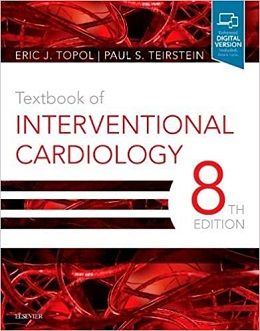
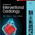
Textbook of Interventional Cardiology, 8ed + Video
From new research and equipment to new procedures, applications, and approaches, the field of interventional cardiology is one of the fastest-changing areas in medicine. Increasing data and recent technological advances have resulted in exciting changes – and an even greater need for cutting-edge, authoritative guidance on current practice. Textbook of Interventional Cardiology, 8th Edition, covers the theories, trends, and applications of diagnostic and interventional cardiology that cardiologists, cardiac surgeons, vascular surgeons, referring physicians, and advanced practitioners need to know.
- Focuses on the latest treatment protocols for managing patients at every level of complexity.
- Includes all-new chapters on Coronary Stenting, Diagnosis and Treatment of Coronary Microvascular Disease, Percutaneous Transcatheter Valve in Valve Implantation, and Percutaneous Tricuspid Valve Repair.
- Features hundreds of new illustrations, tables, and boxes for visual clarity and quick reference.
- Offers expanded coverage of transcatheter aortic valve interventions with extensive updates on practice implications.
- Discusses hot topics such new atherectomy devices, percutaneous mitral valve replacement, and percutaneous treatment of paravalvular leak.
- Provides the unique insights of expert leaders in the field who have pioneered today’s innovative devices and techniques and lend their own analysis of practical, evidence-based clinical applications.
- Presents the most recent data on how genomics and genetics impact interventional cardiology.
- Provides an in-depth understanding of cardiology, making it well suited for cardiology and interventional cardiology exam preparation.
Contents
۱ Individualized Assessment for Percutaneous or Surgical-Based Revascularization
۲ Evidence-Based Interventional Practice
۳ Diabetes
۴ Noninvasive Evaluation: Functional Testing,Multidetector Computed Tomography, and Stress Cardiac MRI
۵ Intracoronary Pressure and Flow Measurements
۶ Contrast-Induced Acute Kidney Injury and the Role of Chronic Kidney Disease in Percutaneous Coronary Intervention
۷ Radiation Safety During Cardiovascular Procedures
۸ Coronary and Valvular Intervention Prior to Noncardiac Surgery
۹ Sex and Ethnicity Issues in Interventional Cardiology
۱۰ Platelet Inhibitor Agents
۱۱ Anticoagulation in Percutaneous Coronary Intervention
۱۲ Thrombolytic Intervention
۱۳ Other Adjunctive Drugs for Coronary Intervention: Beta-Blockers, Calcium-Channel Blockers, and Angiotensin-Converting Enzyme Inhibitors
۱۴ Coronary Stenting: Practical Considerations, Equipment Selection, Tips and Caveats
۱۵ Coronary Stenting
۱۶ Bioresorbable Coronary Scaffolds
۱۷ Drug-Eluting Balloons
۱۸ Elective Intervention for Stable Angina or Silent Ischemia
۱۹ Intervention for Non-ST-Segment Elevation Acute Coronary Syndromes
۲۰ Percutaneous Coronary Intervention in Acute ST-Segment Elevation Myocardial Infarction
More…
۵۵ Percutaneous Transcatheter Valve-in-Valve Implantation
۵۶ Pulmonary Valve Interventions
۵۷ Percutaneous Tricuspid Valve Repair
۵۸ Hypertrophic Cardiomyopathy
۵۹ Percutaneous Balloon Pericardiotomy for Patients With Pericardial Effusion and Tamponade
۶۰ Transcatheter Therapies for Congenital Heart Disease
۶۱ Stem Cell Therapy for Ischemic Heart Disease
۶۲ Percutaneous Treatment of Paravalvular Leak
۶۳ Interventional Heart Failure
۶۴ Qualitative and Quantitative Coronary Angiography
۶۵ Intravascular Ultrasound
۶۶ The Dynamic Spectrum of Coronary Atheroma: From Atherogenesis to Vulnerable Plaque to Plaque Regression
۶۷ Optical Coherence Tomography
۶۸ Medical Economics in Interventional Cardiology
۶۹ Quality of Care in Interventional Cardiology
۷۰ Volume and Outcome
Video Contents
۱. Video 4.1 Resting AP3 view of the left ventricle revealing normal systolic function
۲. Video 4.2 Resting AP4 view.
۳. Video 4.3 Resting AP2 view.
۴. Video 4.3 Peak stress AP3 view of the left ventricle revealing marked apical hypokinesis, cavity dilatation, and decrease in left ventricular ejection fraction consistent with multivessel and/or left main coronary artery disease.
۵. Video 4.5 Peak stress AP4 view.
۶. Video 4.6 Peak stress AP2 view.
۷. Video 4.7 Recovery AP3 view of the left ventricle revealing improvement of the apical hypokinesis and near resolution of the cavity dilatation and decrease in left ventricular ejection fraction visualized at peak stress.
۸. Video 4.8 Recovery AP4 view.
۹. Video 4.9 Recovery AP2 view.
۱۰. Video 4.10 Coronary angiography revealing significant stenoses of the left main, proximal, and mid left anterior descending and proximal left circumflex coronary arteries.
۱۱. Video 4.11 LAO caudal view
۱۲. Video 4.12 Coronary angiography revealing a significant stenosis of the proximal right coronary artery which is a small vessel.
۱۳. Video 4.13 Cardiac magnetic resonance first pass perfusion imaging at rest in an AP2 view.
۱۴. Video 4.14 Cardiac magnetic resonance first pass perfusion imaging, post adenosine infusion in an AP2 view.
۱۵. Video 4.15 Steady state free precession cine imaging in an AP2 view revealing normal left ventricle systolic function.
۱۶. Video 31.1 A. Fluoroscopy in anteroposterior projection is used to ascertain the relative location of the femoral head.
۱۷. Video 32.1 Abnormal plcthysmooxymetric test.
۱۸. Video 32.2 Right radial puncture and sheath insertion.
۱۹. Video 32.3 Retrieval of catheter on a standard 0.035-inch guidewire.
۲۰. Video 32.4 Coronary catheterization through a lusoria subclavian artery with insufficient support.
More…
۵۰. Video 49.15 Release of the patent foramen ovale device.
۵۱. Video 49.16 Balloon-assisted technique for deployment of atrial septal defect Occluder #2.
۵۲. Video 49.17 Balloon-assisted technique for deployment of atrial septal defect Occluder #3,
۵۳. Video 49.18 Balloon-assisted technique for deployment of atrial septal defect Occluder #4,
۵۴. Video 51.1 Degenerative mitral stenosis on a severely calcified mitral annulus without commissural fusion.
۵۵. Video 51.2 Severe rheumatic mitral stenosis.
۵۶. Video 51.3 Severe rheumatic mitral stenosis after percutaneous mitral commissurotomy (same patient as Video 51.2).
۵۷. Video 51.4 Rheumatic mitral stenosis after percutaneous mitral commissurotomy.
۵۸. Video 51.5 Restenosis after percutaneous mitral commissurotomy with persistent opening of both commissures.
۵۹. Video 51.6 Restenosis after percutaneous mitral commissurotomy with persistent opening of both commissures (same patient as Video 51.5).
۶۰. Video 57.1 Transesophageal echocardiographic sweep at 0 degrees.
۶۱. Video 57.2 Transesophageal echocardiographic commissural view X-plane.
۶۲. Video 59.1 Large pericardial effusion.
۶۳. Video 60.1 Rotational angiogram with injection in the right ventricle.
۶۴. Video 60.2 Rotation angiogram in right ventricle following right pulmonary artery stent placement.
۶۵. Video 60.3 Rotational angiogram in ascending aorta following stent placement.
لینک کوتاه : https://bookbaz.ir/?p=162935
نویسنده : Eric J. Topol MD , Paul S. Teirstein MD
ناشر : Elsevier; 8 edition
سال انتشار : 0323568149
زبان کتاب : انگلیسی
نوع فایل : MP4 + PDF (کیفیت اصلی)
تعداد صفحات : 1390 بدون ایندکس
(ISBN) شابک : 0323568149
قیمت کتاب درآمازون : $190.95
حجم فایل : 320 MB


