
دانلود کتاب نورونولوژی و تصویربرداری عصبی سکته مغزی + ویدئو
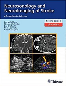
Neurosonology and Neuroimaging of Stroke, 2ed + Video
This is probably the best new and updated book available on the neurosonology and neuroimaging of stroke to add to your medical library. — BIZ INDIA
HIGHLY COMMENDED by the BMA Medical Book Awards for Neurology!
Neurosonology is a first-line modality in the diagnosis and management of cerebrovascular disease and especially of stroke. In this new edition of Neurosonology and Neuroimaging of Stroke, this noninvasive, realtime imaging method has been given expanded coverage, particularly for its clinical utility.
As in the first edition, the new edition offers both a clear overview of the principles of neurosonology and a casebook exploring critical cerebrovascular problems. Ultrasound anatomy, technical aspects of clinical application, and the advantages and limitations of ultrasound are reviewed and contrasted to conventional, magnetic resonance, and computed tomography angiography. Forty-five selected cases from the authors’ extensive collections at Charite – Universitatsmedizin Berlin and the Center of Neurology in Bad Segeberg, Germany, are then discussed. The patient histories and working diagnoses are followed by detailed assessments of the extra- and intracranial color-coded duplex sonographic findings and additional diagnostic procedures. The relevant clinical aspects are presented in a compact, comprehensible way, and for the majority of the cases videos are available in the Thieme MediaCenter, providing further in-depth understanding of the full potential of the method.
Features:
- Complete extra- and intracranial arterial and venous ultrasound examination
- New techniques: ultrasound fusion imaging, ultrafast ultrasound, contrast application
- More than 1,300 high-quality illustrations, including full-color duplex images
- Fifteen newly selected cases on conditions such as subarachnoid hemorrhage and dural fistula, as well as rare stroke causes including sickle cell disease and reversible cerebral vasoconstriction syndrome
- Revision of many cases from the first edition
- More than 60 new video clips (for a total of 130) available at the Thieme MediaCenter, bringing ultrasound anatomy and challenging cases to your monitor!
Neurosonology and Neuroimaging of Stroke, Second Edition, offers neurologists, neuroradiologists, and all physicians treating patients with cerebrovascular disease an authoritative introduction and guide to this powerful diagnostic tool.
Contents
Chapter 1: Flow and Ultrasound Basics
Chapter 2: Vascular Anatomy and Structure of Ultrasound Examination
Chapter 3: Intracranial Hemodynamics and Functional Tests
Chapter 4: Pathogenesis of Stroke
Chapter 5: Vascular Pathology
Chapter 6: Angiographic Techniques in Neuroradiology
Case 1: Right Extracranial Internal Carotid Artery Stenosis
Case 2: Free-floating Thrombus of the Left Internal Carotid Artery
Case 3: Left Common Carotid Artery Occlusion
Case 4: Left Temporal Arteriovenous Malformation
Case 5: Left Ml Middle Cerebral Artery Stenosis
Case 6: Left P2 Posterior Cerebral Artery Stenosis
Case 7: Cerebral Circulatory Arrest
Case 8: Basilar Artery Occlusion in Bilateral Intracranial V4 Vertebral Artery Stenosis
Case 9: Moyamoya Disease
Case 10: Thrombolysis of Ml Middle Cerebral Artery Occlusion
Case 11: Secondary Occlusion in Left-sided Extracranial Internal Carotid Artery Dissection
Case 12: Extracranial Bilateral Internal Carotid Artery and Right Vertebral Artery Occlusion, and Left Vertebral Artery Stenosis
Case 13: Right Internal Carotid Artery Stenosis in Fibromuscular Dysplasia and Granulomatosis with Polyangiitis (formerly Wegener’s Granulomatosis)
Case 14: Isolated Left Carotid Siphon Stenosis
Case 15: Near-occlusion of the Right and High-grade Stenosis of the Left Extracranial Internal Carotid Artery
Case 16: Giant Cell Arteritis with Bilateral Intracranial V4 Vertebral Artery Stenosis
Case 17: Ascending Left Middle Cerebral Artery Occlusion in an HIV-positive Patient
Case 18: Traumatic Bilateral Internal Carotid and Vertebral Artery Dissection with Right-sided Embolic Middle Cerebral Artery Occlusion
Case 19: Bilateral Extracranial Vertebral Artery Dissection with Distal Occlusion of the Right Vertebral Artery
Case 20: Right Internal Carotid Artery Dissection with Fast Recanalization
Case 21: Mid-basilar Artery Occlusion Due to Intracranial Dissection
Case 22: Right Mid-part Ml Middle Cerebral Artery Occlusion with Prominent Early Temporal Branch and Patent Foramen Ovale
Case 23: Takayasu’s Arteritis with Right-sided Subclavian Steal
Case 24: Dissection of the Right Extracranial Internal Carotid Artery and Left Ml Middle Cerebral Artery
Case 25: Progressive Right Ml Middle Cerebral Artery Occlusion Treated with Extracranial-Intracranial Bypass Surgery
Case 26: Extracranial Left Vertebral Artery Dissecting Aneurysm Following Basilar Artery Stenting
Case 27: Diffuse Cerebral Angiomatosis
Case 28: Subclavian Steal in Left Subclavian Artery and Right Internal Carotid Artery Occlusion Leading to Extracranial-Intracranial Bypass Surgery
Case 29: Cerebral Venous Thrombosis
Case 30: Muttilocular Extra- and Intracranial Stenoses and Occlusions
Case 31: Dissection of the Left Internal Carotid Artery C6 Segment
Case 32: Right Temporal Hemorrhage in Pial Arteriovenous Malformation
Case 33: Subarachnoid Hemorrhage after Rupture of Left Supraophthalmic Internal Carotid Artery Aneurysm
Case 34: Right-sided Occipital Dural Arteriovenous Fistula
Case 35: Left Distal Vertebral Artery Occlusion and Right Vertebral Artery Hypoplasia with Retrograde Basilar Artery Flow
Case 36: Postpartum Angiopathy (Reversible Cerebral Vasoconstriction Syndrome)
Case 37: Left Anterior Choroidal Artery Infarction in Left Supraophthalmic Internal Carotid Artery Occlusion
Case 38: Left-sided Amaurosis in Left Central Retinal Artery Occlusion
Case 39: Combined Chronic Right-sided Extracranial Internal Carotid Artery and Middle Cerebral Artery Occlusion
Case 40: Left Internal Carotid Artery Occlusion and Ipsilateral Arteriovenous Malformation in Inflammatory Bowel Disease
Case 41: Right Internal Carotid Artery as the Remaining Patent Brain-supplying Artery
Case 42: Left Internal Carotid Artery Aplasia as an Incidental Diagnosis in Optic Neuritis in Lupus Erythematosus
Case 43: Sickle Cell Disease
Case 44: Transient Global Amnesia with Left Hippocampal Diffusion-weighted Lesion and Asymptomatic Right Middle Cerebral Artery Infarction in High-grade Right Ml Stenosis
Case 45: Bilateral Proximal Vertebral Artery Stenosis and Bilateral Middle Cerebral Artery Aneurysm
Video Contents
Video A1.1: Ultrasound perfusion imaging of a healthy person
Video A1.2: Ultrasound perfusion imaging in old middle cerebral artery territorial infarction
Video A2.1: Common, internal, and external carotid arteries during tapping of the superficial temporal artery
Video A2.2: Common, internal, and external carotid arteries and superior thyroid artery
Video A2.3: External carotid artery and lingual artery during functional testing
Video A2.4: Superficial temporal artery and occipital artery
Video A2.5: Axillary artery and brachial artery during functional testing
Video A2.6: From the common carotid artery to the V2 segment of the vertebral artery
Video A2.7: Common carotid artery and vertebral artery
Video A2.8: From the V2 segment of the vertebral artery to the subclavian artery
Video A2.9: From the subclavian artery to the V3 segment of the vertebral artery
Video A2.10: Application of echo-contrast agent to improve the transtemporal insonation quality
Video A2.11: Overview of arteries in the five axial insonation planes (transtemporal insonation)
Video A2.12: Overview of the five axial insonation planes (transtemporal insonation, fusion imaging with MRI)
Video A2.13: Overview of arteries in the five axial insonation planes (transtemporal insonation, fusion imaging with MRI)
Video A2.14: Overview of arteries in the anterior and posterior coronal planes (transtemporal insonation)
Video A2.15: Transtemporal axial insonation of the internal carotid artery (proximal and distal segments)
Video A2.16: Transtemporal coronal insonation focusing on the internal carotid artery, middle cerebral artery, and basilar artery (fusion imaging with MRI)
Video A2.17: Middle meningeal artery and C6 segment of the internal carotid artery
Video A2.18: Internal carotid artery siphon and ophthalmic artery at rest and during digital tapping of the bulb
Video A2.19: Ophthalmic artery
Video A2.20: Optic nerve sheath, central retinal artery and vein
Video A2.21: Midbrain plane (fusion imaging with MRI)
Video A2.22: Middle cerebral artery (from M1 to M3)
Video A2.23: Middle cerebral artery and posterior cerebral artery (fusion imaging with MRI)
Video A2.24: Internal carotid artery, middle cerebral artery, and anterior cerebral artery
And More…
۱۲۹ Videos
لینک کوتاه : https://bookbaz.ir/?p=97735
نویسنده : José Manuel Valdueza , Stephan Schreiber
ناشر : Thieme; 2nd edition
سال انتشار : 2017
زبان کتاب : انگلیسی
نوع فایل : MP4 + PDF (کیفیت اصلی)
تعداد صفحات : 632
(ISBN) شابک : 3131418729
قیمت کتاب درآمازون : $224.99
حجم فایل : 780 MB
کتاب های مرتبط:
 دانلود کتاب اختلالات خواب آکسفورد
Oxford Textbook of Sleep Disorders, 1ed
 دانلود کتاب اکوکاردیوگرافی عملی نوزاد
Practical Neonatal Echocardiography, 1ed
 دانلود کتاب آناتومی بالینی، بافت شناسی، جنین شناسی و نوروآناتومی
Clinical Anatomy Histology Embryology and Neuroanatomy, 1ed
 دانلود کتاب آسیب مغزی جنین و نوزاد
Fetal and Neonatal Brain Injury, 5ed
 دانلود کتاب فیزیک تصویربرداری پزشکی هندی
Hendee’s Physics of Medical Imaging, 5ed
 دانلود کتاب ناتوانی عصبی و سلامت کودک جامعه آکسفورد
Neurodisability and Community Child Health, 2ed
 دانلود کتاب سیستم شنوایی: آناتومی، فیزیولوژی و همبستگی بالینی
The Auditory System: Anatomy, Physiology, and Clinical Correlates, 2ed
 دانلود کتاب مشکلات رایج در بیماریهای مولتیپل اسکلروزیس و میلین زدا CNS
Common Pitfalls in Multiple Sclerosis and CNS Demyelinating Diseases, 1ed
 دانلود کتاب تشخیص افتراقی در تصویربرداری عصبی: ستون فقرات
Differential Diagnosis in Neuroimaging: Spine, 1ed
 دانلود کتاب سرطان پستان: تشخیص زودرس با ماموگرافی
Breast Cancer: Early Detection with Mammography: Casting Type Calcifications Sign of a Subtype with Deceptive Features, 1ed
 دانلود کتاب SPECT و SPECT/CT: راهنمای بالینی
SPECT and SPECT/CT: A Clinical Guide, 1ed
 دانلود کتاب نورونولوژی و تصویربرداری عصبی سکته مغزی
Neurosonology and Neuroimaging of Stroke, 2ed
 دانلود کتاب پرسش و پاسخ تصویربرداری گوارشی رادکِیس
RadCases Plus Q&A Gastrointestinal Imaging, 2ed
 دانلود کتاب تکنیک های اصلی در جراحی مغز و اعصاب عملی + ویدئو
Core Techniques in Operative Neurosurgery, 2ed + Video
 دانلود کتاب راهنمای پایندگی چشم پزشکی عصبی
The Neuro-Ophthalmology Survival Guide, 2ed
 دانلود کتاب تصویربرداری اسکلتی عضلانی
Musculoskeletal Imaging, 2ed
 دانلود مجموعه ویدئویی Medmastery’s Ultrasound 2023
 دانلود کتاب راهنمای مطالعات اجرایی عصب بوشباچر
Buschbacher’s Manual of Nerve Conduction Studies, 3ed
 دانلود کتاب EMG آسان: راهنمای انجام مطالعات هدایت عصبی و الکترومیوگرافی
Easy EMG: A Guide to Performing Nerve Conduction Studies and Electromyography, 3ed
 دانلود کتاب مغز انسان نولته: مقدمه ای بر آناتومی عملکردی آن
Nolte’s The Human Brain: An Introduction to its Functional Anatomy, 8ed
 دانلود کتاب عصب شناسی مریت
Merritt’s Neurology, 13ed
 دانلود کتاب هفت بایپس: اصول و تکنیک های ریواسکولاریزیشن
 Seven Bypasses: Tenets and Techniques for Revascularization, 1ed
 دانلود کتاب تصویربرداری پراش اشعه ایکس: فناوری و کاربردها
X-Ray Diffraction Imaging: Technology and Applications, 1ed
 دانلود کتاب راهنمای سمیت عصبی
Handbook of Neurotoxicity, 2ed
 دانلود کتاب تصویربرداری رزونانس مغناطیسی (۳ جلدی)
Magnetic Resonance Imaging Handbook (MRI), 3-Vol, 1ed
 دانلود کتاب آرتروسکوپی پیشرفته AANA: شانه، آرنج و مچ دست، ران، زانو، پا و مچ پا (۵ جلدی)
AANA Advanced Arthroscopy: Shoulder, Elbow and Wrist, Hip, Knee, Foot and Ankle, 5-Vol, 1ed
 دانلود کتاب موارد تصویربرداری اسکلتی عضلانی تهران زاده
Musculoskeletal Imaging Cases (McGraw-Hill Radiology), 1ed
 دانلود کتاب متاستازهای مغز از تومورهای اولیه (۳ جلدی)
Brain Metastases from Primary Tumors, 3-Vol, 1ed
 دانلود کتاب اطلس ویدیویی پایش نوروفیزیولوژیک در جراحی تومورهای نفوذی مغز + ویدئو
Video Atlas of Neurophysiological Monitoring in Surgery of Infiltrating Brain Tumors, 1ed + Video
 دانلود کتاب معاینه بالینی عصبی
Neurological Clinical Examination, 4ed
 دانلود کتاب پرینت سه بعدی برای رادیولوژیست
۳D Printing for the Radiologist, 1ed
 دانلود کتاب خون رسانی مجدد مغزی: تکنیک های جراحی بایپس در خارج به داخل جمجمه
Cerebral Revascularization: Techniques in Extracranial-to-Intracranial Bypass Surgery, 1ed
 دانلود کتاب دیسپلازی استخوان: اطلس اختلالات ژنتیکی رشد اسکلت
Bone Dysplasias: An Atlas of Genetic Disorders of Skeletal Development, 4ed
 دانلود کتاب چشم پزشکی عصبی مصور + ویدئو
Neuro-Ophthalmology Illustrated, 3ed + Video
 دانلود کتاب ۳ تفاوت برتر در رادیولوژی عروقی و مداخله ای
 Top 3 Differentials in Vascular and Interventional Radiology, 1ed
 دانلود کتاب اطلس ویدئویی روشهای نورواندوواسکولار
 Video Atlas of Neuroendovascular Procedures, 1ed
 دانلود کتاب راهنمای تصویربرداری عصبی نورو-انکولوژی
Handbook of Neuro-Oncology Neuroimaging, 2ed
 دانلود کتاب تصویربرداری سر و گردن (سری موارد رادیولوژی)
RadCases Head and Neck Imaging, 1ed
 دانلود کتاب اطلس ویدئویی جراحی ستون فقرات
 Video Atlas of Spine Surgery, 1ed
 دانلود کتاب راهنمای الکتروفیزیولوژی کلینیک مایو
Mayo Clinic Electrophysiology Manual, 1ed
 دانلود کتاب مرور بورد مراقبت های حاد و عصبی کلینیک مایو
Mayo Clinic Critical and Neurocritical Care Board Review, 1ed
 دانلود کتاب بررسی موردی رادیولوژی: ستون فقرات
Radiology Case Review Series: Spine, 1ed
 دانلود کتاب اولتراسوند تشخیصی اسکلتی عضلانی و راهنمای تزریق
Diagnostic Musculoskeletal Ultrasound and Guided Injection, 1ed
 دانلود کتاب اطلس ویدئویی جراحی آنوریسم داخل جمجمه
Video Atlas of Intracranial Aneurysm Surgery, 1ed
 دانلود کتاب بیومگنتیک ها: اصول و برنامه های برانگیختن بیومگنتیک و تصویربرداری
Biomagnetics: Principles and Applications of Biomagnetic Stimulation and Imaging, 1ed
 دانلود کتاب اولتراسوند دست و اندام فوقانی + ویدئو
Ultrasound of the Hand and Upper Extremity, 1ed + Video
 دانلود کتاب مشاهدات بحرانی در رادیولوژی برای دانشجویان پزشکی
Critical Observations in Radiology for Medical Students, 1ed
 دانلود کتاب کتاب مبانی فیزیک تصویربرداری و رادیوبیولوژی سلمان
Selman’s The Fundamentals of Imaging Physics and Radiobiology, 10ed
 دانلود کتاب خود ارزیابی مغز و اعصاب: همگام با مغز و اعصاب برادلی
Neurology Self-Assessment: A Companion to Bradley’s Neurology in Clinical Practice, 1ed
 دانلود کتاب مرور پرسش و پاسخ اولتراسوند برای بورد
Ultrasound Q&A Review for the Boards, 1ed
 دانلود کتاب هدایت حین عمل MRI جراحی مغز و اعصاب
Intraoperative MRI-Guided Neurosurgery, 1ed
 دانلود کتاب سیستم عصبی یکپارچه: روش تشخیص سیستماتیک مبتنی بر مورد
The Integrated Nervous System: A Systematic Diagnostic Case-Based Approach, 2ed
 دانلود کتاب آسیب های مغزی تروماتیک: روش های ارزیابی بالینی و قانونی اعصاب روان
Traumatic Brain Injury: Methods for Clinical and Forensic Neuropsychiatric Assessment, 3ed
 دانلود کتاب تکنیک های جراحی مغز و اعصاب اشمیدک و سویت (۲ جلدی) + ویدئو
Schmidek and Sweet Operative Neurosurgical Techniques, 2-Vol, 7ed + Video
 دانلود کتاب تصویربرداری در جراحی شکم
Imaging in Abdominal Surgery, 1ed
 دانلود کتاب تصویربرداری در گوارش
Imaging in Gastroenterology, 1ed
 دانلود کتاب اصول سی تی اسکن بدن
Fundamentals of Body CT, 5ed
 دانلود کتاب تصویربرداری برای دانشجویان
Imaging for Students, 4ed
 دانلود کتاب تصویربرداری رزونانس مغناطیسی میکروسکوپی
Microscopic Magnetic Resonance Imaging, 1ed
 دانلود کتاب کشیک مغز و اعصاب
 On Call Neurology, 4ed
 دانلود کتاب تصویربرداری تشخیصی: مغز
Diagnostic Imaging: Brain, 3ed
دانلود کتاب اطلس ویدیویی پایش نوروفیزیولوژیک در جراحی تومورهای نفوذی مغزVideo Atlas of Neurophysiological Monitoring in Surgery of Infiltrating Brain Tumors, 1ed
 دانلود کتاب اصول و تمرین پزشکی خواب (۲ جلدی) + ویدئو
Principles and Practice of Sleep Medicine, 2-Vol, 7ed + Video
 دانلود کتاب سری کارشناسی ستون فقرات AO: تغییر شکل ستون فقرات بزرگسالان (جلد ۴)
AOSpine Masters Series, Volume 4: Adult Spinal Deformities, 1ed
 دانلود کتاب اعصاب جمجمه: آناتومی، پاتولوژی، تصویربرداری
Cranial Nerves: Anatomy, Pathology, Imaging, 1ed
 دانلود کتاب نوروبیونیک ها: مهندسی پزشکی پروتز عصبی
Neurobionics: The Biomedical Engineering of Neural Prostheses, 1ed
 دانلود کتاب تصویربرداری از مغز با MRI و CT: رویکرد الگوی تصویری
Brain Imaging with MRI and CT: An Image Pattern Approach, 1ed
 دانلود کتاب موارد تصویربرداری پستان
Breast Imaging Cases
 دانلود کتاب تصویربرداری تشخیصی: پزشکی زایمان
Diagnostic Imaging: Obstetrics, 3ed
 دانلود کتاب ملزومات سونوگرافی شکمی و لگنی
Essentials of Abdomino-Pelvic Sonography, 1ed
 دانلود کتاب تفسیر رادیوگرافی قفسه سینه در بیماران قلبی کودک
Chest Radiographic Interpretation in Pediatric Cardiac Patients, 1ed
 دانلود کتاب تصویربرداری تشخیصی کودکان کافی (ویرایش ۲۰۱۹، ۲ جلدی)
Caffey’s Pediatric Diagnostic Imaging, 2-Volume Set, 13ed
 دانلود کتاب اطلس بیهوشی منطقه ای هدایت اولتراسوند + ویدئو
Atlas of Ultrasound-Guided Regional Anesthesia, 3ed + Video
 دانلود کتاب آناتومی تصویربرداری: سر و گردن
Imaging Anatomy: Head and Neck, 1ed
 دانلود کتاب تصویربرداری تشخیصی دستگاه گوارش
Diagnostic Imaging: Gastrointestinal, 4ed
 دانلود کتاب تصویربرداری تشخیصی زایمان
Diagnostic Imaging: Obstetrics, 4ed
 دانلود کتاب تصویربرداری تشخیصی سر و گردن
Diagnostic Imaging: Head and Neck, 4ed
 دانلود کتاب تصویربرداری تشخیصی انکولوژی
Diagnostic Imaging: Oncology, 2ed
 دانلود کتاب راهنمای جراحی مغز و اعصاب کودکان
Handbook of Pediatric Neurosurgery, 1ed
 دانلود کتاب تروما اعصاب و مراقبت بحرانی از ستون فقرات
 Neurotrauma and Critical Care of the Spine, 2ed
 دانلود کتاب تصویربرداری قلب و عروق ESC
The ESC Textbook of Cardiovascular Imaging, 3ed
 دانلود کتاب MRI در یک نگاه
MRI at a Glance, 2ed
 دانلود کتاب صدمات عصب کلاین و هادسون: صدمات عصب، انترپمنت و تومور
Kline and Hudson’s Nerve Injuries: Operative Results for Major Nerve Injuries, Entrapments and Tumors, 2ed
 دانلود کتاب سونوگرافی مداخله ای: راهنمای عملی و اطلس
Interventional Ultrasound: A Practical Guide and Atlas, 1ed
 دانلود کتاب فیزیک ضروری در تصویربرداری برای پرتونگار کلارک
Clark’s Essential Physics in Imaging for Radiographers, 1ed
 دانلود کتاب تصویربرداری ادراری تناسلی (سری موارد رادیولوژی)
Genitourinary Imaging (Radcase), 1ed
 دانلود کتاب تشخیص افتراقی در تصویربرداری سونوگرافی
 Differential Diagnosis in Ultrasound Imaging, 2ed
 دانلود کتاب اطلس درمان عصبی با بیحسی موضعی
 Atlas of Neural Therapy With Local Anesthetics, 3ed
 دانلود کتاب به دام افتادن اعصاب محیطی: تشخیص و مدیریت بالینی
Peripheral Nerve Entrapments: Clinical Diagnosis and Management, 1ed
 دانلود کتاب تصویربرداری تشخیصی: زنان و زایمان
Diagnostic Imaging: Gynecology, 2ed
 دانلود کتاب تصویربرداری زنان و زایمان: تشخیص و مراقبت از جنین
Obstetric Imaging: Fetal Diagnosis and Care, 2ed
 دانلود کتاب اندوسکوپی ترانس نازال قاعده جمجمه و جراحی مغز
Transnasal Endoscopic Skull Base and Brain Surgery, 1ed
 دانلود کتاب نوروپاتولوژی تکاملی
Developmental Neuropathology, 2ed
 دانلود کتاب موقعیت در رادیوگرافی کلارک
Clark’s Positioning in Radiography, 13ed
 دانلود کتاب مدیریت جامع شوانوما وستیبولار
 Comprehensive Management of Vestibular Schwannoma, 1ed
 دانلود کتاب تصویربرداری تشخیصی قفسه سینه
Diagnostic Imaging: Chest, 3ed
 دانلود کتاب تصویربرداری تشخیصی زنان
Diagnostic Imaging: Gynecology, 3ed
 دانلود کتاب آنژیوگرافی مغزی کاربردی: آناتومی و پاتولوژی عروقی نرمال
Applied Cerebral Angiography: Normal Anatomy and Vascular Pathology, 1ed
 دانلود کتاب تصویربرداری قفسه سینه و قلب و عروق
Chest and Cardiovascular Imaging, 3ed
 دانلود کتاب کالبدشناسی اعصاب بالینی واکسمن
Clinical Neuroanatomy, 27ed
 دانلود کتاب روش های افتراقی در میکروجراحی مغز سیگر
Differential Approaches in Microsurgery of the Brain, 1ed
 دانلود کتاب تشخیص افتراقی در توموگرافی کامپیوتری
Differential Diagnosis in Computed Tomography, 2ed
 دانلود کتاب آناتومی تصویربرداری: قفسه سینه، شکم، لگن
Imaging Anatomy: Chest, Abdomen, Pelvis, 2ed
 دانلود کتاب رادیولوژی اضطراری (سری موارد رادیولوژی)
Radcases Emergency Radiology, 1ed
 دانلود کتاب اطلس سر، گردن و مغز روتون: تصاویر دو بعدی و سه بعدی
Rhoton’s Atlas of Head, Neck, and Brain: 2D and 3D Images, 1ed
 دانلود کتاب اطلس توپوگرافی و پاتوتوپوگرافی آناتومی سر و گردن
Atlas of Topographical and Pathotopographical Anatomy of the Head and Neck, 1ed
 دانلود کتاب رادیولوژی اعصاب تشخیصی و مداخله ای
Diagnostic and Interventional Neuroradiology, 1ed
 دانلود کتاب اولتراسوند: مرور کلی
Ultrasound: A Core Review, 1ed
 دانلود کتاب اختلالات مرتبط با ورزش: تشخیص و مدیریت
Sports-Related Concussion: Diagnosis and Management, 2ed
 دانلود کتاب درمان های مولکولی و سلولی برای بیماری های حرکتی نورون
Molecular and Cellular Therapies for Motor Neuron Diseases, 1ed
 دانلود کتاب مجموعه تصاویر پزشکی نتر: سیستم اسکلتی عضلانی (جلد ششم، ۳ جلدی)
The Netter Collection of Medical Illustrations: Musculoskeletal System Package, 2ed
 دانلود کتاب اطلس جیبی آناتومی مقطعی: ستون فقرات، اندام، مفاصل
 Pocket Atlas of Sectional Anatomy: Spine Extremities Joints, 3ed
 دانلود کتاب یادگیری رادیولوژی هِرینگ: شناخت مبانی
Learning Radiology: Recognizing the Basics, 3ed
 دانلود کتاب مغز و اعصاب کودکان
 Handbook of Pediatric Neurology, 1ed
 دانلود کتاب رادیولوژی ۱۰۱: مبانی و اصول تصویربرداری
Radiology 101: The Basics & Fundamentals of Imaging, 4ed
 دانلود کتاب تصویربرداری عمومی اسکلتی عضلانی
Basic Musculoskeletal Imaging, 1ed
 دانلود کتاب داروها در عصب شناسی
 Drugs in Neurology, 1ed
 دانلود کتاب اندوسونوگرافی
 Endosonography, 4ed
 دانلود کتاب تعویض ترنس کاتتر دریچه آئورت
Transcatheter Aortic Valve Implantation, 1ed
دانلود کتاب تکنیک های اصلی در جراحی مغز و اعصاب عملیCore Techniques in Operative Neurosurgery, 2ed
 دانلود کتاب پرسش و پاسخ پزشکی هسته ای رادکِیس
RadCases Plus Q&A Nuclear Medicine, 2ed
 دانلود کتاب تصویربرداری بیماری های بینابینی ریه (سری رادیولوژی کلینیکو)
Clinico Radiological Series: Imaging of Interstitial Lung Diseases, 1ed
 دانلود کتاب اطلس آموزشی تصویربرداری شکم
Teaching Atlas of Abdominal Imaging, 1ed
 دانلود کتاب اطلس رنگی خون رسانی مجدد مغز: آناتومی، تکنیک، موارد بالینی
Color Atlas of Cerebral Revascularization: Anatomy, Techniques, Clinical Cases, 1ed
 دانلود کتاب توموگرافی پرتو مخروطی کامپیوتری در ارتودنسی: موارد مصرف، بینش و نوآوری
Cone Beam Computed Tomography in Orthodontics: Indications, Insights, and Innovations, 1ed
 دانلود کتاب تصویربرداری پزشکی آکسفورد
Oxford Handbook of Medical Imaging, 1ed
 دانلود کتاب رژیم غذایی کتوژنیک و متابولیک درمانی: گسترش نقش در سلامت و بیماری
Ketogenic Diet and Metabolic Therapies: Expanded Roles in Health and Disease, 1ed
 دانلود کتاب سری مرور موردی رادیولوژی: تصویربرداری قفسه سینه
 Radiology Case Review Series: Thoracic Imaging, 1ed
 دانلود کتاب راهنمای رادیو مابولیزاسیون: فیزیک، بیولوژی، پزشکی هسته ای و تصویربرداری
Handbook of Radioembolization: Physics, Biology, Nuclear Medicine, and Imaging, 1ed
 دانلود کتاب تعمیرات X-Ray: راهنمای جامع نصب و سرویس تجهیزات رادیوگرافی
X-Ray Repair: A Comprehensive Guide to the Installation and Servicing of Radiographic Equipment, 3ed
 دانلود کتاب دوز، فواید و خطر در تصویربرداری پزشکی
 Dose, Benefit, and Risk in Medical Imaging, 1ed
 دانلود کتاب جامع فیزیک پزشکی (۱۰ جلدی)
Comprehensive Biomedical Physics, 10-Vol, 1ed
 دانلود کتاب ۳ تفاوت برتر در رادیولوژی مغز و اعصاب
 Top 3 Differentials in Neuroradiology, 1ed
 دانلود کتاب مرور بالینی مغز و اعصاب مبتنی بر تصویر
Neurology Image-Based Clinical Review, 1ed
 دانلود کتاب چشم انداز محاسباتی و پردازش تصویری پزشکی
 Computational Vision and Medical Image Processing V: VipIMAGE 2015
 دانلود کتاب سری بررسی موردی رادیولوژی: تصویربرداری پستان
 Radiology Case Review Series: Breast Imaging, 1ed
 دانلود کتاب تمرین مراقبت نورولوژیک
 The Practice of Neurocritical Care, 1ed
 دانلود کتاب رادیوگرافی PREP (مرور برنامه و آمادگی آزمون)
Radiography PREP (Program Review and Exam Preparation), 8ed
 دانلود کتاب CT قلبی بالینی: آناتومی و عملکرد + ویدئو
Clinical Cardiac CT: Anatomy and Function, 2ed + Video
 دانلود کتاب اصول علوم اعصاب برای علوم و اختلالات ارتباطاتی
Neuroscience Fundamentals for Communication Sciences and Disorders, 1ed
 دانلود کتاب سونوگرافی کودکان
 Pediatric Ultrasound, 1ed
 دانلود کتاب روشهای اسکلتی عضلانی هدایت شده با سونوگرافی در پزشکی ورزشی
Ultrasound Guided Musculoskeletal Procedures in Sports Medicine, 1ed
 دانلود کتاب مانیتورینگ مراقبت حاد مغز و اعصاب
Neurocritical Care Monitoring, 1ed
 دانلود کتاب MRI بافتها همراه با T2 یا T2*s کوتاه
MRI of Tissues with Short T2s or T2*s, 1ed
 دانلود کتاب تصویربرداری تومور قفسه سینه (سری رادیولوژی کلینیکو)
Clinico Radiological Series: Imaging Of Chest Tumors, 1ed
 دانلود کتاب توسعه دیجیتال ریه: از اولین سی تی ریه تا هوش مصنوعی بالینی
Developing the Digital Lung: From First Lung CT to Clinical AI, 1ed
 دانلود کتاب سونوگرافی مراقبت اورژانسی + ویدئو
Emergency Point-of-Care Ultrasound, 2ed + Video
 دانلود کتاب تصویربرداری از استخوان ها و مفاصل: یک روش مختصر و چند وجهی
Imaging of Bones and Joints: A Concise, Multimodality Approach, 1ed
 دانلود کتاب تصویربرداری و تجسم در اتاق عمل مدرن: راهنمای جامع برای پزشکان
Imaging and Visualization in The Modern Operating Room: A Comprehensive Guide for Physicians, 2015th
 دانلود کتاب آناتومی مقطعی برای متخصصان تصویربرداری
 Sectional Anatomy for Imaging Professionals, 3ed