
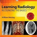
دانلود کتاب یادگیری رادیولوژی: شناخت اصول + ویدئو
Learning Radiology: Recognizing the Basics, 4ed + Video
The leading introductory radiology text for medical students and others who are required to read and interpret common radiologic images, Learning Radiology, 4th Edition, stresses an easy-to-follow pattern recognition approach that teaches how to differentiate normal and abnormal images. Dr. William Herring’s clear, conversational writing style employs a touch of humor to explain what you need to know to effectively interpret medical images of all modalities. From the basics of patient safety, dose reduction, and radiation protection to the latest information on ultrasound, MRI, and CT, this concise, user-friendly text provides a complete, up-to-date introduction to radiology needed by today’s students.
- Teaches how to arrive at a diagnosis by following a pattern recognition approach, and logically overcome difficult diagnostic challenges with the aid of decision trees.
- Features an easy-to-read bulleted format, high-quality illustrations, useful tables, and teaching boxes, as well as special content on Diagnostic Ptifalls; Really Important Points; Weblinks; and Take-Home Points.
- Includes three new chapters: Vascular, Pediatric, and Point-of-Care Ultrasound; Using Image-Guided Interventions in Diagnosis and Treatment (Interventional Radiology); Recognizing the Imaging Findings of Breast Disease.
- Helps ensure mastery of the material with additional online content, bonus images, and USMLE-style Q&A that provide effective chapter review and quick practice for your exams.
- Shares the extensive knowledge and experience of esteemed author Dr. William Herring―a skilled radiology teacher and the host of his own specialty website, www.learningradiology.com.
- Offers quick review and instruction for medical students, residents, and fellows, as well as those in related fields such as nurse practitioners and physician assistants.
- Includes an Enhanced eBook version with purchase. Your enhanced eBook allows you to access all of the text, figures, and references from the book on a variety of devices.
Contents
۱ Recognizing Anything
۲ Recognizing a Technically Adequate Chest Radiograph
۳ Recognizing Normal Pulmonary Anatomy
۴ Recognizing Normal Cardiac Anatomy
۵ Recognizing Airspace Versus Interstitial Lung Disease
۶ Recognizing the Causes of an Opacified Hemithorax
۷ Recognizing Atelectasis
۸ Recognizing a Pleural Effusion
۹ Recognizing Pneumonia
۱۰ Recognizing the Correct Placement of Lines and Tubes And Their Potential Complications
۱۱ Recognizing Other Diseases of the Chest
۱۲ Recognizing Adult Heart Disease
۱۳ Recognizing the Normal Abdomen and Pelvis
۱۴ Recognizing the Normal Abdomen and Pelvis on Computed Tomography
۱۵ Recognizing Bowel Obstruction and Ileus
۱۶ Recognizing Extraluminal Gas in the Abdomen
۱۷ Recognizing Abnormal Calcifications and Their Causes
۱۸ Recognizing Gastrointestinal, Hepatobiliary, and Urinary Tract Abnormalities
۱۹ Ultrasonography
۲۰ Vascular, Pediatric, and Point-of-Care Ultrasound
۲۱ Magnetic Resonance Imaging:
۲۲ Recognizing Nontraumatic Abnormalities of the Appendicular Skeleton Including Arthritis
۲۳ Recognizing Nontraumatic Abnormalities of the Spine
۲۴ Recognizing Trauma to the Bony Skeleton
۲۵ Recognizing the Imaging Findings of Trauma to the Chest
۲۶ Recognizing the Imaging Findings of Trauma to the Abdomen and Pelvis
۲۷ Recognizing Some Common Causes of Intracranial Pathology
۲۸ Recognizing Pediatric Diseases
۲۹ Using Image-Guided Interventions in Diagnosis and Treatment
۳۰ Recognizing the Findings in Breast Imaging
Video Contents
۱. Spinning gantry of CT scanner
۲. Virtual bronchoscopy
۳. Color Doppler scan of carotid artery
۴. Normal swallowing function captured by ?uoroscopy
۵. Fluoroscopy used for angiography
۶. Spinning PET scan
۷. Maximum intensity projections of pulmonary vasculature
۸. Catheter angiogram of right coronary artery
۹. MRI, four-chamber view of the heart
۱۰. ۳D CT coronary angiogram
۱۱. Video swallow, aspiration
۱۲. Tertiary esophageal waves
۱۳. Lipoma seen on CT colonography
۱۴. Hemangioma of the liver
۱۵. Duplex color sonography of the carotid artery
۱۶. Doppler effect (audio only)
۱۷. Twisted vascular pedicle in testicular torsion
۱۸. Sonohysterography
۱۹. Cine of normal, viable fetus
۲۰. Inguinal hernia containing bowel and ?uid that is more prominent during cough
۲۱. Contrast-enhanced ultrasound of the kidney demonstrating an enhancing renal mass
۲۲. Color Doppler carotid with waveform
۲۳. Pseudoaneurysm arising from the brachial artery after catheterization
۲۴. Normal sliding motion of the pleura
۲۵. Absence of sliding motion of the pleura due to a pneumothorax
۲۶. Pneumothorax
۲۷. Pericardial effusion
۲۸. Elevated central venous pressure
۲۹. Chance fracture: T10
۳۰. Fractures of pelvis and ribs
جهت مشاهده ویرایش ۲۰۲۴ این کتاب کلیک کنید
لینک کوتاه : https://bookbaz.ir/?p=155839
نویسنده : William Herring MD FACR
ناشر : Elsevier; 4 edition
سال انتشار : 2020
زبان کتاب : انگلیسی
نوع فایل : MP4 + PDF (کیفیت اصلی)
تعداد صفحات : 504
(ISBN) شابک : 0323567290
قیمت کتاب درآمازون : $54.99
حجم فایل : 200 MB
کتاب های مرتبط:
 دانلود کتاب فرسایش کاتتر آریتمی های قلبی + ویدئو
Catheter Ablation of Cardiac Arrhythmias, 3ed + Video
 دانلود کتاب تمرین رادیولوژی مداخله ای
The Practice of Interventional Radiology, 1ed
 دانلود کتاب بررسی طب پیشگیری و پزشکی اجتماعی (شامل آمار زیستی)
Review of Preventive & Social Medicine (Including Biostatistics), 7ed
 دانلود کتاب تصویربرداری بیماری عروق مغزی: راهنمای عملی
Imaging of Cerebrovascular Disease: A Practical Guide, 1ed
 دانلود کتاب بررسی موردی رادیولوژی: تصویربرداری از مغز
 Radiology Case Review Series: Brain Imaging, 1ed
 دانلود کتاب آزمون بورد استادی USMLE گام ۳
Master the Boards USMLE Step 3, 3ed
 دانلود کتاب یادگیری رادیولوژی: شناخت مبانی + ویدئو
Learning Radiology: Recognizing the Basics, 5ed + Video
 دانلود کتاب پرسش و پاسخ رادیولوژی مداخله ای رادکِیس
RadCases Q&A Interventional Radiology, 2ed
 دانلود کتاب تصویربرداری قلبی (سری موارد رادیولوژی)
Cardiac Imaging (RadCases), 1ed
 دانلود کتاب بورد USMLE مرحله ۲ اخلاق پزشکی مدکوئست + ویدئو
MedQuest USMLE Step 2 High–Yield Ethics, 6ed + Video
 دانلود کتاب رادیولوژی اورژانسی: مطالعات موردی
Emergency Radiology: Case Studies
 دانلود کتاب راهنمای مرور رادیولوژی نسخه آسیای جنوبی
Radiology Review Manual South Asian Edition, 8ed
 دانلود کتاب برتری در آزمون بورد ارتوپدی
 Acing the Orthopedic Board Exam, 1ed
 دانلود کتاب MRI استخوان و تومورهای بافت نرم و ضایعات تومور مانند: تشخیص افتراقی و اطلس
MRI of Bone and Soft Tissue Tumors and Tumorlike Lesions: Differential Diagnosis and Atlas, 1ed
 دانلود کتاب آسان سازی X-Ray قفسه سینه
Chest X-Ray Made Easy, 4ed
 دانلود کتاب علائم رادیولوژی مغز و اعصاب
 Neuroradiology Signs, 1ed
 دانلود کتاب یادداشت های درسی: روانپزشکی
 Lecture Notes: Psychiatry, 11ed
 دانلود کتاب تصویربرداری در اختلالات عصبی
 Imaging in Neurodegenerative Disorders, 1ed
 دانلود کتاب استاد بخش: فلش کارت جراحی
Master the Wards: Surgery Flashcards, 1ed
 دانلود کتاب تصویربرداری تبادل انتقال اشباع شیمیایی
Chemical Exchange Saturation Transfer Imaging, 1ed
 دانلود کتاب خودآزمایی و مرور پزشکی اورژانس پیش آزمون
Emergency Medicine PreTest Self-Assessment and Review, 4ed
 دانلود کتاب هندبوک تکنیک ام آر آی
Handbook of MRI Technique, 4ed
 دانلود کتاب آندوسکوپی تشخیصی (سری فیزیک پزشکی و مهندسی پزشکی)
Diagnostic Endoscopy (Series in Medical Physics and Biomedical Engineering), 1ed
 دانلود کتاب سری بررسی موردی رادیولوژی: تصویربرداری اسکلتی عضلانی
 Radiology Case Review Series: MSK Imaging, 1ed
 دانلود کتاب فارماکولوژی رنگ و دیل
Rang & Dale’s Pharmacology, 9ed
 دانلود کتاب تصویربرداری قفسه سینه و قلب و عروق
Chest and Cardiovascular Imaging, 3ed
 دانلود کتاب تصویربرداری از مغز با MRI و CT: رویکرد الگوی تصویری
Brain Imaging with MRI and CT: An Image Pattern Approach, 1ed
 دانلود کتاب پرسش و پاسخ تصویربرداری قفسه سینه رادکِیس
RadCases Plus Q&A Thoracic Imaging, 2ed
 دانلود کتاب کراش استپ ۳ بروشرت
Brochert’s Crush Step 3: The Ultimate USMLE Step 3 Review, 4ed
 دانلود کتاب مرور بورد پزشکی داخلی کلینیک مایو
Mayo Clinic Internal Medicine Board Review, 11ed
 دانلود کتاب پیشرفت هایی در درمان سرطان
Advances in Cancer Therapy
 دانلود کتاب موارد جراحی (سری ملی پزشکی)
NMS Surgery Casebook, 2ed
 دانلود کتاب بررسی موردی رادیولوژی: رادیولوژی مداخله ای
Radiology Case Review Series: Interventional Radiology, 1ed
 دانلود کتاب نقد و بررسی پزشکی خانواده سوانسون
Swanson’s Family Medicine Review, 7ed
 دانلود کتاب سری بررسی موردی رادیولوژی: تصویربرداری پستان
 Radiology Case Review Series: Breast Imaging, 1ed
 دانلود کتاب مرور بورد کاردیولوژی مداخله ای SCAI
SCAI Interventional Cardiology Board Review, 2ed
 دانلود کتاب راهنمای پزشکی کامل خانگی BMA
BMA Complete Home Medical Guide, 4ed
 دانلود کتاب قلب و عروق مداخله ای (ویرایش ۲۰۱۶)
Textbook of Interventional Cardiology, 7ed
 دانلود کتاب موارد پرونده های پزشکی کودکان
 Case Files Pediatrics, 5ed
 دانلود کتاب اولتراسوند تشخیصی اسکلتی عضلانی و راهنمای تزریق
Diagnostic Musculoskeletal Ultrasound and Guided Injection, 1ed
 دانلود کتاب توسعه دیجیتال ریه: از اولین سی تی ریه تا هوش مصنوعی بالینی
Developing the Digital Lung: From First Lung CT to Clinical AI, 1ed
 دانلود کتاب تصویربرداری پستان (سری تشخیص مستقیم در رادیولوژی)
Breast Imaging (Direct Diagnosis in Radiology: DX-Direct!), 1ed
 دانلود کتاب راهنمای آموزش سونوگرافی: مبانی انجام و تفسیر سونوگرافی
Ultrasound Teaching Manual: The Basics of Performing and Interpreting Ultrasound Scans, 4ed
 دانلود کتاب تصویربرداری قلبی (سری تشخیص مستقیم در رادیولوژی)
Cardiac Imaging (Direct Diagnosis in Radiology: DX-Direct!), 1ed
 دانلود کتاب بررسی فشرده پرسش و پاسخ طب داخلی بریگهام
The Brigham Intensive Review of Internal Medicine Question and Answer Companion, 2ed
 دانلود کتاب خونریزی و سکته مغزی ایسکمیک: پزشکی، تصویربرداری، روش های جراحی و مداخله ای
Hemorrhagic and Ischemic Stroke: Medical, Imaging, Surgical and Interventional Approaches, 1ed
 دانلود کتاب اصول سونوگرافی اورژانسی
Fundamentals of Emergency Ultrasound, 1ed
 دانلود کتاب بورد USMLE مرحله ۲ هماتولوژی مدکوئست + ویدئو
MedQuest USMLE Step 2 High–Yield Hematology, 6ed + Video
 دانلود کتاب بیماری های آئورت: اطلس تصویربرداری تشخیصی بالینی
Aortic Diseases: Clinical Diagnostic Imaging Atlas, 1ed
 دانلود کتاب بلوک عصب محیطی و آناتومی برای بیهوشی منطقه ای هدایت سونوگرافی هادزیک + ویدئو
Hadzic’s Peripheral Nerve Blocks and Anatomy for Ultrasound-Guided Regional Anesthesia, 2ed + Video
 دانلود کتاب سونوگرافی در زنان و زایمان کالن
Ultrasonography in Obstetrics and Gynecology, 5ed
 دانلود کتاب اصول و ابزار سونوگرافی + ویدئو
Sonography Principles and Instruments, 9ed + Video
 دانلود کتاب راهنمای آموزش سونوگرافی: مبانی انجام و تفسیر اسکن سونوگرافی
Ultrasound Teaching Manual: The Basics of Performing and Interpreting Ultrasound Scans, 3ed
 دانلود کتاب رادیوتراپی کودکان
 Pediatric Radiation Oncology, 5ed
 دانلود کتاب یادگیری رادیولوژی: شناخت اصول
Learning Radiology: Recognizing the Basics, 4ed
 دانلود کتاب تصویربرداری اسکلتی عضلانی + ویدئو
Musculoskeletal Imaging, 2ed + Video
 دانلود کتاب اصول اولتراسوند اسکلتی عضلانی
Fundamentals of Musculoskeletal Ultrasound, 3ed
 دانلود کتاب رادیولوژی دستگاه گوارش گور و لوین + ویدئو
Textbook of Gastrointestinal Radiology, 5ed + Video
 دانلود کتاب مداخلات با راهنمای تصویری
 Image-Guided Interventions, 3ed
 دانلود کتاب تومورهای سیستم عصبی مرکزی: طبقه بندی تومورهای سازمان جهانی بهداشت
Central Nervous System Tumours: WHO Classification of Tumours, 5ed
 دانلود کتاب سونوگرافی تشخیصی بالینی: زنان و زایمان + کتاب کار
Diagnostic Medical Sonography: Obstetrics & Gynecology + Workbook, 3ed
 دانلود کتاب تصویربرداری بیماری های بینابینی ریه (سری رادیولوژی کلینیکو)
Clinico Radiological Series: Imaging of Interstitial Lung Diseases, 1ed
 دانلود کتاب تصویربرداری تومور قفسه سینه (سری رادیولوژی کلینیکو)
Clinico Radiological Series: Imaging Of Chest Tumors, 1ed
 دانلود کتاب رادیولوژی دهان و فک و صورت: اصول اساسی و تفسیر
Oral and Maxillofacial Radiology: Basic Principles and Interpretation, 1ed
 دانلود کتاب فلش کارت های آناتومی نتر
Netter’s Anatomy Flash Cards, 5ed
دانلود کتاب بورد USMLE مرحله ۲ رادیولوژی مدکوئست + ویدئوMedQuest USMLE Step 2 High–Yield Radiology, 6ed + Video
 دانلود کتاب EXPERTddx: رادیولوژی اسکلتی – عضلانی
 EXPERTddx: Musculoskeletal: Published by Amirsys
 دانلود کتاب یادگیری رادیولوژی هِرینگ: شناخت مبانی + ویدئو
Learning Radiology: Recognizing the Basics, 3ed + Video
 دانلود کتاب تشخیص افتراقی در توموگرافی کامپیوتری
Differential Diagnosis in Computed Tomography, 2ed
 دانلود کتاب راهنمای پزشکی هریسون
Harrisons Manual of Medicine, 20ed
 دانلود کتاب علم رادیولوژیک برای تکنولوژیست ها: فیزیک، بیولوژی و حفاظت
Radiologic Science for Technologists: Physics, Biology, and Protection, 11ed
 دانلود کتاب CT و MRI کامل بدن (۲ جلدی)
CT and MRI of the Whole Body, 2-Vol, 5ed
 دانلود کتاب دوره پزشکی اورژانس کودکان
 Pediatric Emergency Medicine Course: PEMC, 2ed
 دانلود کتاب مورد پرونده های قلب و عروق
 Case Files Cardiology, 1ed
 دانلود کتاب EXPERTddx: رادیولوژی شکم
 EXPERTddx: Abdomen: Published by Amirsys
 دانلود کتاب فیزیک ضروری در تصویربرداری برای پرتونگار کلارک
Clark’s Essential Physics in Imaging for Radiographers, 1ed
 دانلود کتاب تصویربرداری ادراری تناسلی (سری موارد رادیولوژی)
Genitourinary Imaging (Radcase), 1ed
 دانلود کتاب تشخیص افتراقی در تصویربرداری سونوگرافی
 Differential Diagnosis in Ultrasound Imaging, 2ed
 دانلود کتاب تصویربرداری CT آشکارساز چندگانه (۲ جلدی)
Multi-Detector CT Imaging Handbook, 2-Vol, 1ed
 دانلود کتاب رادیولوژی اضطراری (سری موارد رادیولوژی)
Radcases Emergency Radiology, 1ed
 دانلود کتاب رادیولوژی ۱۰۱: مبانی و اصول تصویربرداری
Radiology 101: The Basics & Fundamentals of Imaging, 4ed
 دانلود کتاب راهنمای روش های رادیولوژی چپمن و ناکیِلنی
Chapman & Nakielny’s Guide to Radiological Procedures, 6ed
 دانلود کتاب پرتو درمانی کودکان: برنامه ریزی و درمان + اطلس مغز
Pediatric Radiotherapy Planning and Treatment + Updates, 1ed
 دانلود کتاب پرتودرمانی بالینی انکولوژی گاندرسون و تپر + ویدئو
Gunderson and Tepper’s Clinical Radiation Oncology, 5ed + Video
 دانلود کتاب پرتودرمانی بالینی سرطان: علائم، تکنیک ها و نتایج
Clinical Radiation Oncology: Indications, Techniques, and Results, 3ed
 دانلود کتاب نکات ضروری در پزشکی درد آکسفورد
Essential Notes in Pain Medicine, 1ed
 دانلود کتاب عمل جراحی عمومی فارکهارسون
Farquharson’s Textbook of Operative General Surgery, 10ed
 دانلود کتاب تصویربرداری آسیب های مغزی
 Imaging of Traumatic Brain Injury, 1ed
 دانلود مجموعه ویدئویی آموزش رادیولوژی Lecturio Radiology
 دانلود کتاب موارد تصویربرداری اسکلتی عضلانی
Musculoskeletal Imaging Cases
 دانلود کتاب استفاده از آنتی بیوتیک های کوچر: بررسی بالینی داروهای ضد باکتری، ضد قارچی، ضد انگل و ضد ویروسی (۳ جلدی)
Kucers’ The Use of Antibiotics: A Clinical Review of Antibacterial, Antifungal, Antiparasitic, and Antiviral Drugs, 3-Vol, 7ed
 دانلود کتاب اطلس بیهوشی منطقه ای هدایت اولتراسوند + ویدئو
Atlas of Ultrasound-Guided Regional Anesthesia, 3ed + Video
 دانلود کتاب بافت شناسی: یک کتاب ضروری
Histology: An Essential Textbook, 1ed
دانلود کتاب بورد USMLE مرحله ۲ ایمنی بیمار مدکوئست + ویدئوMedQuest USMLE Step 2 High–Yield Patient Safety, 6ed + Video
 دانلود کتاب راهنمای عملی سونوگرافی در زنان و زایمان
Practical Guide to Ultrasound in Obstetrics and Gynecology, 1ed
 دانلود کتاب MRI بافتها همراه با T2 یا T2*s کوتاه
MRI of Tissues with Short T2s or T2*s, 1ed
 دانلود کتاب فیزیک پرتودرمانی خان
Khan’s The Physics of Radiation Therapy, 5ed
 دانلود کتاب EXPERTddx: امواج اولتراسوند
 EXPERTddx: Ultrasound: Published by Amirsys
 دانلود کتاب تصویربرداری پزشکی آکسفورد
Oxford Handbook of Medical Imaging, 1ed
 دانلود کتاب صدمات اعصاب و عصب: تاریخچه، جنین شناسی، آناتومی، تصویربرداری و تشخیص (جلد اول)
Nerves and Nerve Injuries: Vol 1: History, Embryology, Anatomy, Imaging, and Diagnostics
 دانلود کتاب تصویربرداری رزونانس مغناطیسی مغز و ستون فقرات (دو جلدی)
Magnetic Resonance Imaging of the Brain and Spine, 2-Vol, 4ed
 دانلود کتاب نقد و بررسی فیزیک و ریاضی MCAT کاپلان
MCAT Physics and Math Review (Kaplan Test Prep), 3ed
 دانلود کتاب بررسی موردی MRI بدن کلینیک مایو
Mayo Clinic Body MRI Case Review, 1ed
 دانلود کتاب سونوگرافی عملی: راهنمای تصویری
Practical Ultrasound: An Illustrated Guide, 2ed
 دانلود کتاب دانشنامه شیمی هم کنشگرانه: علم سطح و الکتروشیمی (۷ جلدی)
Encyclopedia of Interfacial Chemistry: Surface Science and Electrochemistry, 7-Vol, 1ed
 دانلود کتاب آناتومی – کتاب درسی ضروری
Anatomy – An Essential Textbook, 3ed
 دانلود کتاب راهنمای مراقبت های ویژه پزشکی ایروین و ریپه
Irwin & Rippe’s Manual of Intensive Care Medicine, 6ed
 دانلود کتاب راهنمای بالینی اورولوژی پن
Penn Clinical Manual of Urology, 2ed
 دانلود کتاب توموگرافی پرتو مخروطی کامپیوتری: تشخیص و برنامه دهان و فک و صورت
Cone Beam Computed Tomography: Oral and Maxillofacial Diagnosis and Applications
 دانلود مفاصل و رباط های کوئیک اِستادی
Joints & Ligaments, Quickstudy: Academic
 دانلود کتاب یادگیری استدلال بالینی
 Learning Clinical Reasoning, 2ed
 دانلود کتاب بیومگنتیک ها: اصول و برنامه های برانگیختن بیومگنتیک و تصویربرداری
Biomagnetics: Principles and Applications of Biomagnetic Stimulation and Imaging, 1ed
 دانلود کتاب تصویربرداری دستگاه گوارش: مورد نقد و بررسی
Gastrointestinal Imaging: Case Review Series, 3ed
 دانلود کتاب آمار پزشکی آکسفورد
Oxford Handbook of Medical Statistics, 1ed
 دانلود کتاب تروما ستون فقرات: یک رویکرد تصویربرداری
Spinal Trauma: An Imaging Approach, 1ed
 دانلود کتاب رگهای واریس: راهنمای عملی در رادیولوژی مداخله ای
Varicose Veins: Practical Guides in Interventional Radiology, 1ed
 دانلود کتاب جراحی پلاستیک و بازسازی پستان
Plastic and Reconstructive Surgery of the Breast, 2ed
 دانلود کتاب فرهنگ لغت دایره المعارفی پزشکی تابر
Taber’s Cyclopedic Medical Dictionary, 23ed
 دانلود کتاب رادیوتراپی سرطان سر و گردن: علائم و تکنیک ها
Radiotherapy for Head and Neck Cancers: Indications and Techniques, 5ed
 دانلود کتاب بررسی آناتومی مقطعی CT و MRI
 Cross Sectional Anatomy CT & MRI, 1ed
 دانلود کتاب نورورادیولوژی جنین، نوزادان و کودکان
Fetal, Neonatal and Pediatric Neuroradiology, 1ed
 دانلود کتاب کمک به تشخیص افتراقی رادیولوژی چپمن و ناکیِلنی
Chapman & Nakielny’s Aids to Radiological Differential Diagnosis, 6ed
 دانلود کتاب CT و MRI از شکم و لگن: فایل آموزشی
CT & MRI of the Abdomen and Pelvis: A Teaching File, 3ed
 دانلود کتاب تصویربرداری تشخیصی نوزادان و کودکان ولز (جلد اول)
Diagnostic Imaging of Infants and Children, Vol-1, 1ed
 دانلود کتاب یادداشت های پزشکی USMLE مرحله ۱ ۲۰۲۰: مجموعه ۷ کتاب
USMLE Step 1 Lecture Notes 2020: 7-Book Set , 1ed
 دانلود کتاب گزارش صبحگاهی پزشکی کودکان
Pediatrics Morning Report: Beyond the Pearls, 1ed
 دانلود کتاب درسی چشم پزشکی
Textbook of Ophthalmology, 1ed
 دانلود کتاب انکولوژی تابشی بالینی گاندرسون و تِپِر
Gunderson & Tepper’s Clinical Radiation Oncology, 4ed
 دانلود کتاب دانشنامه پزشکی گیل (۹ جلدی)
Gale Encyclopedia of Medicine, 9-Vol, 2015th
 دانلود کتاب تصویربرداری پستان (سری موارد رادیولوژی)
Breast Imaging (RadCases), 1ed
 دانلود کتاب تصویربرداری برای دانشجویان
Imaging for Students, 4ed
 دانلود کتاب ارزیابی مراقبت در بارداری و سلامت زنان: مانیتورینگ و سونوگرافی الکترونیکی جنین
Point-of-Care Assessment in Pregnancy and Women’s Health: Electronic Fetal Monitoring and Sonography, 1ed
 دانلود کتاب تصویربرداری رزونانس مغناطیسی در اختلالات حرکتی
 Magnetic Resonance Imaging in Movement Disorders, 1ed
 دانلود کتاب ملزومات رادیولوژی متلر
Mettler, Essentials of Radiology, 3ed
 دانلود کتاب اصول جراحی و مرور بورد شوارتز
Schwartz’s Principles of Surgery ABSITE and Board Review, 10ed
 دانلود کتاب تشخیص و درمان پزشکی کارنت ۲۰۲۰
CURRENT Medical Diagnosis and Treatment 2020, 59ed
 دانلود کتاب ملزومات زایمان گابه: بارداری های طبیعی و مشکل دار
Gabbe’s Obstetrics Essentials: Normal & Problem Pregnancies, 1ed
 دانلود کتاب اطلس عمل جراحی زولینگر
Zollinger’s Atlas of Surgical Operations, 9th Edition
 دانلود کتاب آناتومی مقطعی انسان: اطلس بخش های بدن، تصاویر CT و MRI
Human Sectional Anatomy: Atlas of Body Sections, CT and MRI Images, 4ed
 دانلود کتاب استراتژی ها، تمرین و مرور ثبت ملی بهیار کاپلان
National Registry Paramedic Examination Strategies, Practice & Review, 1ed
 دانلود کتاب اصول جراحی شوارتز
 Schwartz’s Principles of Surgery, 10ed
 دانلود کتاب عمل جراحی ضروری: تمرین برتر جراحی در جراحی عمومی
Essential Surgical Practice: Higher Surgical Training in General Surgery, 5ed
 دانلود کتاب دانشنامه جراحی و آزمایش های پزشکی گیل (۴ جلدی)
Gale Encyclopedia Of Surgery And Medical Tests, 4-Vol, 3ed
 دانلود کتاب خود ارزیابی برای MCEM بخش C
Self-assessment for the MCEM Part C, 1ed
 دانلود کتاب تصویربرداری شکم: سری کارشناس رادیولوژی
Abdominal Imaging: Expert Radiology Series, 2ed
 دانلود کتاب رادیولوژی بیماری های قفسه سینه
 Radiology of Chest Diseases, 3ed
 دانلود کتاب تصویربرداری سر و گردن (۲ جلدی)
Head and Neck Imaging, 2-Vol, 5ed
 دانلود کتاب سونوگرافی کودکان
Pediatric Sonography, 5ed
 دانلود کتاب بورد و بخش برای USMLE مراحل ۲ و ۳
Boards & Wards for USMLE Steps 2 & 3, 5ed
 دانلود کتاب یادگیری رادیولوژی هِرینگ: شناخت مبانی
Learning Radiology: Recognizing the Basics, 3ed