
دانلود کتاب آناتومی تصویربرداری: قفسه سینه، شکم، لگن
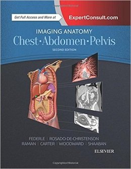
Imaging Anatomy: Chest, Abdomen, Pelvis, 2ed
Designed to help you quickly learn or review normal anatomy and confirm variants, Imaging Anatomy: Chest, Abdomen, Pelvis provides detailed views of anatomic structures in successive imaging slices in each standard plane of imaging. Axial, coronal, sagittal, and 3D reconstructions accompany highly accurate and detailed medical drawings, assisting you in making an accurate diagnosis. Comprehensive coverage of the chest, abdomen, and pelvis, combined with an orderly, easy-to-follow structure, make this unique title unmatched in its field.
- Includes all relevant imaging modalities, ۳D reconstructions, and highly accurate and detailed medical drawings that illustrate the fine points of the imaging anatomy
- Depicts common anatomic variants and covers common pathological processes as a part of its comprehensive coverage
- Provides a detailed overview of airway and interstitial network anatomy-the basis for understanding and diagnosing interstitial lung disease
- Features representative pathologic examples to highlight the effect of disease on human anatomy
- Includes plain radiography, the latest generation of multi-planar advanced cross-sectional MR and CT, ultrasound for pelvis/renal/liver/gallbladder, barium for GI tract, and much more
- Offers state of the art, detailed pelvic floor imaging and perianal/perirectal fistula imaging using high-resolution CT and MR, including 3T MR
- Expert Consult eBook version included with purchase. This enhanced eBook experience allows you to search all of the text, figures, images, and references from the book on a variety of devices.
Review
About the Author
Contents
Chapter 1. Chest Overview
Chapter 2. Lung Development
Chapter 3. Airway Structure
Chapter 4. Vascular Structure
Chapter 5. Interstitial Network
Chapter 6. Lungs
Chapter 7. Hila
Chapter 8. Airways
Chapter 9. Pulmonary Vessels
Chapter 10. Pleura
Chapter 11. Mediastinum
Chapter 12. Systemic Vessels
Chapter 13. Heart
Chapter 14. Coronary Arteries and Cardiac Veins
Chapter 15. Pericardium
Chapter 16. Chest Wall
Chapter 17. Embryology of Abdomen
Chapter 18. Abdominal Wall
Chapter 19. Diaphragm
Chapter 20. Peritoneal Cavity
Chapter 21. Vessels,. Lymphatic System and Nerves, Abdominal
Chapter 22. Esophagus
Chapter 23. Gastroduodenal
Chapter 24. Small Intestine
Chapter 25. Colon
Chapter 26. Spleen
Chapter 27. Liver
Chapter 28. Biliary System
Chapter 29. Pancreas
Chapter 30. Retroperitoneum
Chapter 31. Adrenal
Chapter 32. Kidney
Chapter 33. Ureter and Bladder
Chapter 34. Vessels,. Lymphatic System and Nerves, Pelvic
لینک کوتاه : https://bookbaz.ir/?p=58625
نویسنده : Michael P Federle MD FACR
ناشر : Elsevier; 2 edition
سال انتشار : 2017
زبان کتاب : انگلیسی
نوع فایل : PDF (نسخه اصلی)
تعداد صفحات : 1192
(ISBN) شابک : 032347781X
قیمت کتاب درآمازون : $249.95
حجم فایل : 211 MB
کتاب های مرتبط:
 دانلود کتاب تصویربرداری زنان و زایمان کوپل: سری کارشناس رادیولوژی
Obstetric Imaging: Expert Radiology Series, 1ed
 دانلود کتاب بیماری های آئورت: اطلس تصویربرداری تشخیصی بالینی
Aortic Diseases: Clinical Diagnostic Imaging Atlas, 1ed
 دانلود کتاب سری بررسی موردی رادیولوژی: تصویربرداری اسکلتی عضلانی
 Radiology Case Review Series: MSK Imaging, 1ed
 دانلود کتاب آناتومی رادیولوژیک کاربردی
Applied Radiological Anatomy, 2ed
 دانلود کتاب نورورادیولوژی: طیف و تکامل بیماری
Neuroradiology: Spectrum and Evolution of Disease, 1ed
 دانلود کتاب پزشکی هسته ای و PET/CT: فن آوری و تکنیک
Nuclear Medicine and PET/CT: Technology and Techniques, 7ed
 دانلود کتاب کمک به تشخیص افتراقی رادیولوژی چپمن و ناکیِلنی
Chapman & Nakielny’s Aids to Radiological Differential Diagnosis, 6ed
 دانلود کتاب اولتراسوند: مرور کلی
Ultrasound: A Core Review, 1ed
 دانلود کتاب پرسش و پاسخ پزشکی هسته ای رادکِیس
RadCases Plus Q&A Nuclear Medicine, 2ed
 دانلود کتاب سونوگرافی داپلر در پزشکی زنان و زایمان
Doppler Ultrasound in Gynecology and Obstetrics, 1ed
 دانلود کتاب رادیولوژی ۱۰۱: مبانی و اصول تصویربرداری
Radiology 101: The Basics & Fundamentals of Imaging, 4ed
 دانلود کتاب سر و گردن ExpertDDX
ExpertDDX: Head and Neck, 2ed
 دانلود کتاب سری مرور موردی رادیولوژی: تصویربرداری قفسه سینه
 Radiology Case Review Series: Thoracic Imaging, 1ed
 دانلود کتاب آناتومی تصویربرداری: سر و گردن
Imaging Anatomy: Head and Neck, 1ed
 دانلود کتاب سونوگرافی کودکان
 Pediatric Ultrasound, 1ed
 دانلود کتاب راهنمای بالینی IR: راهنمای مختصر برای رادیولوژی مداخله ای
 Pocketbook of Clinical IR: A Concise Guide to Interventional Radiology, 1ed
 دانلود کتاب تصویربرداری تشخیصی: ادراری تناسلی
Diagnostic Imaging: Genitourinary, 3ed
 دانلود کتاب تصویربرداری تشخیصی مغز
 Diagnostic Imaging: Brain, 4ed
 دانلود کتاب آناتومی مقطعی برای متخصصان تصویربرداری
 Sectional Anatomy for Imaging Professionals, 3ed
 دانلود کتاب مداخله انکولوژی (راهنمای عملی در مداخلات رادیولوژی)
Interventional Oncology (Practical Guides in Interventional Radiology), 1ed
 دانلود کتاب نورورادیولوژی: تشخیص های افتراقی کلیدی و سوالات بالینی
Neuroradiology: Key Differential Diagnoses and Clinical Questions, 2ed
 دانلود کتاب رادیوتراپی آنکولوژی برای تومورهای CNS کودکان
Radiation Oncology for Pediatric CNS Tumors, 1ed
 دانلود کتاب ۳ تفاوت برتر در تصویربرداری دستگاه گوارش
 Top 3 Differentials in Gastrointestinal Imaging, 1ed
 دانلود کتاب راهنمای خود ارزیابی رادیوبیولوژی
Radiobiology Self-Assessment Guide, 1ed
 دانلود کتاب تصویربرداری تخصصی HRCT ریه
Specialty Imaging: HRCT of the Lung, 2ed
 دانلود کتاب پرتودرمانی بالینی سرطان: علائم، تکنیک ها و نتایج
Clinical Radiation Oncology: Indications, Techniques, and Results, 3ed
 دانلود کتاب رادیوتراپی انکولوژی کودکان
Pediatric Radiation Oncology, 6ed
 دانلود کتاب اطلس پزشکی هسته ای بالینی
Atlas of Clinical Nuclear Medicine, 3ed
 دانلود کتاب سونوگرافی نقطه مراقبت + ویدئو
Point of Care Ultrasound, 2ed + Video
 دانلود کتاب اطلس آسیب شناسی بافت نرم و استخوان: با بافت شناسی، سیتولوژی و رادیولوژی
Atlas of Soft Tissue and Bone Pathology: With Histologic, Cytologic, and Radiologic Correlations, 1ed
 دانلود کتاب تصویربرداری در اورولوژی
Imaging in Urology, 1ed
 دانلود کتاب تصویربرداری پراش اشعه ایکس: فناوری و کاربردها
X-Ray Diffraction Imaging: Technology and Applications, 1ed
 دانلود کتاب CT قلبی بالینی: آناتومی و عملکرد
Clinical Cardiac CT: Anatomy and Function, 2ed
 دانلود کتاب همراه نورورادیولوژی: روش ها، دستورالعمل ها و اصول تصویربرداری
Neuroradiology Companion: Methods, Guidelines, and Imaging Fundamentals, 5ed
 دانلود کتاب تصویربرداری در گوش و حلق و بینی
Imaging in Otolaryngology, 1ed
 دانلود کتاب اصول تصویربرداری اسکلتی عضلانی مک کینز
Fundamentals of Musculoskeletal Imaging, 4ed
 دانلود کتاب ملزومات رادیوگرافی و رادیولوژی دندان
Essentials of Dental Radiography and Radiology, 5ed
 دانلود کتاب اشعه ایکس اسکلتی عضلانی برای دانشجویان و کارآموزان پزشکی
Musculoskeletal X-Rays for Medical Students and Trainees, 1ed
 دانلود کتاب تصویربرداری شکم: الزامات اصلی
Abdominal Imaging: The Core Requisites, 1ed
 دانلود کتاب موقعیت ها و تکنیک های رادیوگرافی بانترِیگر
Bontrager’s Handbook of Radiographic Positioning and Techniques, 8ed
 دانلود کتاب تصویربرداری تشخیصی پستان
Diagnostic Imaging: Breast, 3ed
 دانلود کتاب جامع فیزیک پزشکی (۱۰ جلدی)
Comprehensive Biomedical Physics, 10-Vol, 1ed
 دانلود کتاب اطلس جیبی آناتومی مقطعی: سر و گردن
Pocket Atlas of Sectional Anatomy: Head and Neck, 3ed
 دانلود کتاب اطلس آموزشی تصویربرداری شکم
Teaching Atlas of Abdominal Imaging, 1ed
 دانلود کتاب مراقبت از بیمار در رادیوگرافی + چک لیست رویه ها
Patient Care in Radiography, 10ed + Procedures Checklist
 دانلود کتاب اصول سیتی اسکن بدن
Fundamentals of Body CT, 4ed
 دانلود کتاب رادیوتراپی سرطان – مروری بر اساس سوال
Radiation Oncology – A Question Based Review, 2ed
 دانلود کتاب سیستم عروقی (سری سونوگرافی پزشکی تشخیصی)
The Vascular System (Diagnostic Medical Sonography Series), 2ed
 دانلود کتاب اصول سی تی اسکن بدن
Fundamentals of Body CT, 5ed
 دانلود کتاب تصویربرداری تشخیصی کودکان کافی (ویرایش ۲۰۱۹، ۲ جلدی)
Caffey’s Pediatric Diagnostic Imaging, 2-Volume Set, 13ed
 دانلود کتاب تصویربرداری در جراحی شکم
Imaging in Abdominal Surgery, 1ed
 دانلود کتاب تصویربرداری در گوارش
Imaging in Gastroenterology, 1ed
 دانلود کتاب رادیولوژی اعصاب تشخیصی و مداخله ای
Diagnostic and Interventional Neuroradiology, 1ed
 دانلود کتاب یادگیری رادیولوژی: شناخت اصول
Learning Radiology: Recognizing the Basics, 4ed
 دانلود کتاب موارد تصویربرداری پستان
Breast Imaging Cases
 دانلود کتاب تصویربرداری تشخیصی: پزشکی زایمان
Diagnostic Imaging: Obstetrics, 3ed
 دانلود کتاب هدایت حین عمل MRI جراحی مغز و اعصاب
Intraoperative MRI-Guided Neurosurgery, 1ed
 دانلود کتاب ملزومات سونوگرافی شکمی و لگنی
Essentials of Abdomino-Pelvic Sonography, 1ed
 دانلود کتاب تفسیر رادیوگرافی قفسه سینه در بیماران قلبی کودک
Chest Radiographic Interpretation in Pediatric Cardiac Patients, 1ed
 دانلود کتاب اطلس بیهوشی منطقه ای هدایت اولتراسوند + ویدئو
Atlas of Ultrasound-Guided Regional Anesthesia, 3ed + Video
 دانلود کتاب تصویربرداری تشخیصی دستگاه گوارش
Diagnostic Imaging: Gastrointestinal, 4ed
 دانلود کتاب تصویربرداری تشخیصی زایمان
Diagnostic Imaging: Obstetrics, 4ed
 دانلود کتاب تصویربرداری تشخیصی سر و گردن
Diagnostic Imaging: Head and Neck, 4ed
 دانلود کتاب تصویربرداری تشخیصی انکولوژی
Diagnostic Imaging: Oncology, 2ed
 دانلود کتاب MRI در یک نگاه
MRI at a Glance, 2ed
 دانلود کتاب سونوگرافی مداخله ای: راهنمای عملی و اطلس
Interventional Ultrasound: A Practical Guide and Atlas, 1ed
 دانلود کتاب فیزیک ضروری در تصویربرداری برای پرتونگار کلارک
Clark’s Essential Physics in Imaging for Radiographers, 1ed
 دانلود کتاب تصویربرداری ادراری تناسلی (سری موارد رادیولوژی)
Genitourinary Imaging (Radcase), 1ed
 دانلود کتاب تشخیص افتراقی در تصویربرداری سونوگرافی
 Differential Diagnosis in Ultrasound Imaging, 2ed
 دانلود کتاب تصویربرداری تشخیصی: مغز
Diagnostic Imaging: Brain, 3ed
 دانلود کتاب تصویربرداری تشخیصی: زنان و زایمان
Diagnostic Imaging: Gynecology, 2ed
 دانلود کتاب تصویربرداری زنان و زایمان: تشخیص و مراقبت از جنین
Obstetric Imaging: Fetal Diagnosis and Care, 2ed
 دانلود کتاب تصویربرداری اسکلتی عضلانی
Musculoskeletal Imaging, 2ed
 دانلود کتاب موقعیت در رادیوگرافی کلارک
Clark’s Positioning in Radiography, 13ed
 دانلود کتاب تصویربرداری تشخیصی قفسه سینه
Diagnostic Imaging: Chest, 3ed
 دانلود کتاب تصویربرداری تشخیصی زنان
Diagnostic Imaging: Gynecology, 3ed
 دانلود کتاب ملزومات پزشکی هسته ای و تصویربرداری مولکولی
Nuclear Medicine and Molecular Imaging: The Requisites, 5ed
 دانلود کتاب تشخیص افتراقی در تصویربرداری عصبی: ستون فقرات
Differential Diagnosis in Neuroimaging: Spine, 1ed
 دانلود کتاب تعمیرات X-Ray: راهنمای جامع نصب و سرویس تجهیزات رادیوگرافی
X-Ray Repair: A Comprehensive Guide to the Installation and Servicing of Radiographic Equipment, 3ed
 دانلود کتاب دوز، فواید و خطر در تصویربرداری پزشکی
 Dose, Benefit, and Risk in Medical Imaging, 1ed
 دانلود کتاب تصویربرداری قفسه سینه و قلب و عروق
Chest and Cardiovascular Imaging, 3ed
 دانلود کتاب تصویربرداری از مغز با MRI و CT: رویکرد الگوی تصویری
Brain Imaging with MRI and CT: An Image Pattern Approach, 1ed
 دانلود کتاب تشخیص افتراقی در توموگرافی کامپیوتری
Differential Diagnosis in Computed Tomography, 2ed
 دانلود کتاب رادیولوژی اضطراری (سری موارد رادیولوژی)
Radcases Emergency Radiology, 1ed
 دانلود کتاب پرسش و پاسخ تصویربرداری گوارشی رادکِیس
RadCases Plus Q&A Gastrointestinal Imaging, 2ed
دانلود کتاب CT قلبی بالینی: آناتومی و عملکرد + ویدئوClinical Cardiac CT: Anatomy and Function, 2ed + Video
 دانلود کتاب یادگیری رادیولوژی هِرینگ: شناخت مبانی
Learning Radiology: Recognizing the Basics, 3ed
 دانلود کتاب تصویربرداری عمومی اسکلتی عضلانی
Basic Musculoskeletal Imaging, 1ed
 دانلود کتاب تصویربرداری ماموگرافی: راهنمای عملی
Mammographic Imaging: A Practical Guide, 3ed
 دانلود کتاب اندوسونوگرافی
 Endosonography, 4ed
 دانلود کتاب تعویض ترنس کاتتر دریچه آئورت
Transcatheter Aortic Valve Implantation, 1ed
 دانلود کتاب تصویربرداری بیماری های بینابینی ریه (سری رادیولوژی کلینیکو)
Clinico Radiological Series: Imaging of Interstitial Lung Diseases, 1ed
 دانلود کتاب MRI بافتها همراه با T2 یا T2*s کوتاه
MRI of Tissues with Short T2s or T2*s, 1ed
 دانلود کتاب توموگرافی پرتو مخروطی کامپیوتری در ارتودنسی: موارد مصرف، بینش و نوآوری
Cone Beam Computed Tomography in Orthodontics: Indications, Insights, and Innovations, 1ed
 دانلود کتاب تصویربرداری پزشکی آکسفورد
Oxford Handbook of Medical Imaging, 1ed
 دانلود کتاب سرطان پستان: تشخیص زودرس با ماموگرافی
Breast Cancer: Early Detection with Mammography: Casting Type Calcifications Sign of a Subtype with Deceptive Features, 1ed
 دانلود کتاب راهنمای رادیو مابولیزاسیون: فیزیک، بیولوژی، پزشکی هسته ای و تصویربرداری
Handbook of Radioembolization: Physics, Biology, Nuclear Medicine, and Imaging, 1ed
 دانلود کتاب نورونولوژی و تصویربرداری عصبی سکته مغزی
Neurosonology and Neuroimaging of Stroke, 2ed
 دانلود کتاب اطلس جیبی آناتومی مقطعی: ستون فقرات، اندام، مفاصل
 Pocket Atlas of Sectional Anatomy: Spine Extremities Joints, 3ed
 دانلود کتاب چشم انداز محاسباتی و پردازش تصویری پزشکی
 Computational Vision and Medical Image Processing V: VipIMAGE 2015
 دانلود کتاب سری بررسی موردی رادیولوژی: تصویربرداری پستان
 Radiology Case Review Series: Breast Imaging, 1ed
 دانلود کتاب SPECT و SPECT/CT: راهنمای بالینی
SPECT and SPECT/CT: A Clinical Guide, 1ed
 دانلود کتاب رادیوگرافی PREP (مرور برنامه و آمادگی آزمون)
Radiography PREP (Program Review and Exam Preparation), 8ed
 دانلود کتاب روشهای اسکلتی عضلانی هدایت شده با سونوگرافی در پزشکی ورزشی
Ultrasound Guided Musculoskeletal Procedures in Sports Medicine, 1ed
 دانلود کتاب تصویربرداری تومور قفسه سینه (سری رادیولوژی کلینیکو)
Clinico Radiological Series: Imaging Of Chest Tumors, 1ed
 دانلود کتاب ۳ تفاوت برتر در رادیولوژی مغز و اعصاب
 Top 3 Differentials in Neuroradiology, 1ed
 دانلود کتاب مرور بالینی مغز و اعصاب مبتنی بر تصویر
Neurology Image-Based Clinical Review, 1ed
 دانلود کتاب توسعه دیجیتال ریه: از اولین سی تی ریه تا هوش مصنوعی بالینی
Developing the Digital Lung: From First Lung CT to Clinical AI, 1ed
 دانلود کتاب سونوگرافی مراقبت اورژانسی + ویدئو
Emergency Point-of-Care Ultrasound, 2ed + Video
 دانلود کتاب تصویربرداری از استخوان ها و مفاصل: یک روش مختصر و چند وجهی
Imaging of Bones and Joints: A Concise, Multimodality Approach, 1ed
 دانلود کتاب تصویربرداری و تجسم در اتاق عمل مدرن: راهنمای جامع برای پزشکان
Imaging and Visualization in The Modern Operating Room: A Comprehensive Guide for Physicians, 2015th
 دانلود کتاب تصویربرداری پستان (سری تشخیص مستقیم در رادیولوژی)
Breast Imaging (Direct Diagnosis in Radiology: DX-Direct!), 1ed
 دانلود کتاب تصویربرداری قلبی (سری تشخیص مستقیم در رادیولوژی)
Cardiac Imaging (Direct Diagnosis in Radiology: DX-Direct!), 1ed
 دانلود کتاب سونوگرافی تشخیصی بالینی: شکم و ساختار ظاهری + کتاب کار
Diagnostic Medical Sonography: Abdomen and Superficial Structures + Workbook, 3ed
 دانلود کتاب راهنمای جیبی برای رادیوگراف های کلارک
Clark’s Pocket Handbook for Radiographers, 2ed
 دانلود کتاب سونوگرافی تشخیصی هیگن-انسرت (۲ جلدی)
Textbook of Diagnostic Sonography, 2-Vol, 7ed
دانلود کتاب نورونولوژی و تصویربرداری عصبی سکته مغزی + ویدئوNeurosonology and Neuroimaging of Stroke, 2ed + Video
 دانلود کتاب اولتراسوند مراقبت بحرانی
 Critical Care Ultrasound, 1ed
 دانلود کتاب دانشنامه فیزیک پزشکی (۲ جلدی)
Encyclopaedia of Medical Physics, 2-Vol, 2ed
 دانلود کتاب تصویربرداری آناتومی: زانو، مچ پا، پا
Imaging Anatomy: Knee, Ankle, Foot, 2ed
 دانلود کتاب ۱۰۱ راه حل سی تی اسکن شکمی
 ۱۰۱Ct Abdomen Solutions, 1ed
 دانلود کتاب حفاظت در برابر تشعشع در رادیوگرافی پزشکی
Radiation Protection in Medical Radiography, 7ed
 دانلود کتاب پرفشاری ورید باب: تصویربرداری، تشخیص و مدیریت اندوواسکولار
Portal Hypertension: Imaging, Diagnosis, and Endovascular Management, 3ed
 دانلود کتاب رادیولوژی اضطراری بالینی
Clinical Emergency Radiology, 2ed
 دانلود کتاب تصویربرداری اشباع وزنی و اشباع تانسور
Diffusion Weighted and Diffusion Tensor Imaging, 1ed
 دانلود کتاب ملزومات رادیولوژی تشخیصی گرینگر و آلیسون
Grainger & Allison’s Diagnostic Radiology Essentials, 2ed
 دانلود کتاب مبانی آناتومی مقطعی: روش تصویربرداری
Fundamentals of Sectional Anatomy: An Imaging Approach, 2ed
 دانلود کتاب فیزیک MR بالینی از طریق تصاویر گرفته شده
The Physics of Clinical MR Taught Through Images, 4ed
 دانلود کتاب تصویربرداری تشخیصی رادیولوژی پزشکی هسته ای
 Radiology-Nuclear Medicine Diagnostic Imaging, 1ed
 دانلود کتاب نور پزشکی (نور درمانی)
Handbook of Photomedicine, 1ed
 دانلود کتاب MRI استخوان و تومورهای بافت نرم و ضایعات تومور مانند: تشخیص افتراقی و اطلس
MRI of Bone and Soft Tissue Tumors and Tumorlike Lesions: Differential Diagnosis and Atlas, 1ed
 دانلود کتاب پزشکی هسته ای در تشخیص و درمان بالینی (۲ جلدی)
Nuclear Medicine in Clinical Diagnosis and Treatment, 2-Vol, 3ed
 دانلود کتاب ایمنی تابش در انکولوژی تابشی
Radiation Safety in Radiation Oncology, 1ed
 دانلود کتاب آسان سازی X-Ray قفسه سینه
Chest X-Ray Made Easy, 4ed
 دانلود کتاب راهنمای غیر رسمی در رادیولوژی: ۱۰۰ تمرین اشعه ایکس قفسه سینه
The Unofficial Guide to Radiology: 100 Practice Chest X Rays, 1ed
 دانلود کتاب ۳ تفاوت برتر در رادیولوژی: بررسی موردی
Top 3 Differentials in Radiology: A Case Review, 2ed
 دانلود کتاب ضروریات انکولوژی رادیوتراپی بالینی
Essentials of Clinical Radiation Oncology, 1ed
 دانلود کتاب دیسپلازی استخوان: اطلس اختلالات ژنتیکی رشد اسکلت
Bone Dysplasias: An Atlas of Genetic Disorders of Skeletal Development, 4ed
دانلود کتاب یادگیری رادیولوژی: شناخت اصول + ویدئوLearning Radiology: Recognizing the Basics, 4ed + Video
 دانلود کتاب رادیوتراپی والتر و میلر
Walter and Miller’s Textbook of Radiotherapy, 8ed
 دانلود کتاب رادیولوژی دستگاه گوارش گور و لوین + ویدئو
Textbook of Gastrointestinal Radiology, 5ed + Video
 دانلود کتاب پرتودرمانی عملی: فیزیک و تجهیزات
Practical Radiotherapy: Physics and Equipment, 3ed
 دانلود کتاب فیزیک پزشکی هسته ای بالینی با متلب
Clinical Nuclear Medicine Physics with MATLAB, 1ed
 دانلود کتاب آزمون اصلی را بترکانید (۲ جلدی)
Crack The Core Exam, 2-Vol, 9ed
 دانلود کتاب تشخیص افتراقی در تصویربرداری کودکان
Differential Diagnosis in Pediatric Imaging, 1ed
 دانلود کتاب تصویربرداری هیبرید در پزشکی قلب و عروق
Hybrid Imaging in Cardiovascular Medicine, 1ed
 دانلود کتاب رادیوسرجری استریوتاکتیک و پرتو درمانی استریوتاکتیک بدن
Stereotactic Radiosurgery and Stereotactic Body Radiation Therapy, 1ed
 دانلود کتاب راهنمای سی تی قلب و عروق EACVI
EACVI Handbook of Cardiovascular CT, 1ed
 دانلود کتاب پرتو درمانی کودکان: برنامه ریزی و درمان + اطلس مغز
Pediatric Radiotherapy Planning and Treatment + Updates, 1ed
 دانلود کتاب پزشکی هسته ای: سری بررسی موردی
Nuclear Medicine: Case Review Series, 2ed