
دانلود کتاب اصول و تمرین جراحی سوراخ کلید مغز + ویدئو
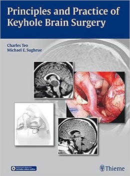
Principles and Practice of Keyhole Brain Surgery, 1ed + Video
Developed 20 years ago by leading innovators in the field, the keyhole concept of brain surgery has become an integral part of the practice of neurosurgery. This timely and comprehensive book covers the thinking, philosophy, and techniques of modern keyhole brain surgery, including a realistic assessment of its benefits and limitations. Written by expert practitioners and highlighted by vivid surgical illustrations and procedural videos, Principles and Practice of Keyhole Brain Surgery functions as an experienced mentor working side by side with neurosurgeons as they master the techniques.
Special Features:
- Introduces the basic principles of the keyhole approach, including the practical, technical, and logistical aspects of planning procedures and operating through small openings
- Beautifully illustrated with nearly 900 endoscopic images, diagrams, surgical drawings, and operative photographs, many showing step-by-step procedures
- Details the pivotal role of the endoscope in keyhole brain surgery and its ability to provide multiple angles of visualization, including a useful catalog of clinical situations where the endoscope has proven most effective
- Demonstrates contemporary keyhole approaches (e.g. the eyebrow/sub-frontal approach) in procedures for supratentorial intra-axial brain tumors, tumors of the cribriform plate and orbit, parasellar masses, craniopharyngiomas, tumors of the middle fossa and cavernous sinus and many other conditions in the cranial base
- Offers more than 100 procedural videos on the Thieme’s MediaCenter, narrated by the authors and aligned to chapters in the book for an unparalleled learning resource
Providing all the information necessary to achieve surgical goals through well placed, smaller openingswith the added benefits of shorter procedures, fewer wound complications and better patient outcomesPrinciples and Practice of Keyhole Brain Surgery is essential for every neurosurgeon in practice today.
Contents
۱ An Introduction to the Keyhole Concept
۲ Cognitive Principles of Planning Keyhole Approaches
۳ Technical Principles of Operating in Keyhole Craniotomies
۴ The Role of Endoscopy in Keyhole Surgery
۵ An Atlas of Intracranial Endoscopy
۶ Steps in Common Keyhole Approaches
۷ Endonasal Surgery and Its Current Role in Neurosurgical Paradigms
۸ Keyhole Surgery for Supratentorial Intra-Axial Tumors
۹ Keyhole Approaches to Tumors of the Cribriform Plate and Orbit
۱۰ Keyhole Approaches to Parasellar Masses and the Interpeduncular Space
۱۱ Craniopharyngiomas
۱۲ Keyhole Approaches to Tumors of the Middle Fossa, Cavernous Sinus, Tentorium and Lateral Midbrain
۱۳ The Cerebellopontine Angle
۱۴ Tackling the Petrous Apex through a Keyhole
۱۵ Keyhole Surgery of the Tectum and Pineal Region
۱۶ Keyhole Surgery for Foramen Magnum,Fourth Ventricular,and Midline Cerebellar Tumors
Videos Contents
۲.۱ The relative merits of microscope and endoscope
۳.۱ The importance of frequent repositioning of the microscope
۳.۲ The keyhole principle demonstrated
۳.۳ The technique of anticipatory microscope positioning
۳.۴ The three-point technique
۳.۵ Working in a properly planned keyhole craniotomy
۳.۶ Draining CSF and its impact on visualization
۴.۱ Proper introduction of the endoscope
۴.۲ Safe angle changes in intracranial endoscopy
۴.۳ Safe techniques for introducing instruments endoscopically
۴.۴ Inspection of the IAC with the endoscope
۴.۵ The use of angled instruments in endoscopic surgery
۴.۶ Endoscopic clarification
۷.۱ Endonasal resection of a brainstem tumor
۷.۲ Endonasal resection of a lower clival chordoma
۷.۳ Endonasal transpterygoid approach to Meckel cave
۷.۴ Endonasal transpterygoid approach to Meckel cave
۸.۱ The use of the endoscope to improve resection in a glioma
۸.۲ Resection of the frontal lobe via a minicraniotomy
۸.۳ Microsurgical appearance of the caudate nucleus
۸.۴ Appearance of the cavity after complete removal of the frontal lobe
۸.۵ Appearance of the cavity after complete removal of the frontal lobe
۸.۶ Resection of the temporal lobe via a minicraniotomy
۸.۷ Resection of a localized insular glioma (Case 1)
۸.۸ Resection of a localized insular glioma (Case 2)
۸.۹ Resection of a complex multilobular insular glioma
۸.۱۰ Postresection appearance of a cingulate gyrus glioma
۸.۱۱ Resection of a cingulate cavernoma via a contralateral interhemispheric approach
۸.۱۲ The use of the endoscope to improve the ispilateral view in a cingulate glioma
۸.۱۳ Interhemispheric approach to a thalamic glioma
۸.۱۴ Resection of a thalamic glioma via an interhemispheric approach
۸.۱۵ The postresection view of a thalamic glioma via a transcortical approach
۹.۱ Endonasal removal of an orbital apex tumor
۱۰.۱ Tuberculum meningioma resected via the eyebrow approach
۱۰.۲ The use of the endoscope in the eyebrow approach
۱۰.۳ Tuberculum meningioma resected via the eyebrow approach
۱۰.۴ Resection of a medial uncus lesion via the eyebrow approach
۱۰.۵ Resection of an epidermoid of the interpeduncular space via the eyebrow
۱۰.۶ Resection of a tumor of the cerebral peduncle
۱۰.۷ Resection of a large tumor of the cerebral peduncle via the eyebrow approach
۱۰.۸ Introduction of the endoscope into the opticocarotid triangle
۱۰.۹ Endoscopic view down the dorsum sellae through the eyebrow approach
۱۰.۱۰ Transcallosal-tenoforniceal approach to the third ventricle
۱۰.۱۱ Removal of a colloid cyst from the third ventricle
۱۰.۱۲ Ventriculoscopic resection of a hypothalamic hamartoma (Case 1)
۱۰.۱۳ Ventriculoscopic resection of a hypothalamic hamartoma (Case 2)
۱۰.۱۴ Ventriculoscopic resection of intraventricular nodules
۱۰.۱۵ Resection of a large central skull base tumor via the eyebrow approach
۱۱.۱ Endonasal resection of a massive craniopharyngioma
۱۱.۲ Initial steps in the endonasal endoscopic approach to craniopharyngiomas
۱۱.۳ Venous versus carotid arterial bleeding in endonasal surgery
۱۱.۴ Endonasal resection of a craniopharyngioma
۱۱.۵ Endonasal resection of a craniopharyngioma
۱۱.۶ Endonasal resection of a craniopharyngioma
۱۱.۷ Endoscopic repair of the postresection defect
۱۱.۸ Craniopharyngioma resection via the eyebrow approach
۱۱.۹ Endoscopic inspection after a transcranial craniopharyngioma resection
۱۱.۱۰ Endoscopic inspection after a transcranial craniopharyngioma resection (Case 1)
۱۱.۱۱ Endoscopic inspection after a transcranial craniopharyngioma resection (Case 2)
۱۱.۱۲ Transcallosal-tenofornicea resection of a craniopharyngioma
۱۲.۱ Mini-pterional resection of a clinoid meningioma
۱۲.۲ Endoscopic inspection of the Meckel cave
۱۲.۳ Endonasal resection of a cavernous sinus pituitary tumor
۱۲.۴ Cavernous sinus meningioma resection via a mini-pterional craniotomy
۱۲.۵ Mini-pterional resection of a parasellar meningioma
۱۲.۶ Mini-subtemporal approach to the lateral midbrain
۱۲.۷ Transtemporal transchoroidal fissure approach to a lateral midbrain cavernoma
۱۲.۸ Resection of a massive pontine cavernoma via the mini-subtemporal approach
۱۲.۹ The use of endoscopy to work around the edges of the tentorium
۱۳.۱ Delivery of a large vestibular schwannoma via the mini-retrosigmoid approach
۱۳.۲ Access to the CSF spaces in the mini-retrosigmoid approach
۱۳.۳ Access to the CSF spaces in a large CPA tumor
۱۳.۴ The use of the endoscope to avoid IAC drilling in some cases
۱۳.۵ Resection of a vestibular schwannoma with hearing preservation
۱۳.۶ Resection of a jugular foramen meningioma
۱۳.۷ Removal of a petroclival meningioma via the mini-retrosigmoid approach
۱۳.۸ Mini-retrosigmoid approach to the tectum
۱۳.۹ Mini-retrosigmoid resection of a posterior fossa epidermoid
۱۳.۱۰ Mini-retrosigmoid resection of a pontine lesion
۱۳.۱۱ Mini-retrosigmoid resection of a jugular foramen tumor
۱۳.۱۲ Endoscopic inspection and waxing of aircells following IAC drillout
۱۳.۱۳ Endoscopic inspection and resection of a petroclival meningioma in Meckel cave
۱۳.۱۴ Endoscopic inspection and resection of a meningioma in Meckel cave
۱۳.۱۵ Removal of a pontine glioma via the mini-retrosigmoid approach
۱۴.۱ Endonasal drainage of a petrous apex granuloma
۱۴.۲ Visualization of basilar perforators in a tumor
۱۴.۳ Eyebrow approach to a petroclival meningioma
۱۴.۴ Mini-pterional approach to a petroclival meningioma
۱۴.۵ Eyebrow approach to a petroclival meningioma
۱۵.۱ Anterior interhemispheric approach to a posterior third ventricular tumor
۱۵.۲ Anterior interhemispheric approach to a posterior third ventricular tumor
۱۵.۳ Mini-occipital/transtentorial approach to the pineal region
۱۵.۴ Mini-occipital/transtentorial approach to a pineal tumor
۱۵.۵ Mini-occipital/transtentorial approach to a tectal tumor
۱۵.۶ The use of the endoscope in the pineal region
۱۵.۷ Endoscopic inspection of the cerebellomesencephalic fissure
۱۵.۸ Microscopic versus endoscopic views of the pineal region
۱۵.۹ Endoscopic inspection and resection of a tectal tumor
۱۵.۱۰ Endoscopic inspection after removal of a tectal tumor
۱۵.۱۱ Endoscopic view into the third ventricle via a mini-occipital/transtentorial approach
۱۶.۱ Endoscopic view into the fourth ventricle through the foramen of Luschka
۱۶.۲ Mini-suboccipital resection of a fourth ventricular tumor (Case 1)
۱۶.۳ Mini-suboccipital resection of a fourth ventricular tumor (Case 2)
۱۶.۴ Mini-suboccipital resection of a superior fourth ventricular tumor
۱۶.۵ Mini-suboccipital resection of a medullary tumor
۱۶.۶ Removal of a tumor from the foramen of Luschka
۱۶.۷ Endoscopic removal of a tumor from the foramen of Luschka
۱۶.۸ Endoscopic views through the foramen of Luschka
۱۶.۹ Mini-suboccipital resection of a lateral schwannoma at the foramen magnum
لینک کوتاه : https://bookbaz.ir/?p=28945
نویسنده : Charles Teo , Michael E. Sughrue
ناشر : Thieme; 1st edition
سال انتشار : 2015
زبان کتاب : انگلیسی
نوع فایل : PDF + MP4
تعداد صفحات : 277
(ISBN) شابک : 3131758511
قیمت کتاب درآمازون : $187.87
حجم فایل : 1400 MB
کتاب های مرتبط:
 دانلود کتاب تغییر شکل ستون فقرات: ملزومات
Spinal Deformities: The Essentials, 2ed
 دانلود کتاب یک رویکرد تشریحی برای جراحی ستون فقرات کم تهاجمی + ویدئو
An Anatomic Approach to Minimally Invasive Spine Surgery, 2ed + Video
 دانلود کتاب از دست دادن حافظه، آلزایمر و زوال عقل: راهنمای عملی برای پزشکان + ویدئو
Memory Loss, Alzheimer’s Disease and Dementia: A Practical Guide for Clinicians, 3ed + Video
 دانلود کتاب اصول و عملکرد جراحی رباتیک
Principles and Practice of Robotic Surgery, 1ed
 دانلود کتاب موضوعات اصلی در جراحی عمومی و اورژانس: همگام با تمرین متخصص جراحی
Core Topics in General & Emergency Surgery: A Companion to Specialist Surgical Practice, 5ed
 دانلود کتاب جراحی عروقی و اندوواسکولار: همگام با تمرین متخصص جراحی
Vascular and Endovascular Surgery: A Companion to Specialist Surgical Practice, 5ed
 دانلود کتاب جراحی پستان: همگام با تمرین متخصص جراحی
Breast Surgery: A Companion to Specialist Surgical Practice, 5ed
 دانلود کتاب کالبدشناسی اعصاب بالینی ضروری
Essential Clinical Neuroanatomy, 1ed
 دانلود کتاب جراحی غدد درون ریز: همگام با تمرین متخصص جراحی
Endocrine Surgery: A Companion to Specialist Surgical Practice, 5ed
 دانلود کتاب جراحی کبد و پانکراس: همگام با تمرین متخصص جراحی
Hepatobiliary and Pancreatic Surgery: A Companion to Specialist Surgical Practice, 5ed
 دانلود کتاب پیوند اعضا: همگام با تمرین متخصص جراحی
Transplantation: A Companion to Specialist Surgical Practice, 5ed
 دانلود کتاب اصول و تمرین پزشکی خواب (۲ جلدی)
Principles and Practice of Sleep Medicine, 2-Vol, 7ed
 دانلود کتاب روش های بالینی ضروری دِن
Dehn’ Essential Clinical Procedures, 3ed
 دانلود کتاب راینولوژی: بیماریهای بینی و سینوس و قاعده جمجمه
Rhinology: Diseases of the Nose, Sinuses, and Skull Base, 1ed
 دانلود کتاب فیزیولوژی سلولی و فیزیولوژی عصبی موزبی
Mosby’s Cellular Physiology and Neurophysiology, 3ed
 دانلود کتاب اطلس ویدیویی پایش نوروفیزیولوژیک در جراحی تومورهای نفوذی مغز
Video Atlas of Neurophysiological Monitoring in Surgery of Infiltrating Brain Tumors, 1ed
 دانلود کتاب اصول جراحی مغز و اعصاب
Principles of Neurological Surgery, 4ed
 دانلود کتاب اطلس تکنیک های جراحی قفسه سینه
 Atlas of Thoracic Surgical Techniques, 1ed
 دانلود کتاب جراحی سابیستون: مبانی بیولوژیکی عمل جراحی مدرن
Sabiston Textbook of Surgery: The Biological Basis of Modern Surgical Practice, 21ed
 دانلود کتاب جراحی ترانس نازال آندوسکوپیک قاعده جمجمه و مغز
Transnasal Endoscopic Skull Base and Brain Surgery, 2ed
 دانلود کتاب درمان کنونی تروما و مراقبت های حیاتی جراحی
Current Therapy of Trauma and Surgical Critical Care, 3ed
 دانلود کتاب تصمیم گیری و اجتناب از عوارض در فلپ پوست
 Making Decisions and Avoiding Complications in Skin Flaps, 1ed
 دانلود کتاب جراحی پلاستیک و ترمیمی: روش ها و تکنیک ها
Plastic and Reconstructive Surgery: Approaches and Techniques, 1ed
 دانلود کتاب شکستگی و در رفتگی کودکان
Pediatric Fractures and Dislocations, 1ed
 دانلود کتاب ملزومات جراحی عمومی
Essentials of General Surgery, 5ed
 دانلود کتاب جراحی کبد ، مجرای صفراوی و پانکراس بلومگارت (۲ جلدی)
Blumgart’s Surgery of the Liver, Biliary Tract and Pancreas, 2-Vol, 5ed
 دانلود کتاب نوروپاتولوژی یاکنیس: آسیب شناسی عملکرد بالا
Neuropathology: A Volume in the High Yield Pathology Series, 1ed
 دانلود کتاب روش های جراحی ارتوپدی
 Orthopaedic Surgical Approaches, 2ed
 دانلود کتاب فناوری جراحی: اصول و تمرین + کتاب کار
Surgical Technology: Principles and Practice + Workbook, 6ed
 دانلود کتاب اطلس کاشت تراپی برای مدیریت درد
 Atlas of Implantable Therapies for Pain Management, 2ed
 دانلود کتاب اختلال شناختی و زوال عقل در بیماری پارکینسون
Cognitive Impairment and Dementia in Parkinson’s Disease, 2ed
 دانلود کتاب صدمات نوک انگشتان: تشخیص، مدیریت و بازسازی
Fingertip Injuries: Diagnosis, Management and Reconstruction, 2015th
 دانلود کتاب تصویربرداری رزونانس مغناطیسی در اختلالات حرکتی
 Magnetic Resonance Imaging in Movement Disorders, 1ed
 دانلود کتاب تمرین مراقبت حاد و اورژانسی مغز و اعصاب
The Practice of Emergency and Critical Care Neurology, 2ed
 دانلود کتاب اصول جراحی شوارتز
 Schwartz’s Principles of Surgery, 10ed
 دانلود کتاب جراحی قفسه سینه و مری پیرسون (۲ جلدی)
Pearson’s Thoracic and Esophageal Surgery, 2-Vol, 3ed
 دانلود کتاب مغز و اعصاب بالینی کودکان فنیچل
Fenichel’s Clinical Pediatric Neurology, 7ed
 دانلود کتاب دانشنامه جراحی و آزمایش های پزشکی گیل (۴ جلدی)
Gale Encyclopedia Of Surgery And Medical Tests, 4-Vol, 3ed
 دانلود کتاب علوم اعصاب اساسی برای کاربرد های بالینی و عمومی هِینز
Haines’ Fundamental Neuroscience for Basic and Clinical Applications, 4ed
 دانلود کتاب راهنمای مطالعه برای پرستاری جراحی داخلی: ارزیابی و مدیریت مشکلات بالینی
Study Guide for Medical-Surgical Nursing: Assessment and Management of Clinical Problems, 9ed
 دانلود کتاب ارتوپدی کودکان تاچیان (۳ جلدی)
Tachdjian’s Pediatric Orthopaedics, 3-Vol, 5ed
 دانلود کتاب جراحی واسکولار و اندوواسکولار: همگام با عمل جراحی متخصص
Vascular and Endovascular Surgery: A Companion to Specialist Surgical Practice, 6ed
 دانلود کتاب جراحی سرطان مری شکمی: همگام با تمرین متخصص جراحی
Oesophagogastric Surgery: A Companion to Specialist Surgical Practice, 5ed
 دانلود کتاب مشاوره حل بحران (اصول اصلی مغز و اعصاب حاد)
Solving Critical Consults (Core Principles of Acute Neurology), 1ed
 دانلود کتاب اطلس ویدئویی جراحی ستون فقرات
 Video Atlas of Spine Surgery, 1ed
 دانلود کتاب تصویربرداری آسیب های مغزی
 Imaging of Traumatic Brain Injury, 1ed
 دانلود کتاب اصول و عملکرد اختلالات حرکتی
Principles and Practice of Movement Disorders, 3ed
 دانلود کتاب اطلس ویدئویی جراحی کبد، صفراوی و پانکراس
 Video Atlas: Liver, Biliary & Pancreatic Surgery, 1ed
 دانلود کتاب اطلس ویدئویی جراحی آنوریسم داخل جمجمه
Video Atlas of Intracranial Aneurysm Surgery, 1ed
 دانلود کتاب اطلس ویدئویی جراحی کم تهاجمی پیشرفته
Video Atlas of Advanced Minimally Invasive Surgery, 1ed
 دانلود کتاب سرطان سر و گردن: یک رویکرد چند رشته ای
Head and Neck Cancer: A Multidisciplinary Approach, 4ed
 دانلود کتاب تصویربرداری رزونانس مغناطیسی مغز و ستون فقرات (دو جلدی)
Magnetic Resonance Imaging of the Brain and Spine, 2-Vol, 4ed
 دانلود کتاب جراحی صرع در کودکان: ارزیابی قبل از عمل و درمان جراحی
Pediatric Epilepsy Surgery: Preoperative Assessment and Surgical Treatment, 2ed
 دانلود کتاب جراحی کولورکتال: همگام با عمل جراحی متخصص
Colorectal Surgery: A Companion to Specialist Surgical Practice, 6ed
 دانلود کتاب مرور بورد مقدماتی جراحی مغز و اعصاب
 Neurosurgery Primary Board Review, 1ed
 دانلود کتاب اصول و تمرین جراحی سوراخ کلید مغز
 Principles and Practice of Keyhole Brain Surgery, 1ed
 دانلود کتاب زندگی نباتی پس از تروماتیک
 Post-Traumatic Vegetative State, 1ed
 دانلود کتاب تکنیک های جراحی مغز و اعصاب اشمیدک و سویت (۲ جلدی) + ویدئو
Schmidek and Sweet Operative Neurosurgical Techniques, 2-Vol, 7ed + Video
 دانلود کتاب راهنمای جراحی مغز و اعصاب گرینبرگ (ویرایش ۲۰۱۶)
Greenberg’s Handbook of Neurosurgery, 8ed
 دانلود کتاب بازسازی پیشرفته: زانو
 Advanced Reconstruction: Knee, 1ed
 دانلود کتاب اصول جراحی و مرور بورد شوارتز
Schwartz’s Principles of Surgery ABSITE and Board Review, 10ed
 دانلود کتاب جراحی زیر پوستی مجاری ادراری فوقانی
Percutaneous Surgery of the Upper Urinary Tract, 1ed
 دانلود کتاب جراحی آندوسکوپی سینوس: آناتومی، بازسازی سه بعدی و تکنیک جراحی
Endoscopic Sinus Surgery: Anatomy, Three-Dimensional Reconstruction, and Surgical Technique, 4ed
 دانلود کتاب جراحی زیبایی بینی دالاس (۲ جلدی)
Dallas Rhinoplasty: Nasal Surgery by the Masters, 2-Vol, 3ed
 دانلود کتاب روانشناسی اعصاب بالینی و علوم اعصاب رفتاری
Textbook of Clinical Neuropsychiatry and Behavioral Neuroscience, 3ed
 دانلود کتاب جراحی اطفال: تشخیص و مدیریت
Pediatric Surgery: Diagnosis and Management, 1ed
 دانلود کتاب جراحی اندوسکوپیک مغز و اعصاب
Neuroendoscopic Surgery, 1ed
 دانلود کتاب کرانیوفارنژیوما: تشخیص، درمان و پیامد جامع
Craniopharyngiomas: Comprehensive Diagnosis, Treatment and Outcome, 1ed
 دانلود کتاب اجازه برای دیالیز: روشهای جراحی و رادیولوژیک
Access for Dialysis: Surgical and Radiologic Procedures, 2ed
 دانلود کتاب الکترومیوگرافی ضروری
Essential Electromyography, 1ed
 دانلود کتاب مغز و اعصاب در عمل بالینی برادلی + ویدئو
Bradley’s Neurology in Clinical Practice, 2-Vol, 7ed + Video
 دانلود کتاب جراحی کودکان اشکرفت
Ashcraft’s Pediatric Surgery, 7ed
 دانلود کتاب مبانی تکنیک های جراحی میکروسکوپی و بای پس + ویدئو
Microsurgical Basics and Bypass Techniques, 1ed + Video
 دانلود کتاب جراحی عمومی انکولوژی پیچیده
Textbook of Complex General Surgical Oncology, 1ed
 دانلود کتاب جراحی آندوسکوپی ستون فقرات
Endoscopic Spine Surgery, 2ed
 دانلود کتاب جراحی پلاستیک و بازسازی پستان بوستویک + ویدئو
Bostwick’s Plastic and Reconstructive Breast Surgery, 4ed + Video
 دانلود کتاب پاتولوژی جراحی روسای و اکرمن (۲ جلدی، ویرایش ۲۰۱۸)
Rosai and Ackerman’s Surgical Pathology, 2-Vol, 11ed
 دانلود کتاب اطلس رنگی جراحی ساقه مغز
 Color Atlas of Brainstem Surgery, 1ed
 دانلود کتاب موارد آتالارینژیک: نمونه کارهای بالینی دانشگاه سینسیناتی
Otolaryngology Cases: The University of Cincinnati Clinical Portfolio, 2ed
 دانلود کتاب از دست دادن حافظه، آلزایمر و دمانس: راهنمای عملی برای پزشکان + ویدئو
Memory Loss, Alzheimer’s Disease, and Dementia: A Practical Guide for Clinicians, 2ed + Video
 دانلود کتاب استراتژی های جراحی قاعده جمجمه
 Skull Base Surgery: Strategies, 1ed
دانلود کتاب جراحی ترانس نازال آندوسکوپیک قاعده جمجمه و مغز + ویدئوTransnasal Endoscopic Skull Base and Brain Surgery, 2ed + Video
 دانلود کتاب راهنمای جراحی
 The Surgical Handbook, 1ed
 دانلود کتاب جراحی ستون فقرات بنزل (۲ جلدی)
Benzel’s Spine Surgery, 2-Vol, 5ed
 دانلود کتاب اطلس جراحی عمومی رباتیک
Atlas of Robotic General Surgery, 1ed
 دانلود کتاب تکنیک های عملی جراحی ستون فقرات
 Operative Techniques: Spine Surgery, 3ed
 دانلود کتاب جراحی مغز و اعصاب کودکان کوهن
Pediatric Neurosurgery: Tricks of the Trade, 1ed
 دانلود کتاب جراحی ستون فقرات حداقل تهاجمی
Minimally Invasive Spine Surgery, 1ed
 دانلود کتاب جراحی پلاستیک: جراحی زیبایی (جلد دوم)
Plastic Surgery: Volume 2: Aesthetic Surgery, 3ed
 دانلود کتاب مراقبت حاد جراحی: اصول و تمرین
Acute Care Surgery: Principles and Practice
 دانلود کتاب جراحی پلاستیک نلیگان: پستان + ویدئو (جلد ۵)
Plastic Surgery: Breast, Vol-5, 3ed + Video
 دانلود کتاب مهره های ستون فقرات گردن + ویدئو
Textbook of the Cervical Spine, 1ed + Video
 دانلود کتاب اختلالات کوروئید
 Choroidal Disorders, 1ed
دانلود کتاب جراحی پلاستیک و بازسازی پستان بوستویکBostwick’s Plastic and Reconstructive Breast Surgery, 4ed
 دانلود کتاب چالش های عروقی در جراحی قاعده جمجمه
Vascular Challenges in Skull Base Surgery, 1ed
 دانلود کتاب جراحی آندوسکوپی پایه جمجمه جانبی: اصول، آناتومی، رویکردها
Endoscopic Lateral Skull Base Surgery: Principles, Anatomy, Approaches, 1ed
 دانلود کتاب مغز انسان در عکس ها و نمودارها نولته
Nolte’s The Human Brain in Photographs and Diagrams, 5ed
 دانلود کتاب جراحی و انکولوژی سر و گردن استل و ماران
Stell & Maran’s Textbook of Head and Neck Surgery and Oncology, 5ed
 دانلود کتاب جراحی پلاستیک: اصول (جلد اول)
Plastic Surgery: Volume 1: Principles, 3ed
 دانلود کتاب ساده سازی جراحی بالینی
 Clinical Surgery Made Easy, 1ed
 دانلود کتاب تکنیک های اصلی در جراحی ارتوپدی: ارتوپدی انکولوژی و بازسازی های پیچیده
Master Techniques in Orthopaedic Surgery: Orthopaedic Oncology and Complex Reconstruction, 1ed
 دانلود کتاب اطلس روشهای هدایت تصویری ستون فقرات
 Atlas of Image-Guided Spinal Procedures, 2ed
 دانلود کتاب جراحی آندوسکوپی حدقه چشم: آناتومی، پاتولوژی و مدیریت
Endoscopic Surgery of the Orbit: Anatomy, Pathology, and Management, 1ed
 دانلود کتاب راهنمای جراحی آندوسکوپی سینوس و قاعده جمجمه
Manual of Endoscopic Sinus and Skull Base Surgery, 2ed
 دانلود کتاب جراحی آندوسکوپی اندونازال قاعده جمجمه کودکان
Pediatric Endoscopic Endonasal Skull Base Surgery, 1ed
 دانلود کتاب جراحی عروق و درمان اندوواسکولار رادرفورد (۲ جلدی)
Rutherford’s Vascular Surgery and Endovascular Therapy, 2-Vol, 10ed
 دانلود کتاب جراحی قفسه سینه سابیستون و اسپنسر (۲ جلدی)
Sabiston and Spencer Surgery of the Chest 10th Edition
 دانلود کتاب راهنمای پایندگی چشم پزشکی عصبی + ویدئو
The Neuro-Ophthalmology Survival Guide, 2ed + Video
 دانلود کتاب نتیجه نامطلوب در جراحی پلاستیک: اجتناب و درمان (۲ جلدی)
The Unfavorable Result in Plastic Surgery: Avoidance and Treatment, 2-Vol, 4ed
 دانلود کتاب وصل کردن فلپ ها: آناتومی، روش و کاربرد های بالینی (۲ جلدی) + ویدئو
Perforator Flaps: Anatomy, Technique, & Clinical Applications, 2-Vol, 2ed + Video
دانلود کتاب استراتژی های جراحی قاعده جمجمه + ویدئوSkull Base Surgery: Strategies, 1ed + Video
 دانلود کتاب جراحی قاعده جمجمه جانبی: اطلس کلینیک هاوس
Lateral Skull Base Surgery: The House Clinic Atlas, 1ed
 دانلود کتاب تومور و سرطان: سیستم عصبی مرکزی و محیطی
Tumors and Cancers: Central and Peripheral Nervous Systems, 1ed
 دانلود کتاب تغذیه و شیوه زندگی در بیماری های خودایمنی عصبی: مولتیپل اسکلروز
Nutrition and Lifestyle in Neurological Autoimmune Diseases: Multiple Sclerosis, 1ed
 دانلود کتاب مواد مخدر، اعتیاد و مغز
Drugs, Addiction, and the Brain, 1ed
 دانلود کتاب آسیب شناسی و جراحی در اطراف شریان مهره
Pathology And Surgery Around The Vertebral Artery, 2011th ed
 دانلود کتاب بیماری قلبی در نوزادان، کودکان و نوجوانان ماس و آدامز: شامل جنین و جوانان (۲ جلدی)
Moss & Adams’ Heart Disease in Infants, Children, and Adolescents: Including the Fetus and Young Adult, 2-Vol, 8ed
 دانلود کتاب علوم اعصاب شناختی
 The Cognitive Neurosciences, 6ed
 دانلود کتاب جراحی پلاستیک و ترمیمی
Textbook of Plastic & Reconstructive Surgery, 1ed
 دانلود کتاب نوروبیولوژی پزشکی
Medical Neurobiology, 2ed
 دانلود کتاب درمان جراحی کنونی + ویدئو
Current Surgical Therapy, 14ed + Video
 دانلود کتاب موارد چالش برانگیز در جراحی ستون فقرات
 Challening Cases in Spine Surgery, 1ed
 دانلود کتاب دیسک بین مهره ای کمر
The Lumbar Intervertebral Disc, 1ed
 دانلود کتاب ستون فقرات راتمن-سیمون و هرکوییتز (۲ جلدی) + ویدئو
Rothman-Simeone and Herkowitz’s The Spine, 2-Vol, 7ed + Video
 دانلود کتاب دانشنامه پزشکی گیل (۹ جلدی)
Gale Encyclopedia of Medicine, 9-Vol, 2015th
 دانلود کتاب آناتومی جراحی قلب ویلکاکس
Wilcox’s Surgical Anatomy of the Heart, 4ed
 دانلود کتاب عصب شناسی و پزشکی عمومی امینوف
Aminoff’s Neurology and General Medicine, 6ed
 دانلود کتاب پاتو فلش: فلش کارت های پاتوفیزیولوژی
 Patho Phlash!: Pathophysiology Flash Cards, 1ed
 دانلود کتاب مرور بورد شفاهی جراحی مغز و اعصاب
 Neurosurgery Oral Board Review, 3ed
 دانلود کتاب اختلالات خواب در نورولوژی: یک رویکرد عملی
Sleep Disorders in Neurology: A Practical Approach, 2ed
 دانلود کتاب خونریزی و سکته مغزی ایسکمیک: پزشکی، تصویربرداری، روش های جراحی و مداخله ای
Hemorrhagic and Ischemic Stroke: Medical, Imaging, Surgical and Interventional Approaches, 1ed
 دانلود کتاب ضروریات علوم اعصاب مدرن
Essentials of Modern Neuroscience, 1ed
 دانلود کتاب اطلس نقشه نگاری مغز
The Cartographic Atlas of the Brain, 1ed
 دانلود کتاب پس از سکته مغزی: ۵۰۰ نکته برای زندگی خوب
After a Stroke: 500 Tips for Living Well, 2ed
 دانلود کتاب نقشه برداری مغز: نشانه ها و تکنیک ها
Brain Mapping: Indications and Techniques, 1ed
دانلود کتاب اصول جراحی مغز و اعصاب + ویدئوPrinciples of Neurological Surgery, 4ed + Video
 دانلود کتاب تکنیک های جراحی پلاستیک زیبایی: جوان سازی صورت با فیلر ها
Techniques in Aesthetic Plastic Surgery Series: Facial Rejuvenation with Fillers, 1ed
 دانلود کتاب اصول و تمرین جراحی
 Principles and Practice of Surgery, 6ed
 دانلود کتاب بررسی نهایی برای بورد مغز و اعصاب (ویرایش ۲۰۱۷)
Ultimate Review for the Neurology Boards, 3ed
 دانلود کتاب اطلس علوم اعصاب نتر + ویدئو
Netter’s Atlas of Neuroscience, 3ed + Video
 دانلود کتاب مباحث اصلی در جراحی عمومی و اورژانسی: همگام با عمل جراحی متخصص
Core Topics in General & Emergency Surgery: A Companion to Specialist Surgical Practice, 6ed
 دانلود کتاب جراحی مغز و اعصاب کودکان کوهن: ترفندهای حرفه ای + ویدئو
Pediatric Neurosurgery: Tricks of the Trade + Video, 1ed
 دانلود کتاب تکنیک های اصلی در جراحی: جراحی مری
 Master Techniques in Surgery: Esophageal Surgery, 1ed
 دانلود کتاب بهترین شواهد جراحی ستون فقرات: ۲۰ مورد کاردینال
Best Evidence for Spine Surgery: 20 Cardinal Cases, 1ed
 دانلود کتاب روش های کم تهاجمی و سرپایی در جراحی پلاستیک صورت
Minimally Invasive and Office-Based Procedures in Facial Plastic Surgery, 1ed
 دانلود کتاب پرستاری تنفسی در یک نگاه
Respiratory Nursing at a Glance, 1ed
 دانلود کتاب مرور جراحی: راهنمای مطالعه علوم پایه و بالینی یکپارچه
The Surgical Review: An Integrated Basic and Clinical Science Study Guide, 4ed
 دانلود کتاب گلیوما + ویدئو
The Glioma Book, 1ed + Video
 دانلود کتاب تمرین کوتاه جراحی بیلی و لاو
Bailey & Love’s Short Practice of Surgery, 28ed
 دانلود کتاب جراحی اندوسکوپیک قاعده جمجمه: راهنمای جامع با موارد گویا
Endoscopic Skull Base Surgery: A Comprehensive Guide with Illustrative Cases