
دانلود کتاب اطلس درموسکوپی
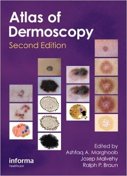
An Atlas of Dermoscopy, 2ed
Building on a successful first edition, this revised and extended Atlas of Dermoscopy demonstrates the state of the art of how to use dermoscopy to detect and diagnose lesions of the skin, with a special emphasis on malignant skin tumours. With well over 1,500 photographs, drawings, and tables, the book has extensive clinical correlation with dermoscopic images, so readers can appreciate the added benefits of dermoscopy by comparing the clinical morphology seen with the naked eye with the corresponding dermoscopic morphology; extensive illustrations from the image collections of internationally recognized experts, who have years of experience refining their techniques; and extensive schematic drawings to help readers single out the key structures and patterns to recognize in the dermoscopic images.
The second edition has important new material on such topics as observed differences between polarized and non-polarized dermoscopy, newly recognized structures and patterns, refined and revised suggestions for pattern analysis, dermoscopy of the hair and nails, and how to integrate dermoscopy into general clinical practice. It also covers dermoscopically equivocal, false negative, and false positive lesions; discusses further indications for dermoscopy beyond skin cancer; and details common checklists of criteria and algorithms used to diagnose skin lesions.
Features
- Discusses differences in dermoscopic criteria depending on anatomical location and computer-assisted diagnosis
- Stresses the dermoscopist’s role in prevention and early detection
- Explores the latest uses for dermoscopy, including diagnosing scabies, evaluating paranychial capillaries, and observing vascular aspects of cutaneous diseases
- Covers important melanocytic, non-melanocytic, benign, and malignant pigmented skin lesions
- Contains more than 1,500 figures, tables, and schematics
Review
“…The atlas is unputdownable. It also features something that many dermoscopy books lack: all of the key descriptive features of the images are included in a box just beneath the picture. There is no need to search for information….The chapters are authored by the most prominent experts in the field….
…I am confident that the informative text and spectacular images will trigger continued enthusiasm.”
―Journal of the American Academy of Dermatology
About the Author
Ashfaq Marghoob: Director of Memorial Sloan-Kettering Cancer Center’s regional skin cancer clinic in Hauppauge, Long Island, USA.
Ralph Braun: Department of Dermatology, University of Zurich, Switzerland.
Josep Malvehy: Director, Melanoma Unit, University Hospital Clinic, Barcelona, Spain. Board member of International Dermoscopy Society
Contents
Chapter 1 Introduction
Chapter 2 Principles of dermoscopy and dermoscopic equipment
Chapter 3 Histopathologic tissue correlations of dermoscopic structures
Chapter 4 Two-step algorithm: Differentiating melanocytic from nonmelanocytic lesions
Chapter 5a Basal cell carcinoma
Chapter 5b Actinic keratosis, Bowen’s disease, keratoacanthoma, and squamous cell carcinoma
Chapter 5c Solar lentigines, seborrheic keratoses, and lichen planus-like keratosis
Chapter 5d Vascular lesions
Chapter 5e Dermatofibromas
Chapter 6a Analytic features of melanoma vs benign nevi on nonglabrous skin: Colors and structures
Chapter 6b Pattern analysis
Chapter 6c ABCD rule
Chapter 6d Menzies method
Chapter 6e Seven-point checklist and the seven rules not to miss melanorma incognito
Chapter 6f CASH algorithm
Chapter 6g Three-point checklist
Chapter 6h Revised version of pattern analysis
Chapter 7a Congenital melanocytic nevi
Chapter 7b Dysplastic [atypical) nevi
Chapter 7c Banal nevi and growing nevi
Chapter 7d Blue nevi and variants
Chapter 7e Spitz and Reed nevi
Chapter 7f Recurrent [persistent) nevi
Chapter Sa Superficial spreading melanoma
Chapter 8b Acrolentiginous melanoma
Chapter 8c Nodular melanoma
Chapter 8d Lentigo maligna melanoma
Chapter 8e Amelanotic and hypomelanotic melanoma
Chapter 8f Equivocal lesions, false positive, and false negative
Chapter 8g Desmoplastic melanoma and metastatic melanoma
Chapter 9a Palms and soles
Chapter 9b Nails
Chapter 9c Face
Chapter 9d Hair and scalp [trichoscopy)
Chapter 9e Mucosal lesions
Chapter 10 Exceptions to the two-step dermoscopy algorithm
Chapter 11 Dermoscopy in general dermatology
Chapter 12 Vascular structures
Chapter 13 Diagnostic accuracy of dermoscopy
Chapter 14 Followup of melanocytic skin lesions with digital dermoscopy
Chapter 15 Teledermoscopy and computer-assisted diagnosis
Chapter 16 Beyond dermoscopy
Chapter 17 Concluding remarks
لینک کوتاه : https://bookbaz.ir/?p=26041
نویسنده : Ashfaq A Marghoob , Josep Malvehy
ناشر : CRC Press; 2 edition
سال انتشار : 2012
زبان کتاب : انگلیسی
نوع فایل : PDF
تعداد صفحات : 396
(ISBN) شابک : 0415458951
قیمت کتاب درآمازون : $242.03
حجم فایل : 37 MB
کتاب های مرتبط:
 دانلود کتاب دانشنامه درماتولوژی (۶ جلدی)
Encyclopedia of Dermatology, 6-Vol, 1ed
 دانلود کتاب جراحی پوست کانتور
Dermatologic Surgery, 1ed
 دانلود کتاب روش های عملی در جراحی پلاستیک ، زیبایی و ترمیمی (ویرایش ۲۰۱۶)
Operative Procedures in Plastic, Aesthetic and Reconstructive Surgery, 1ed
 دانلود کتاب راهنمای درمان پوستی
 Manual of Dermatologic Therapeutics, 8ed
 دانلود کتاب پزشکی زیبایی: رشد تمرینات شما
Aesthetic Medicine: Growing Your Practice, 1ed
 دانلود کتاب درماپاتولوژی الستون
Dermatopathology, 3ed
 دانلود کتاب درموسکوپی: راهنمای خود ارزیابی مصور
Dermoscopy: An Illustrated Self-Assessment Guide, 2ed
 دانلود کتاب درماتولوژی برای ارائه دهنده مراقبت های اولیه
Dermatology for the Primary Care Provider, 1ed
 دانلود کتاب راهنمای عملی کاشت مو + ویدئو
Practical Guide to Hair Transplantation, 1ed + Video
 دانلود کتاب درماتولوژی در پزشکی عمومی فیتزپاتریک
Fitzpatrick’s Dermatology in General Medicine, 2-Vol, 8ed
 دانلود کتاب هنر ترمیم و جوانسازی پوست اوباگی
The Art of Skin Health Restoration and Rejuvenation, 2ed
 دانلود کتاب درماتولوژی برای تمرین پیشرفته پزشکان
Dermatology for Advanced Practice Clinicians, 1ed
 دانلود کتاب آلوپسی
 Alopecia, 1ed
 دانلود کتاب درماتوپاتولوژی پرایمر بیماری های التهابی
Dermatopathology Primer of Inflammatory Diseases, 1ed
 دانلود کتاب بورد بررسی تخصص درماتولوژی مک هیل
McGraw-Hill Specialty Board Review Dermatology, 3ed
 دانلود کتاب اطلس ویدئویی جراحی پلاستیک زیبایی غیوران
Aesthetic Plastic Surgery Video Atlas, 1ed
 دانلود کتاب درماتولوژی زیبایی: محصولات و رویه ها
Cosmetic Dermatology: Products and Procedures, 3ed
 دانلود کتاب ایمونوهیستوشیمی کاربردی در ارزیابی نئوپلاسم پوست
Applied Immunohistochemistry in the Evaluation of Skin Neoplasms, 1ed
 دانلود کتاب فناوری های نوظهور در کانتورینگ صورت و بدن
Emerging Technologies in Face and Body Contouring, 1ed
 دانلود کتاب اطلس رنگی بیماری های دهان و دندان: تشخیص و درمان
Color Atlas of Oral Diseases: Diagnosis and Treatment, 4ed
 دانلود کتاب آنتی اکسیدان ها و پوست
 Antioxidants and the Skin, 2ed
 دانلود کتاب راهنمای دوز سم بوتولینوم
 Botulinum Toxin Dosing Manual, 1ed
 دانلود کتاب آرایشی بهداشتی دارویی: روش های درماتولوژی آرایشی بهداشتی
Cosmeceuticals: Procedures in Cosmetic Dermatology Series, 3ed
 دانلود کتاب درمان های ناخن: تمرین بالینی فعلی
Nail Therapies: Current Clinical Practice, 2ed
 دانلود کتاب بازسازی صورت ترکیبی بعد از جراحی موس
Combination Facial Reconstruction after Mohs Surgery, 1ed
 دانلود کتاب راهنمای جوشش و واکنش دارو لیت
Litt’s Drug Eruption and Reaction Manual, 21ed
 دانلود کتاب راهنمای جراحی زیبایی پوست
 Concise Manual of Cosmetic Dermatologic Surgery, 1ed
 دانلود کتاب پیری پوست (ویرایش ۲۰۱۷)
Textbook of Aging Skin, 2ed
 دانلود کتاب کاشت مو + ویدئو
Hair Transplantation, 5ed + Video
 دانلود کتاب سموم بوتولینوم: کاربردهای زیبایی و بالینی
Botulinum Toxins: Cosmetic and Clinical Applications, 1ed
 دانلود کتاب بازسازی صورت پس از جراحی ماز
Facial Reconstruction Post-Mohs Surgery, 1ed
 دانلود کتاب مراقبت سوختگی کامل + ویدئو
Total Burn Care, 5ed + Video
 دانلود کتاب راهنمای جراحی پلاستیک
 Handbook of Plastic Surgery, 1ed
 دانلود کتاب لارو درمانی: کتابچه درمان زخم به کمک لارو
Maggot Therapy: A Handbook of Maggot-Assisted Wound Healing, 1ed
 دانلود کتاب راهنمای جوش و واکنش دارویی لیت
Litt’s Drug Eruption & Reaction Manual, 24ed
 دانلود کتاب درماتومیوزیت: تشخیص، عوامل خطر و گزینه های درمان
Dermatomyositis: Diagnosis, Risk Factors and Treatment Options, 1ed
 دانلود کتاب جراحی پوست: روند درماتولوژی + ویدئو
Surgery of the Skin: Procedural Dermatology, 3ed + Video
 دانلود کتاب مراقبت های اولیه درماتولوژی
 Textbook of Primary Care Dermatology, 1ed
 دانلود کتاب درمان اختلالات ناخن
 Therapies for Nail Disorders, 1ed
 دانلود کتاب درماتولوژی: یک متن رنگی مصور
Dermatology: An Illustrated Colour Text, 7ed
 دانلود کتاب اطلس درموسکوپی دستگاه تناسلی
Atlas of Genital Dermoscopy, 1ed
 دانلود کتاب اطلس بیماری ولوواژینال در انواع پوست تیره تر
Atlas of Vulvovaginal Disease in Darker Skin Types, 1ed
 دانلود کتاب هیپوپیگمانتاسیون
Hypopigmentation, 1ed
 دانلود کتاب راهنمای جوشش و واکنش دارویی لیت
Litt’s Drug Eruption & Reaction Manual, 30ed
 دانلود کتاب پیشرفتهای اخیر در پزشکی پوست (جلد سوم)
Recent Advances in Dermatology, Vol3
 دانلود کتاب اطلس رنگی درماتولوژی بالینی فیتزپاتریک
Fitzpatrick’s Color Atlas and Synopsis of Clinical Dermatology, 7ed
 دانلود کتاب درمان پوست های رنگی
Treatments for Skin of Color: Expert Consult, 1ed
 دانلود کتاب درماتولوژی کودکان شاکنر (۲ جلدی)
Pediatric Dermatology: Expert Consult, 2-Vol, 4ed
 دانلود کتاب تمرین پزشکی عمومی جان مورتاگ
John Murtagh’s General Practice, 6ed
 دانلود کتاب راهنمای عملی برای روشهای زیبایی سم بوتولینوم
A Practical Guide to Botulinum Toxin Procedures, 1ed
 دانلود کتاب ملزومات پاتولوژی پوست ویدون
Weedon’s Skin Pathology Essentials, 2ed
 دانلود کتاب اطلس پاتولوژی مو با ارتباط بالینی
An Atlas of Hair Pathology with Clinical Correlations, 2ed
 دانلود کتاب آلوپسی: تشخیص و درمان
The Alopecias: Diagnosis and Treatments, 1ed
 دانلود کتاب درماتولوژی زیبایی و آرایشی (ویرایش ۲۰۱۷)
Textbook of Cosmetic Dermatology, 5ed
دانلود کتاب کاشت موHair Transplantation, 5ed
 دانلود کتاب هایپرپیگمنتیشن
 Hyperpigmentation, 1ed
 دانلود کتاب اطلس رنگی و چکیده درماتولوژی کودکان
Color Atlas and Synopsis of Pediatric Dermatology, 3ed
 دانلود کتاب مراقبت های اورژانسی درماتولوژی: تشخیص مبتنی بر علائم
Urgent Care Dermatology: Symptom-Based Diagnosis, 1ed
 دانلود کتاب PRP و میکرونیدلینگ در پزشکی زیبایی + ویدئو
PRP and Microneedling in Aesthetic Medicine, 1ed + Video
 دانلود کتاب روغن های ضروری: برخورد با آلرژی و ترکیب شیمیایی
Essential Oils: Contact Allergy and Chemical Composition, 1ed
 دانلود کتاب حجم دهی صورت با فیلر
Facial Volumization with Fillers, 1ed
 دانلود کتاب پاتولوژی تومورهای ملانوسیتیک
 Pathology of Melanocytic Tumors, 1ed
 دانلود کتاب درماتولوژی بالینی کودکان پالر، مانچینی و هورویتز
Paller and Mancini – Hurwitz Clinical Pediatric Dermatology, 6ed
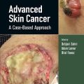 دانلود کتاب سرطان پوست پیشرفته: رویکردی مبتنی بر مورد
Advanced Skin Cancer: A Case-Based Approach, 1ed
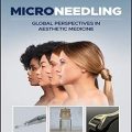 دانلود کتاب میکرونیدلینگ: دیدگاه های جهانی در پزشکی زیبایی
Microneedling: Global Perspectives in Aesthetic Medicine, 1ed
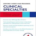 دانلود کتاب ارزیابی و پیشرفت آکسفورد: تخصص های بالینی
 Oxford Assess and Progress: Clinical Specialties, 4ed
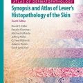 دانلود کتاب اطلس درماتوپاتولوژی: خلاصه و اطلس هیستوپاتولوژی پوست لِور
Atlas of Dermatopathology: Synopsis and Atlas of Lever’s Histopathology of the Skin, 4ed
 دانلود کتاب درموسکوپی عملی
Practical Dermoscopy, 1ed
 دانلود کتاب علم آرادارو در عمل بالینی
Cosmeceutical Science in Clinical Practice, 2ed
 دانلود کتاب درموسکوپی تشخیصی: راهنمای مصور
Diagnostic Dermoscopy: The Illustrated Guide, 1ed
 دانلود کتاب ۲۵۰ مورد در پزشکی بالینی
 ۲۵۰Cases in Clinical Medicine, 4ed
 دانلود کتاب درماتولوژی کودکان کوهن
Cohen’ Pediatric Dermatology, 4ed
 دانلود کتاب درمان بیماریهای پوستی: راهکارهای جامع درمانی
Treatment of Skin Disease: Comprehensive Therapeutic Strategies, 4ed
 دانلود کتاب اطلس اجمالی هیستوپاتولوژی پوست لِوِر
Atlas and Synopsis of Lever’s Histopathology of the Skin, 3ed
 دانلود کتاب درماتولوژی بالینی کودکان هورویتز: اختلالات پوستی دوران کودکی و نوجوانی
Hurwitz Clinical Pediatric Dermatology: Skin Disorders of Childhood and Adolescence, 5ed
 دانلود کتاب لیزر و نورها: سری روش ها در زیبایی آرایشی و بهداشتی پوست
Lasers and Lights: Procedures in Cosmetic Dermatology Series, 3ed
 دانلود کتاب راهنمای پزشکی هریسون
Harrisons Manual of Medicine, 19ed
 دانلود کتاب اطلس رنگی پوست
 Color Atlas of Dermatology, 1ed
 دانلود کتاب اطلس رنگی و اجمالی بیماریهای مقاربتی هندزفیلد
Color Atlas & Synopsis of Sexually Transmitted Diseases, 3ed
 دانلود کتاب تشخیص و درمان اختلالات مو: اطلس مبتنی بر شواهد
Diagnosis and Treatment of Hair Disorders: An Evidence-Based Atlas, 1ed
 دانلود کتاب اطلس اختلالات رنگدانه پوست
Atlas of Pigmentary Disorders, 1ed
 دانلود کتاب اطلس رنگی درماتولوژی زیبایی
Color Atlas of Cosmetic Dermatology, 2ed
 دانلود کتاب درماتوپاتولوژی: تشخیص توسط برداشت اول (ویرایش ۲۰۱۶)
Dermatopathology: Diagnosis by First Impression, 3ed
 دانلود کتاب بیماری های ناخن کودکان
Pediatric Nail Disorders, 1ed
 دانلود کتاب اصول و عمل درماتولوژیک در انکولوژی: شرایط پوست، مو و ناخن در بیماران مبتلا به سرطان
Dermatologic Principles and Practice in Oncology: Conditions of the Skin, Hair, and Nails in Cancer Patients, 1ed
 دانلود کتاب اطلس رنگی و چکیده درماتولوژی بالینی فیتزپاتریک (ویرایش ۲۰۱۷)
Fitzpatrick’s Color Atlas and Synopsis of Clinical Dermatology, 8ed
 دانلود کتاب درماتولوژی برای USMLE
Dermatology for the USMLE, 1ed
 دانلود کتاب اطلس بالینی بیماری های پوست اندرو
Andrews’ Diseases of the Skin Clinical Atlas, 1ed
 دانلود کتاب درماتولوژی: یک متن مصور رنگی
Dermatology: An Illustrated Colour Text, 6ed
 دانلود کتاب سرطان پوست
 Cancer of the Skin, 2ed
 دانلود کتاب درماتولوژی در محیط های سلامت عمومی
Dermatology in Public Health Environments, 1ed
 دانلود کتاب پسوریازیس و آرتریت پسوریاتیک: پاتوفیزیولوژی، درمان مداخله ای و پزشکی مکمل
Psoriasis and Psoriatic Arthritis: Pathophysiology, Therapeutic Intervention, and Complementary Medicine, 1ed
 دانلود کتاب پیوند مو
 Hair Transplantation, 1ed
 دانلود کتاب تصویربرداری در درماتولوژی
 Imaging in Dermatology, 1ed
 دانلود کتاب جذام IAL
Ial Textbook of Leprosy, 2ed
 دانلود کتاب دانشنامه جوان سازی زیبایی از طریق افزایش حجم + ویدئو
Encyclopedia of Aesthetic Rejuvenation Through Volume Enhancement, 1ed + Video
 دانلود کتاب جراحی پلاستیک زیبایی در آسیایی ها: اصول و تکنیک ها (۲ جلدی) + ویدئو
Aesthetic Plastic Surgery in Asians: Principles and Techniques, 2-Vol, 1ed + Video
 دانلود کتاب راهنمای علوم و فناوری های زیبایی
 Handbook of Cosmetic Science and Technology, 4ed
 دانلود کتاب بیماری های ناخن و مدیریت آن باران و داوبر
Baran and Dawber’s Diseases of the Nails and their Management, 5ed
 دانلود کتاب اختلالات ناخن
 Nail Disorders, 1ed
 دانلود کتاب راهنمای عملی برای روش های پرکننده پوستی
 A Practical Guide to Dermal Filler Procedures, 1ed
 دانلود کتاب بیماری واژن: نقض غیر علمی
Vulvar Disease: Breaking the Myths, 1ed
 دانلود کتاب پرکننده های تزریقی: اصول و تمرین
Injectable Fillers: Principles and Practice, 1ed
 دانلود کتاب موارد چالش برانگیز در درماتولوژی: تشخیص پیشرفته و مدیریت تاکتیک
Challenging Cases in Dermatology: Advanced Diagnoses and Management Tactics, 1ed
 دانلود کتاب جامع ویتیلیگو
 Comprehensive Textbook on Vitiligo, 1ed
 دانلود کتاب تریکولوژی IADVL
IADVL Textbook of Trichology, 1ed
 دانلود کتاب درمان بیماری های پوستی: راهبردهای جامع درمانی
Treatment of Skin Disease: Comprehensive Therapeutic Strategies, 6ed
 دانلود کتاب رتینوئیدها در درماتولوژی
Retinoids in Dermatology, 1ed
 دانلود کتاب راهنمای جامع هیدرادنیت چرکی + ویدئو
A Comprehensive Guide to Hidradenitis Suppurativa, 1ed + Video
 دانلود کتاب سم بوتولینوم در پزشکی زیبایی: پروتکل های تزریق و مدیریت عوارض
Botulinum Toxin in Aesthetic Medicine: Injection Protocols and Complication Management, 1ed
 دانلود کتاب کاشت مو
Hair Transplantation, 6ed
 دانلود کتاب اطلس بازسازی آناتومیک پس از جراحی سرطان پوست
Atlas of Anatomic Reconstruction After Skin Cancer Surgery, 1ed
 دانلود کتاب درماتولوژی بالینی
Clinical Dermatology, 5ed
 دانلود کتاب بیماری های پوست اندرو: درماتولوژی بالینی
Andrews’ Diseases of the Skin: Clinical Dermatology, 12ed
 دانلود کتاب پوست و جراحی کوچک براون: متن و اطلس رنگی
Brown’s Skin and Minor Surgery: A Text & Colour Atlas, 5ed
 دانلود کتاب پزشکی تورنتو نوت (نسخه ۲۰۱۵)
Toronto Notes 2015 (Essential Med Notes), 31ed
 دانلود کتاب ملزومات پزشکی دیویدسون
Davidson’s Essentials of Medicine, 2ed
 دانلود کتاب حقایق سریع در درماتولوژی فری: راهنمای عملی برای بیماریها و اختلالات پوستی
Ferri’s Fast Facts in Dermatology: A Practical Guide to Skin Diseases and Disorders, 1ed
 دانلود کتاب آسیب شناسی پوست ویدون
Weedon’s Skin Pathology, 4ed
 دانلود کتاب درماتوسکوپی تومورهای پوست غیر رنگی
Dermatoscopy of Non-Pigmented Skin Tumors: Pink – Think – Blink
 دانلود کتاب ریزش مو و ترمیم
 Hair Loss and Restoration, 2ed
 دانلود کتاب درماتولوژی زیبایی: محصولات و روشها
Cosmetic Dermatology: Products and Procedures, 2ed
 دانلود کتاب آسیب و عفونت پوست و بافت نرم
 Skin and Soft Tissue Injuries & Infections, 1ed
 دانلود کتاب آسیب شناسی پوست مک کی (۲ جلدی)
McKee’s Pathology of the Skin, 2-Vol, 4ed
 دانلود کتاب تصمیم گیری و اجتناب از عوارض در فلپ پوست
 Making Decisions and Avoiding Complications in Skin Flaps, 1ed
 دانلود کتاب درماتولوژی بالینی لانگ
Clinical Dermatology (Lange Medical Books), 1ed
 دانلود کتاب پرولیفراسیون ملانوسیتیک: کتاب جامع ضایعات رنگی
The Melanocytic Proliferations: A Comprehensive Textbook of Pigmented Lesions, 2ed
 دانلود کتاب راهنمای مطالعه تشخیص پزشکی و درمان کارنت
CURRENT Medical Diagnosis and Treatment Study Guide, 2ed
 دانلود کتاب تشخیص افتراقی در درماتولوژی
 Differential Diagnosis in Dermatology, 4ed
 دانلود کتاب راهنمای بالینی برای پوست و مراقبت از زخم
Clinical Guide to Skin and Wound Care, 7ed
 دانلود کتاب راهنمای جوشش و واکنش دارویی لیت
Litt’s Drug Eruption and Reaction Manual, 22ed
 دانلود کتاب اختلالات پوستی ژنتیکی (ویرایش ۲۰۱۷)
Genetic Skin Disorders, 3ed
 دانلود کتاب راهنمای سفر و پزشکی گرمسیری
 The Travel and Tropical Medicine Manual, 5ed
 دانلود کتاب پاتولوژی تشخیصی: درماتوپاتولوژی غیرنئوپلاستیک
Diagnostic Pathology: Nonneoplastic Dermatopathology, 2ed
دانلود کتاب سموم بوتولینوم: کاربردهای زیبایی و بالینی + ویدئوBotulinum Toxins: Cosmetic and Clinical Applications, 1ed + Video
 دانلود کتاب بیماری پوست: تشخیص و درمان
Skin Disease: Diagnosis and Treatment, 4ed
 دانلود کتاب روش های اصلی در جراحی پلاستیک نلیگان + ویدئو
Core Procedures in Plastic Surgery, 1ed + Video
 دانلود کتاب اصول درماتولوژی لوکینگبیل و مارکس
Lookingbill and Marks’ Principles of Dermatology, 6ed
 دانلود کتاب روش های بالینی در لیزر جوان سازی پوست
 Clinical Procedures in Laser Skin Rejuvenation, 1ed
 دانلود کتاب نوروتوکسین و پر کننده در جراحی زیبایی صورت
Neurotoxins and Fillers in Facial Esthetic Surgery, 1ed
 دانلود کتاب اطلس درماتوپاتولوژی: تومور، خال و کیست
Atlas of Dermatopathology: Tumors, Nevi, and Cysts, 1ed
 دانلود کتاب تشخیص های درماتولوژی واژن
Vulval Dermatologic Diagnosis, 1ed
دانلود کتاب PRP و میکرونیدلینگ در پزشکی زیباییPRP and Microneedling in Aesthetic Medicine, 1ed
 دانلود کتاب راهنمای مصور لنفوم پوستی
 Skin Lymphoma: The Illustrated Guide, 5ed
 دانلود کتاب پزشکی اورژانس کودکان
Pediatric Emergency Medicine, 2ed
 دانلود کتاب پرکننده های پوستی: تکنیک های آناتومی و تزریق صورت + ویدئو
Dermal Fillers: Facial Anatomy and Injection Techniques, 1ed + Video
 دانلود کتاب اطلس رنگی درموسکوپی
 Colour Atlas of Dermoscopy, 1ed
 دانلود کتاب پاتولوژی پوست ویدون
Weedon’s Skin Pathology, 5ed
 دانلود کتاب PRF در زیبایی صورت
PRF in Facial Esthetics, 1ed
 دانلود کتاب راهنمای مختصر نیدلینگ پوستی
 The Concise Guide to Dermal Needling, 3ed