
دانلود کتاب اطلس رادیوگرافی رشد اسکلتی
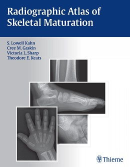
Radiographic Atlas of Skeletal Maturation, 1ed
The value of this atlas is to provide appropriate standards for the maturing skeleton {that} will enhance accuracy and ease interpretation — From the Foreword by Theodore E. Keats, MD, Former Alumni Professor of Radiology, University of Virginia Health Sciences Center
When dealing with the maturing skeleton and its many complex growth alterations, physicians are constantly faced with the question: Is this image normal? TheRadiographic Atlas of Skeletal Maturation succinctly answers that question by providing a comprehensive set of male and female reference images for every age and body part. This allows physicians to quickly hone in on normal ranges for the specific case they are reviewing–particularly useful when called upon to read a pediatric skeletal radiograph in the emergency room or while on call.
Special Features:
- Access to nearly 2,300 high-quality images that provide instant reference to normal views of the skeleton at every developmental milestone-available in both the text and accompanying DVD
- Multiple projections at every age, sex, and body part combination so that the user can match the reference points in the book to the case at hand and arrive at a solid clinical interpretation
- Practical text layout organized by gender and body part that provides quick access to images of normal development at any given age
- A software virtual skeletal survey demonstrates images of younger and older individuals and crystallizes the subtle variations in growth patterns
- Powerful software package with advanced image enhancement tools allows optimization of atlas image details for greater clarity. Compatible with numerous image formats (including DICOM) allowing viewing and editing of outside images
- Convenient growth charts included in the book and DVD
This unique resource, with its vast collection of print and DVD images of normal progressive skeletal development, gives physicians the full range of comparative information they need to interpret pediatric skeletal radiographs in any clinical setting. It is the reference standard for radiologists, pediatricians, orthopedists, emergency room physicians, internists, rehabilitation physicians, and training physicians who are called upon to review a pediatric radiograph and confidently make a diagnosis.
About the Author
Contents
۱. Male Skull
۲. Male Cervical Spine
۳. Male Thoracic Spine
۴. Male Lumbar Spine
۵. Male Chest
۶. Male Shoulder
۷. Male Humerus
۸. Male Elbow
۹. Male Forearm
۱۰. Male Wrist
۱۱. Male Hand
۱۲. Male Pelvis
۱۳. Male Femur
۱۴. Male Knee
۱۵. Male Tibia and Fibula
۱۶. Male Ankle
۱۷. Male Foot
۱۸. Female Skull
۱۹. Female Cervical Spine
۲۰. Female Thoracic Spine
۲۱. Female Lumbar Spine
۲۲. Female Chest
۲۳. Female Shoulder
۲۴. Female Humerus
۲۵. Female Elbow
۲۶. Female Forearm
۲۷. Female Wrist
۲۸. Female Hand
۲۹. Female Pelvis
۳۰. Female Femur
۳۱. Female Knee
۳۲. Female Tibia and Fibula
۳۳. Female Ankle
۳۴. Female Foot
لینک کوتاه : https://bookbaz.ir/?p=82136
نویسنده : S. Lowell Kahn , Christopher M. Gaskin
ناشر : Thieme; 1 edition
سال انتشار : 2012
زبان کتاب : انگلیسی
نوع فایل : PDF
تعداد صفحات : 621
(ISBN) شابک : 1604065710
قیمت کتاب درآمازون : $244.34
حجم فایل : 43 MB
کتاب های مرتبط:
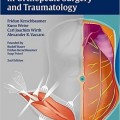 دانلود کتاب روشهای عملی در جراحی ارتوپدی و تروما
Operative Approaches in Orthopedic Surgery and Traumatology, 2ed
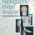 دانلود کتاب آنالیز تصویر رادیوگرافی
 Radiographic Image Analysis, 4ed
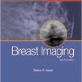 دانلود کتاب تصویربرداری پستان: ضروریات در رادیولوژی
Breast Imaging: The Requisites (Requisites in Radiology), 2ed
 دانلود کتاب تثبیت داخلی در پوکی استخوان
 Internal Fixation in Osteoporotic Bone, 1ed
 دانلود کتاب راهنمای مدیریت شکستگی AO: فیکساتور های داخلی
AO Manual of Fracture Management: Internal Fixators, 1ed
 دانلود کتاب پوکی استخوان مارکوس و فلدمن (۲ جلدی)
Osteoporosis, 2-Volume Set, Fourth Edition
 دانلود کتاب تکنیک های پیشرفته در انتقال تصویر جراحی مغز و ستون فقرات
 Advanced Techniques in Image-Guided Brain and Spine Surgery, 1ed
 دانلود کتاب جراحی ستون فقرات حداقل تهاجمی + ویدئو
Minimally Invasive Spine Surgery, 1ed + Video
 دانلود کتاب اصول تصویربرداری کودکان
 Fundamentals of Pediatric Imaging, 2ed
 دانلود کتاب اطلس آسیب شناسی بافت نرم و استخوان: با بافت شناسی، سیتولوژی و رادیولوژی
Atlas of Soft Tissue and Bone Pathology: With Histologic, Cytologic, and Radiologic Correlations, 1ed
 دانلود کتاب تومورهای استخوان و بافت نرم: بررسی چند رشته با ارائه مورد
Bone and Soft Tissue Tumours: A Multidisciplinary Review With Case Presentations, 1ed
 دانلود کتاب عمل جراحی اضطراری دست (ویرایش ۲۰۱۷)
Emergency Surgery of the Hand, 1ed
 دانلود کتاب ابزار دقیق ستون فقرات: تکنیک های جراحی
Spinal Instrumentation: Surgical Techniques, 1ed
 دانلود کتاب راهنمای ارتوپدی برای GP امروزی
An Orthopaedics Guide for Today’s GP, 1ed
 دانلود کتاب اطلس تعیین سن اسکلت: MRI دست و مچ دست در کودکان
Text-Atlas of Skeletal Age Determination: MRI of the Hand and Wrist in Children, 1ed
 دانلود کتاب جیبی ارتوپدی تروما و روماتولوژی چرچیل
Churchill’s Pocketbook of Orthopaedics, Trauma and Rheumatology, 2ed
 دانلود کتاب بی ثباتی شانه در ورزشکار
 Shoulder Instability in the Athlete, 1ed
 دانلود کتاب تصویربرداری تشخیصی نقایص مادرزادی قلب
Diagnostic Imaging of Congenital Heart Defects, 1ed
 دانلود کتاب کاربردهای بالینی پرینت سه بعدی در جراحی پا و مچ پا
 Clinical Applications of 3D Printing in Foot and Ankle Surgery, 1ed
 دانلود کتاب اندام فوقانی کودکان
 The Pediatric Upper Extremity, 2015th
 دانلود کتاب سونوگرافی تشخیصی روماک (۲ جلدی)
Diagnostic Ultrasound, 2-Volume Set 6th Edition
 دانلود کتاب خط مرزی و عادی اولیه پاتولوژی فین فریشمیت کوهلر/زیمر
Freyschmidt’s ,Koehler/Zimmer, Borderlands of Normal and Early Pathological Fin, 5ed
 دانلود کتاب راهنمای اکوکاردیوگرافی: همگام با کتاب اکوکاردیوگرافی بالینی اوتو
Echocardiography Review Guide: Companion to the Textbook of Clinical Echocardiography, 3ed
 دانلود کتاب MRI اسکلتی عضلانی آسیف سفیودین
Asif Saifuddin’s Musculoskeletal MRI, 2ed
 دانلود کتاب سرطان پستان: هنر و علم تشخیص زود هنگام با ماموگرافی
Breast Cancer: The Art and Science of Early Detection with Mammography, 1ed
 دانلود کتاب سری کارشناسی ستون فقرات AO: ناهنجاری های ستون فقرات کودکان (جلد ۹)
AOSpine Masters Series, Volume 9: Pediatric Spinal Deformities, 1ed
 دانلود کتاب درمان جراحی و پزشکی پوکی استخوان
 Surgical and Medical Treatment of Osteoporosis, 1ed
 دانلود کتاب نورورادیولوژی: تشخیص های افتراقی کلیدی و سوالات بالینی
Neuroradiology: Key Differential Diagnoses and Clinical Questions, 2ed
 دانلود کتاب اشعه ایکس اسکلتی عضلانی برای دانشجویان و کارآموزان پزشکی
Musculoskeletal X-Rays for Medical Students and Trainees, 1ed
 دانلود کتاب ۳ تفاوت برتر در پزشکی هسته ای
 Top 3 Differentials in Nuclear Medicine, 1ed
 دانلود کتاب تصویربرداری تشخیصی: بیماری اسکلتی عضلانی غیر تروماتیک + ویدئو
Diagnostic Imaging: Musculoskeletal Non-Traumatic Disease, 3ed + Video
 دانلود کتاب رادیولوژی حوادث و اورژانس: راهنمای بقا
Accident and Emergency Radiology: A Survival Guide, 3ed
 دانلود کتاب مدیریت شکستگی برای مراقبت های اولیه
Fracture Management for Primary Care, 3ed
 دانلود کتاب تصویربرداری سر و گردن مقدماتی
Introductory Head and Neck Imaging, 1ed
 دانلود کتاب سونوگرافی تشخیصی: سر و گردن
Diagnostic Ultrasound: Head and Neck, 1ed
 دانلود کتاب ارتوپدی اضطراری سیمون + ویدئو
Simon’s Emergency Orthopedics, 7ed + Video
 دانلود کتاب اطلس جیبی اکوکاردیوگرافی
 Pocket Atlas of Echocardiography, 2ed
 دانلود کتاب راهنمای گام به گام اولتراسوند شکمی
 Abdominal Ultrasound: Step by Step, 3ed
 دانلود کتاب بیولوژیک در جراحی ارتوپدی
 Biologics in Orthopaedic Surgery, 1ed
 دانلود کتاب روش های کلارک در تصویربرداری تشخیصی
 Clark’s Procedures in Diagnostic Imaging, 1ed
 دانلود کتاب اطلس آندوسکوپی تشخیصی
Atlas of Diagnostic Endoscopy, 3ed
 دانلود کتاب اسکولیوز ایدیوپاتیک
 Idiopathic Scoliosis, 2ed
 دانلود کتاب ملزومات عملی شدت مدوله پرتو درمانی
Practical Essentials of Intensity Modulated Radiation Therapy, 3ed
 دانلود کتاب تصویربرداری در گوش و حلق و بینی
Imaging in Otolaryngology, 1ed
 دانلود کتاب اپیدمیولوژی بالینی تروما ارتوپدی
Clinical Epidemiology of Orthopaedic Trauma, 2ed
 دانلود کتاب اصول رادیولوژی دهان و فک و صورت
Fundamentals of Oral and Maxillofacial Radiology, 1ed
 دانلود کتاب بیومکانیک ستون فقرات: مفاهیم اساسی، اختلالات نخاعی و درمان
Biomechanics of the Spine: Basic Concepts, Spinal Disorders and Treatments, 1ed
 دانلود کتاب تعویض ترنس کاتتر دریچه آئورت
Transcatheter Aortic Valve Implantation, 1ed
 دانلود کتاب جراحی ارتوپدی جامع چاپمن (۴ جلدی)
Chapman’s Comprehensive Orthopaedic Surgery, 4ed
 دانلود کتاب توموگرافی پرتو مخروطی کامپیوتری در ارتودنسی: موارد مصرف، بینش و نوآوری
Cone Beam Computed Tomography in Orthodontics: Indications, Insights, and Innovations, 1ed
 دانلود کتاب سونوگرافی عروقی عملی: راهنمای مصور
Practical Vascular Ultrasound: An Illustrated Guide, 1ed
 دانلود کتاب EMG آسان: راهنمای انجام مطالعات هدایت عصب و الکترومیوگرافی + ویدئو
Easy EMG: A Guide to Performing Nerve Conduction Studies and Electromyography, 2ed + Video
 دانلود کتاب رویکردهای بیولوژیکی برای ترمیم و بازسازی دیسک ستون فقرات برای پزشکان
Biological Approaches to Spinal Disc Repair and Regeneration for Clinicians, 1ed
 دانلود کتاب تصویربرداری شکم: الزامات اصلی
Abdominal Imaging: The Core Requisites, 1ed
 دانلود کتاب اطلس روشهای ارتوپدی مداخله ای
 Atlas of Interventional Orthopedics Procedures, 1ed
 دانلود کتاب تصویربرداری انکولوژیک: یک رویکرد چند رشته ای
Oncologic Imaging: A Multidisciplinary Approach, 2ed
 دانلود کتاب پوکی استخوان مارکوس و فلدمن (۲ جلدی)
Marcus and Feldman’s Osteoporosis, 5ed
 دانلود کتاب رادیولوژی پا و مچ پا
Foot and Ankle Radiology, 2ed
 دانلود کتاب سری کارشناسی ستون فقرات AO: تغییر شکل ستون فقرات بزرگسالان (جلد ۴)
AOSpine Masters Series, Volume 4: Adult Spinal Deformities, 1ed
 دانلود کتاب اطلس رادیولوژی اورژانس: سیستم عروقی، قفسه سینه، شکم و لگن و سیستم تولید مثل
Atlas of Emergency Radiology: Vascular System, Chest, Abdomen and Pelvis, and Reproductive System 2015th
 دانلود کتاب آندوسونوگرافی + ویدئو
Endosonography, 3ed + Video
 دانلود کتاب سری کارشناسی ستون فقرات AO: عفونت های نخاعی (جلد ۱۰)
AOSpine Masters Series, Volume 10: Spinal Infections, 1ed
 دانلود کتاب ارزیابی فیزیکی ارتوپدی + ویدئو
Orthopedic Physical Assessment, 6ed + Video
 دانلود کتاب یک رویکرد تشریحی برای جراحی ستون فقرات کم تهاجمی
An Anatomic Approach to Minimally Invasive Spine Surgery, 2ed
 دانلود کتاب توانبخشی دست و اندام فوقانی (۲ جلدی) + ویدئو
Rehabilitation of the Hand and Upper Extremity, 2-Vol, 7ed + Video
 دانلود کتاب خلاصه ای از جراحی شانه
 Synopsis of Shoulder Surgery, 1ed
 دانلود کتاب تصویربرداری قفسه سینه (سری تشخیص مستقیم در رادیولوژی)
Thoracic Imaging (Dx-Direct), 1ed
 دانلود کتاب سونوگرافی تشخیصی: راهنما و اطلس مقدماتی
Endoscopic Ultrasound: An Introductory Manual and Atlas, 2ed
 دانلود کتاب ساختار سر و گردن ویکر
Structures of the Head and Neck, 1ed
 دانلود کتاب مداخلات در پزشکی ریه
 Interventions in Pulmonary Medicine, 2ed
 دانلود کتاب اطلس بیهوشی منطقه ای هدایت اولتراسوند
Atlas of Ultrasound-Guided Regional Anesthesia, 3ed
 دانلود کتاب تصویربرداری تشخیصی پستان
Diagnostic Imaging: Breast, 3ed
 دانلود کتاب فناوری اولتراسوند برای پزشکان بالینی
Ultrasound Technology for Clinical Practitioners, 1ed
 دانلود کتاب تصویربرداری دستگاه گوارش (سری تشخیص مستقیم در رادیولوژی)
Gastrointestinal Imaging (Direct Diagnosis in Radiology: DX-Direct!), 1ed
 دانلود کتاب ام آر آی: ملزومات برای فن آوری های نوآورانه
MRI: Essentials for Innovative Technologies
 دانلود کتاب ۱۰۱ راه حل MRI مغز
 ۱۰۱MRI Brain Solutions, 1ed
 دانلود کتاب آرنج تنیس: مدیریت بالینی
Tennis Elbow: Clinical Management, 2015th
 دانلود کتاب تصویربرداری رزونانس مغناطیسی: اصول فیزیکی و زیست شناختی
Magnetic Resonance Imaging: Physical and Biological Principles, 4ed
 دانلود کتاب ارتوپدی جهانی
Global Orthopedics, 2ed
 دانلود کتاب ترمیم غضروف و حفظ مفصل زانو + ویدئو
Cartilage Repair and Joint Preservation of the Knee, 2ed + Video
 دانلود کتاب اولتراسوند تشخیصی اسکلتی عضلانی و راهنمای تزریق
Diagnostic Musculoskeletal Ultrasound and Guided Injection, 1ed
 دانلود کتاب پرتو درمانی والتر و میلر: فیزیک پرتو، درمان و انکولوژی
Walter and Miller’s Textbook of Radiotherapy: Radiation Physics, Therapy and Oncology, 7ed
 دانلود کتاب اطلس جیبی آناتومی مقطعی: سر و گردن
Pocket Atlas of Sectional Anatomy: Head and Neck, 3ed
 دانلود کتاب سونوگرافی مراقبت اورژانسی + ویدئو
Emergency Point-of-Care Ultrasound, 2ed + Video
 دانلود کتاب اطلس آموزشی تصویربرداری شکم
Teaching Atlas of Abdominal Imaging, 1ed
 دانلود کتاب موارد رادیولوژی مغز و اعصاب
Neuroradiology Cases
 دانلود کتاب موارد اکوکاردیوگرافی عملی بالینی
Practical Bedside Echocardiography Cases, 1ed
 دانلود کتاب اطلس ویدئویی جراحی ستون فقرات
 Video Atlas of Spine Surgery, 1ed
 دانلود کتاب راهنمای روش های هیستولوژی برای استخوان و غضروف
Handbook of Histology Methods for Bone and Cartilage, 1ed
 دانلود کتاب تصویربرداری تشخیصی: بیماری غیر تروماتیک اسکلتی عضلانی
Diagnostic Imaging: Musculoskeletal Non-Traumatic Disease, 2ed
 دانلود کتاب تصویربرداری استخوان گیجگاهی
 Imaging of the Temporal Bone, 4ed
 دانلود کتاب مداخله انکولوژی (راهنمای عملی در مداخلات رادیولوژی)
Interventional Oncology (Practical Guides in Interventional Radiology), 1ed
 دانلود کتاب پاتولوژی ارتوپدی عملی: یک روش تشخیصی
Practical Orthopedic Pathology: A Diagnostic Approach, 1ed
 دانلود کتاب Vascular Ultrasound: B-Mode, Color Doppler and Duplex Ultrasound, Contrast-Enhanced Ultrasound, 1ed + Video
 دانلود کتاب تصویربرداری زنان و زایمان کوپل: سری کارشناس رادیولوژی
Obstetric Imaging: Expert Radiology Series, 1ed
 دانلود کتاب بورد و بخش برای USMLE مراحل ۲ و ۳
Boards & Wards for USMLE Steps 2 & 3, 5ed
 دانلود کتاب روش های جراحی در ارتوپدی اطفال تاچیان + ویدئو
Tachdjian’s Procedures in Pediatric Orthopaedics, 1ed + Video
 دانلود کتاب تکنیک های کلیدی در جراحی ارتوپدی
 Key Techniques in Orthopaedic Surgery, 2ed
 دانلود کتاب بیماری های آئورت: اطلس تصویربرداری تشخیصی بالینی
Aortic Diseases: Clinical Diagnostic Imaging Atlas, 1ed
 دانلود کتاب توموسنتز پستان + ویدئو
Breast Tomosynthesis, 1ed + Video
 دانلود کتاب رادیولوژی تشخیصی گرینگر و آلیسون (۲ جلدی)
Grainger & Allison’s Diagnostic Radiology, 2-Vol, 6ed
 دانلود کتاب مرور بالینی مغز و اعصاب مبتنی بر تصویر
Neurology Image-Based Clinical Review, 1ed
 دانلود کتاب سونوگرافی داپلر در پزشکی زنان و زایمان
Doppler Ultrasound in Gynecology and Obstetrics, 1ed
 دانلود کتاب ارتوپدی برای دستیاران پزشک + ویدئو
Orthopaedics for Physician Assistants, 2ed + Video
 دانلود کتاب تروما اسکلتی در کودکان
Green’s Skeletal Trauma in Children, 6ed
 دانلود کتاب سونوگرافی کودکان
 Pediatric Ultrasound, 1ed
 دانلود کتاب تفسیر رادیوگرافی قفسه سینه در بیماران قلبی کودک
Chest Radiographic Interpretation in Pediatric Cardiac Patients, 1ed
 دانلود کتاب موقعیت در رادیوگرافی کلارک
Clark’s Positioning in Radiography, 13ed
 دانلود کتاب کار برای رادیوگرافی دندانی: کتاب کار و راهنمای آزمایشگاهی
Workbook for Dental Radiography: A Workbook and Laboratory Manual, 5ed
 دانلود کتاب مراقبت از بیمار در رادیوگرافی: همراه با معرفی تصویربرداری پزشکی
Patient Care in Radiography: With an Introduction to Medical Imaging, 9ed
 دانلود کتاب راهنمای جیبی رادیوگرافی مریل
Merrill’s Pocket Guide to Radiography, 14ed
 دانلود کتاب تصویربرداری و قرار گرفتن در معرض رادیوگرافی
Radiographic Imaging and Exposure, 5ed
 دانلود کتاب رادیوگرافی PREP (مرور برنامه و آمادگی آزمون)
Radiography PREP (Program Review and Exam Preparation), 8ed
 دانلود کتاب راهنمای روش و صلاحیت رادیوگرافی
 The Radiography Procedure and Competency Manual, 3ed
 دانلود کتاب رادیوگرافی دندان: اصول و تکنیک ها
Dental Radiography: Principles and Techniques, 5ed
 دانلود کتاب اصول تصویربرداری رادیوگرافی: هنر و علم
Principles of Radiographic Imaging: An Art and A Science, 6ed
 دانلود کتاب ملزومات رادیوگرافی و رادیولوژی دندان
Essentials of Dental Radiography and Radiology, 5ed
 دانلود کتاب ارتودنسی در یک نگاه
Orthodontics at a Glance, 1ed
 دانلود کتاب درسی موقعیت و آناتومی مرتبط با رادیوگرافی
 Textbook of Radiographic Positioning and Related Anatomy, 8ed
 دانلود کتاب سونوگرافی تشخیصی بالینی: شکم و ساختار ظاهری + کتاب کار
Diagnostic Medical Sonography: Abdomen and Superficial Structures + Workbook, 3ed
 دانلود کتاب راهنمای جیبی برای رادیوگراف های کلارک
Clark’s Pocket Handbook for Radiographers, 2ed
 دانلود کتاب مراقبت از بیمار در رادیوگرافی + چک لیست رویه ها
Patient Care in Radiography, 10ed + Procedures Checklist
 دانلود کتاب سونوگرافی تشخیصی هیگن-انسرت (۲ جلدی)
Textbook of Diagnostic Sonography, 2-Vol, 7ed
 دانلود کتاب شانه راک وود و ماتسن + ویدئو
Rockwood and Matsen’s The Shoulder, 6ed + Video
 دانلود کتاب تعمیرات X-Ray: راهنمای جامع نصب و سرویس تجهیزات رادیوگرافی
X-Ray Repair: A Comprehensive Guide to the Installation and Servicing of Radiographic Equipment, 3ed
 دانلود کتاب نورونولوژی و تصویربرداری عصبی سکته مغزی + ویدئو
Neurosonology and Neuroimaging of Stroke, 2ed + Video
 دانلود کتاب تشخیص اسکنتی ترکیبی و رادیوگرافی بیماری های استخوانی و مفصلی
Combined Scintigraphic and Radiographic Diagnosis of Bone and Joint Diseases, 5ed
 دانلود کتاب پاتولوژی رادیوگرافی جامع
Comprehensive Radiographic Pathology, 6ed
 دانلود کتاب رادیوگرافی قفسه سینه
 The Chest X-Ray: A Survival Guide, 1ed
 دانلود کتاب اولتراسوند مراقبت بحرانی
 Critical Care Ultrasound, 1ed
 دانلود کتاب دانشنامه فیزیک پزشکی (۲ جلدی)
Encyclopaedia of Medical Physics, 2-Vol, 2ed
 دانلود کتاب پزشکی هسته ای ارتوپدی
Orthopedic Nuclear Medicine, 2ed
 دانلود کتاب ارائه، تصویربرداری و درمان بیماری های اسکلتی عضلانی رایج
Presentation Imaging and Treatment of Common Musculoskeletal Conditions, 1ed
 دانلود کتاب تصویربرداری آناتومی: زانو، مچ پا، پا
Imaging Anatomy: Knee, Ankle, Foot, 2ed
 دانلود کتاب ۱۰۱ راه حل سی تی اسکن شکمی
 ۱۰۱Ct Abdomen Solutions, 1ed
 دانلود کتاب حفاظت در برابر تشعشع در رادیوگرافی پزشکی
Radiation Protection in Medical Radiography, 7ed
 دانلود کتاب پرفشاری ورید باب: تصویربرداری، تشخیص و مدیریت اندوواسکولار
Portal Hypertension: Imaging, Diagnosis, and Endovascular Management, 3ed
 دانلود کتاب رادیولوژی اضطراری بالینی
Clinical Emergency Radiology, 2ed
 دانلود کتاب تصویربرداری اشباع وزنی و اشباع تانسور
Diffusion Weighted and Diffusion Tensor Imaging, 1ed
 دانلود کتاب تصویربرداری ترومای اسکلتی
 Imaging Skeletal Trauma, 4ed
 دانلود کتاب ملزومات رادیولوژی تشخیصی گرینگر و آلیسون
Grainger & Allison’s Diagnostic Radiology Essentials, 2ed
 دانلود کتاب پرسش و پاسخ رادیولوژی اسکلتی عضلانی رادکِیس
RadCases Q&A Musculoskeletal Radiology, 2ed
 دانلود کتاب اصول سی تی اسکن بدن
Fundamentals of Body CT, 5ed
 دانلود کتاب ملزومات پزشکی هسته ای و تصویربرداری مولکولی
Nuclear Medicine and Molecular Imaging: The Requisites, 5ed
 دانلود کتاب آرتروسکوپی پیشرفته AANA: شانه، آرنج و مچ دست، ران، زانو، پا و مچ پا (۵ جلدی)
AANA Advanced Arthroscopy: Shoulder, Elbow and Wrist, Hip, Knee, Foot and Ankle, 5-Vol, 1ed
 دانلود کتاب تصویربرداری رزونانس مغناطیسی در ارتوپدی و پزشکی ورزشی (۲ جلدی)
Magnetic Resonance Imaging in Orthopaedics and Sports Medicine, 2-Vol, 3ed
 دانلود کتاب MRI برای جراحان ارتوپدی
MRI for Orthopaedic Surgeons, 1ed
 دانلود کتاب موارد تصویربرداری اسکلتی عضلانی تهران زاده
Musculoskeletal Imaging Cases (McGraw-Hill Radiology), 1ed
 دانلود کتاب مبانی آناتومی مقطعی: روش تصویربرداری
Fundamentals of Sectional Anatomy: An Imaging Approach, 2ed
 دانلود کتاب فیزیک MR بالینی از طریق تصاویر گرفته شده
The Physics of Clinical MR Taught Through Images, 4ed