
دانلود کتاب اطلس رنگی آناتومی دامپزشکی (۳ جلدی)
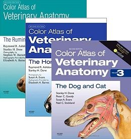
Color Atlas of Veterinary Anatomy, 3-Vol, 2ed
Color Atlas of Veterinary Anatomy, Volume 1, The Ruminants presents a unique photographic record of dissections showing the topographical anatomy of the ruminant. With this book you will be able to see the position and relationships of the bones, muscles, nerves, blood vessels and viscera that go to make up each region of the body and each organ system. Each book in this three volume series is packed with full-color photographs and drawings of dissections prepared specifically for these texts.
- Accessibly and systematically structured with each chapter devoted to a specific body region.
- Important features of regional and topographical anatomy presented using full-color photos of detailed dissections.
- Detailed color line drawings clarify the relationships of relevant structures.
- Presents anatomy in a clinical context.
- Accompanying website with interactive quizzes and the chance to test yourself with self-assessment questions.
- New chapter on radiological anatomy.
- Special notes highlight clinical significance of each section.
Color Atlas of Veterinary Anatomy, Volume 2, The Horse presents a unique photographic record of dissections showing the topographical anatomy of the horse. With this book you will be able to see the position and relationships of the bones, muscles, nerves, blood vessels and viscera that go to make up each region of the body and each organ system. Each book in this 3 volume series is packed with full-color photographs and drawings of dissections prepared specifically for these texts.
-
- Accessibly and systematically structured with each chapter devoted to a specific body region
-
- Important features of regional and topographical anatomy presented using full color photos of detailed dissections
-
- Dissections presented in the standing position
-
- Detailed color line drawings clarify the relationships of relevant structures
- Presents anatomy in a clinical context
This new edition second edition offers important new features, including:
-
- Accompanying website presents over 100 interactive quizzes and self-assessment questions
-
- Many more radiographs throughout
-
- Additional CT and MRI images
- Clinical notes highlight areas of particular clinical significance
Color Atlas of Veterinary Anatomy, Volume 3, The Dog and Cat If you are looking for a book that presents a unique photographic record of dissections showing the topographical anatomy of the dog and cat: this is the atlas for you! Part of a comprehensive 3-volume set that also covers Ruminants (Volume 1) and The Horse (Volume 2), the Color Atlas of the Dog and Cat takes a complete look at virtually every aspect of veterinary anatomy. With this book you will be able to see the position and relationships of bones, muscles, nerves, blood vessels and viscera that go to make up each region of the body and each organ system. Rich with full-color photographs and drawings of dissections prepared specifically for these texts, each book in the series illustrates regional surface features photographed before dissection, then gives high-quality complementary photographs of articulated skeletons.
- Accessibly and systematically structured with each chapter is devoted to a specific body region
- Important features of regional and topographical anatomy presented in full color photos of detailed dissections
- Detailed color line drawings clarify the relationships of relevant structures
- Website offers drag and drop quizzes and the chance to test yourself with mcqs
- Informative captions give additional information necessary for proper interpretation of the images
- Presents anatomy in a clinical context
Contents
۱ The head
۲ The neck
۳ The forelimb
۴ The thorax
۵ The abdomen
۶ The hindlimb
۷ The foot
۸ The pelvis
۹ The udder, scrotum and penis
۱۰ Radiographic anatomy of the head, manus and pes
۱ The Head (including the skin)
۲ The Neck
۳ The Forelimb
۴ The Thorax
۵ The Abdomen
۶ The Hindlimb
۷ The Foot
۸ The Pelvis (including the spine)
۹ Diagnostic Imaging of the Head, Withers, Manus and Pes
۱ Live and skeletal anatomy of whole animal
۲ The head
۳ The neck
۴ The forelimb
۵ The thorax
۶ The abdomen
۷ The hindlimb
۸ The pelvis
۹ The vertebral column
۱۰ The cat: comparative aspects
لینک کوتاه : https://bookbaz.ir/?p=213725
نویسنده : Raymond R. Ashdown
ناشر : Mosby; 2nd edition
سال انتشار : 2010
زبان کتاب : انگلیسی
نوع فایل : PDF
تعداد صفحات : 1176
(ISBN) شابک : -
قیمت کتاب درآمازون : $110.69
حجم فایل : 400 MB
کتاب های مرتبط:
 دانلود کتاب بیماری کلوستریدیا حیوانات
Clostridial Diseases of Animals, 1ed
 دانلود کتاب اصول و عملکرد فناوری دامپزشکی
Principles and Practice of Veterinary Technology 5th Edition
 دانلود کتاب پزشکی گوسفند، بز و گوزن
Sheep, Goat, and Cervid Medicine, 3ed
 دانلود کتاب جراحی دامپزشکی حیوانات کوچک (۲ جلدی)
Veterinary Surgery: Small Animal, 2-Vol, 2ed
 دانلود کتاب اطلس دندانپزشکی در گربه ها و سگ ها
Atlas of Dentistry in Cats and Dogs, 1ed
 دانلود کتاب پزشکی داخلی بالینی حیوانات کوچک
Clinical Small Animal Internal Medicine, 1ed
 دانلود کتاب اورژانس و مراقبت های ویژه گربه
Feline Emergency and Critical Care Medicine, 2ed
 دانلود کتاب جراحی اسب + ویدئو
Equine Surgery, 5ed + Video
 دانلود کتاب آناتومی و فیزیولوژی کاربردی حیوانات اهلی
Functional Anatomy and Physiology of Domestic Animals, 5ed
 دانلود کتاب اصول استئوسنتز اسب AO
AO Principles of Equine Osteosynthesis, 1ed
 دانلود کتاب جراحی تنفسی فوقانی در اسب
Advances in Equine Upper Respiratory Surgery
 دانلود کتاب پزشکی پرندگان و جراحی در عمل: همنشین و ضمائم پرندگان
Avian Medicine and Surgery in Practice: Companion and Aviary Birds, 2ed
 دانلود کتاب راهنمای عملی پرستار دامپزشکی برای بیهوشی حیوانات کوچک
The Veterinary Nurse’s Practical Guide to Small Animal Anaesthesia, 1ed
 دانلود کتاب پزشکی داخلی دامپزشکی اتینگر (۲ جلدی)
Textbook of Veterinary Internal Medicine 8th Edition
 دانلود کتاب جراحی دهان و فک و صورت در سگ و گربه
Oral and Maxillofacial Surgery in Dogs and Cats
 دانلود کتاب پیشرفتهایی در کرانیال رباط صلیبی سگ
 Advances in the Canine Cranial Cruciate Ligament
 دانلود کتاب پزشکی داخلی دامهای بزرگ اسمیت
Large Animal Internal Medicine, 5ed
 دانلود کتاب چشم پزشکی اسب (ویرایش ۲۰۱۷)
Equine Ophthalmology, 3ed
دانلود کتاب جراحی اسب Equine Surgery, 5ed
 دانلود کتاب روشهای سونوگرافی برای متخصص مراقبت از حیوانات کوچک
Point-of-Care Ultrasound Techniques for the Small Animal Practitioner, 2ed
 دانلود کتاب آناتومی دامپزشکی حیوانات اهلی
Veterinary Anatomy of Domestic Animals, 7ed
 دانلود کتاب لنگش در اسبها آدامز و استاشاک
Adams and Stashak’s Lameness in Horses, 7ed
 دانلود کتاب رادیولوژی تشخیصی دامپزشکی
 Textbook of Veterinary Diagnostic Radiology, 7ed
 دانلود کتاب پنج دقیقه معاینه بالینی دامپزشکی بلک ول: درماتولوژی حیوانات کوچک
Blackwell’s Five-Minute Veterinary Consult Clinical Companion: Small Animal Dermatology, 3ed
 دانلود کتاب تشخیص کم هزینه بالینی دامپزشکی
 Low-Cost Veterinary Clinical Diagnostics, 1ed
 دانلود کتاب مدیریت درد برای تکنسین دامپزشکی و پرستاران
Pain Management for Veterinary Technicians and Nurses
 دانلود کتاب مشاور بالینی دامپزشکی کوت: سگ ها و گربه ها
Cote’s Clinical Veterinary Advisor: Dogs and Cats, 4ed
 دانلود کتاب درسی رادیولوژی تشخیصی دامپزشکی
 Textbook of Veterinary Diagnostic Radiology, 6ed
 دانلود کتاب فرهنگ لغت جامع دامپزشکی ساندرز
Saunders Comprehensive Veterinary Dictionary, 5ed
 دانلود کتاب اطلس رنگی بیماری ها و اختلالات اسب نوتنبلت و پاسکوئه
Knottenbelt and Pascoe’s Color Atlas of Diseases and Disorders of the Horse, 2ed
 دانلود کتاب راهنمای دامپزشکی مرک
The Merck Veterinary Manual, 11ed
 دانلود کتاب جراحی حیوانات عجیب و غریب
Surgery of Exotic Animals, 1ed
 دانلود کتاب جراحی حیوانات کوچک
Small Animal Surgery, 5ed
 دانلود کتاب علوم اسب
 Equine Science, 3ed
دانلود کتاب جراحی حیوانات کوچک + ویدئوSmall Animal Surgery, 5ed + Video
 دانلود کتاب پزشکی گاو کاککرافت
Bovine Medicine, 3ed
 دانلود کتاب آسیب شناسی بالینی دامپزشکی
 Veterinary Clinical Pathology: A Case-Based Approach
 دانلود کتاب میکروبیولوژی دامپزشکی
Veterinary Microbiology, 4ed
 دانلود کتاب پزشکی اسب ، جراحی و تولید مثل
Equine Medicine, Surgery and Reproduction, 2ed
 دانلود کتاب گلبول قرمز مرغی: ادیسه فیلوژنتیکی
The Avian Erythrocyte: Its Phylogenetic Odyssey
 دانلود کتاب تکنیک های تشخیصی در درماتولوژی دامپزشکی
Diagnostic Techniques in Veterinary Dermatology, 1ed
 دانلود کتاب دامپزشکی کنستیبل (۲ جلدی)
Veterinary Medicine, 2-Vol, 11ed
 دانلود کتاب راهنمای داروهای دامپزشکی پاپیچ
Papich Handbook of Veterinary Drugs, 5ed
 دانلود کتاب تشخیص لنگش در سگ ها
Diagnosis of Lameness in Dogs, 1ed
 دانلود کتاب نکاتی در مورد پزشکی داخلی سگ
 Notes on Canine Internal Medicine, 4ed
 دانلود کتاب کامل پرستاری دامپزشکی آسپینال
Aspinall’s Complete Textbook of Veterinary Nursing, 3ed
 دانلود کتاب درمان پزشکی حیوانات وحشی و باغ وحش فاولر (جلد هفتم)
Fowler’s Zoo and Wild Animal Medicine Current Therapy, 1ed
 دانلود کتاب پزشکی گربه – بررسی و آزمایش
Feline Medicine – Review and Test, 1ed
 دانلود کتاب MRI اسب موری
Equine MRI, 1ed
 دانلود کتاب اسب مسابقه: راهنمای دامپزشکی
The Racehorse: A Veterinary Manual, 1ed
 دانلود کتاب بیومکانیک و تربیت بدنی اسب
Biomechanics and Physical Training of the Horse, 1ed
 دانلود کتاب عوارض در جراحی حیوانات کوچک
Complications in Small Animal Surgery, 1ed
 دانلود کتاب اطلس رنگی درماتولوژی حیوانات مزرعه
Color Atlas of Farm Animal Dermatology, 2ed
 تفسیر سریع صداهای قلب و ریه: راهنمای سمع قلبی و تنفسی در سگ ها و گربه ها
Rapid Interpretation of Heart and Lung Sounds: A Guide to Cardiac and Respiratory Auscultation in Dogs and Cats, 3ed
 دانلود کتاب نظارت و روش های پیشرفته برای مراقبتهای ویژه و اورژانسی حیوانات کوچک
Advanced Monitoring and Procedures for Small Animal Emergency and Critical Care, 1ed
 دانلود کتاب تولید مثل اسب (۲ جلدی)
Equine Reproduction, 2-Vol, 2ed
 دانلود کتاب راهنمای زهرداران و بی مهرگان مهم پزشکی
Guide to Venomous and Medically Important Invertebrates, 1ed
 دانلود کتاب پزشکی و جراحی خزندگان و دوزیستان میدر
Mader’s Reptile and Amphibian Medicine and Surgery, 3ed
 دانلود کتاب بیماری های عفونی اسب
Infectious Diseases of the Horse, 2ed
 دانلود کتاب اطلس بالینی بیماری چشمی سگ و گربه
Clinical Atlas of Canine and Feline Ophthalmic Disease, 2ed
 دانلود کتاب درد مزمن در پزشکی حیوانات کوچک
Chronic Pain in Small Animal Medicine, 2ed
 دانلود کتاب اطلس رنگی آسیب شناسی اسب
Color Atlas of Equine Pathology, 1ed
 دانلود کتاب تشخیص مولکولی پاتوژن ویروسی حیوانات
Molecular Detection of Animal Viral Pathogens, 1ed
 دانلود کتاب پزشکی بالینی سگ و گربه (ویرایش ۲۰۱۶)
Clinical Medicine of the Dog and Cat, 3ed
 دانلود کتاب پایه پاتولوژیک بیماری دامپزشکی (ویرایش ۲۰۱۷)
Pathologic Basis of Veterinary Disease, 6ed
 دانلود کتاب راهنمای پزشکی اضطراری و مراقبت های حیاتی حیوانات کوچک
Manual of Small Animal Emergency and Critical Care Medicine, 2ed
 دانلود کتاب شکم حاد اسب
The Equine Acute Abdomen, 3ed
 دانلود کتاب اطلس مدیریت و جراحی بازسازی زخم حیوانات کوچک
Atlas of Small Animal Wound Management and Reconstructive Surgery, 4ed
 دانلود کتاب عضلات مهره داران: رشد، همانندی و تکامل
Muscles of Chordates: Development, Homologies, and Evolution, 1ed
 دانلود کتاب رادیولوژی بالینی اسب
 Clinical Radiology of the Horse, 4ed
 دانلود کتاب MRI تشخیصی در سگ ها و گربه ها
Diagnostic MRI in Dogs and Cats, 1ed
 دانلود کتاب پزشکی داخلی حیوانات بزرگ + ویدئو
Large Animal Internal Medicine, 6ed + Video
 دانلود کتاب ارتوپدی و شکستگی حیوانات کوچک برینکر، پیرماتی و فلو
Brinker, Piermattei and Flo’s Handbook of Small Animal Orthopedics and Fracture, 5ed
 دانلود کتاب مرجع رفتار حیوانات
Animal Behavior Desk Reference: A Dictionary of Animal Behavior, Ecology, and Evolution, Third Edition
 دانلود کتاب تشخیص و مدیریت لنگش در اسب (نسخه پیشرفته)
Diagnosis and Management of Lameness in the Horse, 2ed
 دانلود کتاب اطلس رنگی بیماری ها و اختلالات در کره اسب ها
Color Atlas of Diseases and Disorders of the Foal, 1ed
 دانلود کتاب بیماری های خوکی
Diseases of Swine, 10ed
 دانلود کتاب راهنمای روشهای بالینی در سگ، گربه، خرگوش و جوندگان کرو و والشاو
Crow and Walshaw’s Manual of Clinical Procedures in Dogs, Cats, Rabbits and Rodents, 4ed
 دانلود کتاب انکولوژی بالینی حیوانات کوچک: بررسی رنگی خود ارزیابی
Small Animal Clinical Oncology: Self-Assessment Color Review, 1ed
 دانلود کتاب رادیوگرافی سگ و گربه: راهنمای تهیه و تفسیر رادیوگرافی
Radiography of the Dog and Cat: Guide to Making and Interpreting Radiographs, 1ed
 دانلود کتاب پزشکی داخلی حیوانات کوچک
Small Animal Internal Medicine, 6ed
دانلود کتاب پزشکی داخلی حیوانات بزرگLarge Animal Internal Medicine, 6ed
 دانلود کتاب پزشکی و جراحی طیور بکیارد
Backyard Poultry Medicine and Surgery, 2ed
 دانلود کتاب جراحی حیوانات مزرعه
Farm Animal Surgery, 2ed
 دانلود کتاب پزشکی حیوانات خاص برای تکنسین دامپزشکی
Exotic Animal Medicine for the Veterinary Technician, 3ed
 دانلود کتاب اطلس تشخیصی تومورها در سگ و گربه
Atlas for the Diagnosis of Tumors in the Dog and Cat, 1ed
 دانلود کتاب تومورها در حیوانات اهلی
Tumors in Domestic Animals, 5ed
 دانلود کتاب بیماری های گوش و حلق و بینی سگ و گربه
Ear, Nose and Throat Diseases of the Dog and Cat, 1ed
 دانلود کتاب نگهداری و اداره حیوانات وحشی و اهلی
Restraint and Handling of Wild and Domestic Animals, 3ed
 دانلود کتاب ایمونولوژی بالینی اسب
 Equine Clinical Immunology, 1ed
 دانلود کتاب اورژانس های جراحی حیوانات کوچک
 Small Animal Surgical Emergencies, 2ed
 دانلود کتاب اطلس و راهنمای چشم پزشکی حیوانات کوچک
Small Animal Ophthalmic Atlas and Guide, 2ed
 دانلود کتاب پزشکی اورژانسی حیوانات کوچک (۲ جلدی)
Textbook of Small Animal Emergency Medicine, 2-Vol, 1ed
 دانلود کتاب بیماری های عفونی منتقله از بندپایان سگ و گربه
Arthropod-borne Infectious Diseases of the Dog and Cat, 2ed
 دانلود کتاب جراحی قفسه سینه حیوانات کوچک
Small Animal Thoracic Surgery, 1ed
 دانلود کتاب پزشکی کره اسب
 Equine Pediatric Medicine, 2ed
 دانلود کتاب آناتومی و بافت شناسی مقایسه ای (ویرایش ۲۰۱۸)
Comparative Anatomy and Histology: A Mouse, Rat, and Human Atlas, 2ed
 دانلود کتاب سیتولوژی و هماتولوژی تشخیصی سگ و گربه کاول و تایلر
Cowell and Tyler’s Diagnostic Cytology and Hematology of the Dog and Cat, 5ed
 دانلود کتاب زیست شناسی، پزشکی و جراحی فیلها
Biology, Medicine, and Surgery of Elephants
 دانلود کتاب جراحی اسب
 Equine Surgery, 4ed
دانلود کتاب اصول استئوسنتز اسب AO + ویدئوAO Principles of Equine Osteosynthesis, 1ed + Videos
 دانلود کتاب درماتولوژی حیوانات کوچک: اطلس رنگی و راهنمای درمانی
Small Animal Dermatology: A Color Atlas and Therapeutic Guide, 4ed
دانلود کتاب پزشکی داخلی حیوانات کوچک + ویدئوSmall Animal Internal Medicine, 6ed + Video