
دانلود کتاب اطلس سیتوپاتولوژی پانکراس به همراه هیستوپاتولوژیک
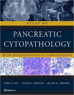
Atlas of Pancreatic Cytopathology with Histopathologic Correlations, 1ed
“Clinical and radiologic examinations cannot reliably distinguish benign or inflammatory pancreatic disease from carcinoma. The increased use of pancreatic fine needle aspiration (FNA) along with advances in imaging techniques and the introduction of endoscopic ultrasound guidance have led to significantly better detection and recognition of pancreatic masses. Consequently, pancreatic cytopathology is integral to accurate pre-operative diagnosis yet it is a challenging diagnostic area with a variety of potential pitfalls and “look-alike” lesions. Skillful recognition and an awareness of the limitations of the procedure are essential in avoiding misdiagnosis of these dangerous lesions.
Atlas of Pancreatic Cytopathology with Histopathologic Correlations fills a void in current pathology literature. With 450 high-resolution images, including images of histopathologic and radiologic features, this practical atlas presents an integrated approach to diagnostic cytopathology that will help physician cytopathologists, cytotechnologists, and pathologists avoid potential pitfalls and “look-alike” lesions. Written by recognized experts in the field, the extensive high-resolution color images of the characteristic features of pancreatic disease are presented with detailed descriptions that cover classic features, diagnostic clues, and potential pitfalls.
Atlas of Pancreatic Cytopathology with Histopathologic Correlations is a valuable resource for the seasoned cytopathologist, general and surgical pathologists, pathology trainees, and cytotechnologists.
Review
This well-organized, photomicrograph-based atlas is an excellent reference for surgical pathologists and cytopathologists as well as gastroenterologists who take an interest in the morphology of their patients’ lesions. –Gastroenterology, 2010
About the Author
لینک کوتاه : https://bookbaz.ir/?p=20842
نویسنده : Syed Z. Ali MD , Yener S. Erozan MD
ناشر : Demos Medical Publishing; First edition
سال انتشار : 2010
زبان کتاب : انگلیسی
نوع فایل : PDF
تعداد صفحات : 227
(ISBN) شابک : 1933864400
قیمت کتاب درآمازون : $134.39
حجم فایل : 28 MB
کتاب های مرتبط:
 دانلود کتاب تصویربرداری دستگاه گوارش (سری تشخیص مستقیم در رادیولوژی)
Gastrointestinal Imaging (Direct Diagnosis in Radiology: DX-Direct!), 1ed
 دانلود کتاب اطلس ویدئویی جراحی کبد، صفراوی و پانکراس
 Video Atlas: Liver, Biliary & Pancreatic Surgery, 1ed
 دانلود کتاب آسیب شناسی پستان دَبس
Breast Pathology: Expert Consult, 1ed
 دانلود کتاب منابع راهنمای درمان برای آسیب شناسی گفتار و زبان
 Treatment Resource Manual for Speech Language Pathology, 5ed
 دانلود کتاب نوروپاتولوژی جراحی کانونی صرع
Surgical Neuropathology of Focal Epilepsies, 1ed
 دانلود کتاب مرور کامل پاتولوژی و هماتولوژی برای NBE
Complete Review of Pathology & Hematology for NBE, 6ed
 دانلود کتاب اطلس پاتولوژی کبد
Atlas of Liver Pathology, 4ed
 دانلود کتاب هیستوپاتولوژی ناخن: اونیکوپاتولوژی
Histopathology of the Nail: Onychopathology, 1ed
 دانلود کتاب آسیب شناسی: MCQs و مرور سریع
Pathology: Quick Review and MCQs, 4ed
 دانلود کتاب جراحی کبد، دستگاه صفراوی و پانکراس بلومگارت (۲ جلدی)
Blumgart’s Surgery of the Liver, Biliary Tract and Pancreas, 2-Vol, 6ed
 دانلود کتاب پاتوفیزیولوژی اختلالات خونی
Pathophysiology of Blood Disorders, 1ed
 دانلود کتاب اطلس آسیب شناسی بافت نرم و استخوان: با بافت شناسی، سیتولوژی و رادیولوژی
Atlas of Soft Tissue and Bone Pathology: With Histologic, Cytologic, and Radiologic Correlations, 1ed
 دانلود کتاب اطلس گوارش یامادا
 Yamada’s Atlas of Gastroenterology, 5ed
 دانلود کتاب بیماری های دستگاه گوارش و کبد اسلینگر و فوردترن (۲ جلدی) + ویدئو
Sleisenger and Fordtran’s Gastrointestinal and Liver Disease, 2-Vol, 11ed + Video
 دانلود کتاب پیشرفت در پاتولوژی جراحی: کارسینوم کولورکتال و تومورهای کرمی ضمیمه
Advances in Surgical Pathology: Colorectal Carcinoma and Tumors of the Vermiform Appendix, 1ed
 دانلود کتاب پاتولوژی تشخیصی کبد و لوزالمعده
Diagnostic Pathology: Hepatobiliary and Pancreas, 2ed
 دانلود کتاب ایمونوهیستوشیمی تشخیصی: ترانوستیک و برنامه های ژنومی
Diagnostic Immunohistochemistry: Theranostic and Genomic Applications, 4ed
 دانلود کتاب تومورهای استخوان و بافت نرم: بررسی چند رشته با ارائه مورد
Bone and Soft Tissue Tumours: A Multidisciplinary Review With Case Presentations, 1ed
 دانلود کتاب سرطان سر و گردن: درمان، توانبخشی و نتایج
Head and Neck Cancer: Treatment, Rehabilitation, and Outcomes, 2ed
 دانلود کتاب اطلس رنگی هیستولوژی گارتنر
Color Atlas and Text of Histology, 7ed
 دانلود کتاب تفسیر نمونه برداری از ضایعات کودکان
Biopsy Interpretation of Pediatric Lesions
 دانلود کتاب اطلس پاتولوژی مدیاستن (آناتومیک)
Atlas of Mediastinal Pathology, 2015th
 دانلود کتاب پانکراتیت حاد: از A تا Z
Acute Pancreatitis: An A-Z, 1ed
 دانلود کتاب سندروم روده کوتاه بزرگسالان: تغذیه، پزشکی و مدیریت جراحی
Adult Short Bowel Syndrome: Nutritional, Medical, and Surgical Management, 1ed
 دانلود کتاب پاتولوژی نئوپلاستیک گوارشی
Neoplastic Gastrointestinal Pathology, 1ed
 دانلود کتاب آسیب شناسی مزوتلیوما بدخیم
Pathology of Malignant Mesothelioma
 دانلود کتاب اصول پاتولوژی پاتوما + ویدئو
Fundamentals of Pathology Pathoma + Video
 دانلود کتاب جراحی کولورکتال: همگام با تمرین متخصص جراحی
Colorectal Surgery: A Companion to Specialist Surgical Practice, 5ed
 دانلود کتاب بافت شناسی: تصویر بزرگ
Histology: The Big Picture, 1ed
 دانلود کتاب اطلس آندوسکوپی تشخیصی
Atlas of Diagnostic Endoscopy, 3ed
 دانلود کتاب درماپاتولوژی الستون
Dermatopathology, 3ed
 دانلود کتاب پاتولوژی استخوان و بافت نرم
Bone and Soft Tissue Pathology, 2ed
 دانلود کتاب آسیب شناسی کالبد شکافی کودکان
Handbook of Pediatric Autopsy Pathology
 دانلود کتاب راهنمای نوروپاتولوژی عمومی اسکرول و پوریِر
Escourolle & Poirier’s Manual of Basic Neuropathology, 5ed
 دانلود کتاب اطلس هماتوپاتولوژی: مورفولوژی، ایمونوفنوتیپ، سیتوژنتیک و روشهای مولکولی
Atlas of Hematopathology: Morphology, Immunophenotype, Cytogenetics, and Molecular Approaches
 دانلود کتاب نقد و بررسی پاتولوژی رابینز و کوتران
Robbins and Cotran Review of Pathology, 4ed
 دانلود کتاب برتری در آزمون بورد دستگاه گوارش
 Acing the GI Board Exam: The Ultimate Crunch-Time Resource, 2ed
 دانلود کتاب اطلس سیتوپاتولوژی غدد بزاقی: با همبستگی هیستوپاتولوژیک
Atlas of Salivary Gland Cytopathology: with Histopathologic Correlations, 1ed
 دانلود کتاب آمادگی آزمون برای USMLE® تیئمه: پرسش و پاسخ پاتولوژی
Thieme Test Prep for the USMLE®: Pathology Q&A, 1ed
 دانلود کتاب اطلس لاپاراسکوپی و توراکوسکوپی کودکان
Atlas of Pediatric Laparoscopy and Thoracoscopy, 2ed
 دانلود کتاب اطلس پاتولوژی رابینز و کوتران
Robbins and Cotran Atlas of Pathology, 4ed
 دانلود کتاب اطلس جراحی عمومی رباتیک
Atlas of Robotic General Surgery, 1ed
 دانلود کتاب تفسیر بیوپسی ریه
Biopsy Interpretation of the Lung, 2ed
 دانلود کتاب بافت شناسی انسانی استیونز و لو
Stevens & Lowe’s Human Histology, 5ed
 دانلود کتاب بررسی تشخیصی آسیب شناسی جراحی استرنبرگ
Sternberg’s Diagnostic Surgical Pathology Review
 دانلود کتاب سندرم روده کوتاه: رویکرد عملی به مدیریت
Short Bowel Syndrome: Practical Approach to Management, 1ed
 دانلود کتاب آندوسونوگرافی + ویدئو
Endosonography, 3ed + Video
 دانلود کتاب کبد: بیولوژی و پاتوبیولوژی
The Liver: Biology and Pathobiology, 6ed
 دانلود کتاب دستگاه گوارش و کبد هریسون
Harrison’s Gastroenterology and Hepatology, 2ed
 دانلود کتاب اطلس رنگی کالبد شکافی واگنر (ویرایش ۲۰۱۷)
Color Atlas of the Autopsy, 2ed
 دانلود کتاب آموزش موفق در آندوسکوپی دستگاه گوارش
Successful Training in Gastrointestinal Endoscopy, 2ed
 دانلود کتاب درماتوپاتولوژی پرایمر بیماری های التهابی
Dermatopathology Primer of Inflammatory Diseases, 1ed
 دانلود کتاب آنژیوگرافی مغزی کاربردی: آناتومی و پاتولوژی عروقی نرمال
Applied Cerebral Angiography: Normal Anatomy and Vascular Pathology, 1ed
 دانلود کتاب بافت شناسی ضروری نتر
Netter’s Essential Histology, 3ed
 دانلود کتاب اطلس ویدیویی جراحی کبد، صفراوی و پانکراس بلومگارت
Blumgart’s Video Atlas: Liver, Biliary & Pancreatic Surgery, 2ed
 دانلود کتاب پاتولوژی تشخیصی: ادراری تناسلی
Diagnostic Pathology: Genitourinary, 2ed
 دانلود کتاب راهنمای مراحل سرطان AJCC (ویرایش ۲۰۱۷)
AJCC Cancer Staging Manual, 8ed
 دانلود کتاب بیوپسی عضلانی: یک رویکرد عملی
Muscle Biopsy: A Practical Approach, 5ed
 دانلود کتاب ملزومات پزشکی گوارش سیتارامان و فریدمن
Sitaraman and Friedman’s Essentials of Gastroenterology, 2ed
 دانلود کتاب اطلس آموزشی تصویربرداری شکم
Teaching Atlas of Abdominal Imaging, 1ed
 دانلود کتاب اطلس سیتوپاتولوژی: رویکردی مبتنی بر الگو
Atlas of Cytopathology: A Pattern Based Approach, 1ed
 دانلود کتاب راهنمای روش های هیستولوژی برای استخوان و غضروف
Handbook of Histology Methods for Bone and Cartilage, 1ed
 دانلود کتاب سیتولوژی پزشکی زنان
 Gynecologic Cytology, 1ed
 دانلود کتاب اطلس ویدئویی جراحی کم تهاجمی پیشرفته
Video Atlas of Advanced Minimally Invasive Surgery, 1ed
 دانلود کتاب اطلس جامع آندوسکوپی با وضوح بالا و تصویربرداری نروباند کوهن
Comprehensive Atlas of High-Resolution Endoscopy and Narrowband Imaging, 2ed
 دانلود کتاب پاتولوژی جراحی لوله گوارش، کبد، مجرای صفراوی و پانکراس اودزه و گلدبلوم
Odze and Goldblum Surgical Pathology of the GI Tract, Liver, Biliary Tract and Pancreas, 3ed
 دانلود کتاب طبقه بندی تومورهای پستان WHO
WHO Classification of Tumours of the Breast, 4ed
 دانلود کتاب تشخیص آزمایشگاهی بیماری ها در آزمایشگاه بالینی
Laboratory Medicine Diagnosis of Disease in Clinical Laboratory, 2ed
 دانلود کتاب پاتولوژی گوارشی: اطلس و متن
Gastrointestinal Pathology: An Atlas and Text, 3ed
 دانلود کتاب پاتولوژی قفسه سینه عملی: بیماری های ریه، قلب و تیموس
Practical Thoracic Pathology: Diseases of the Lung, Heart, and Thymus, 1ed
 دانلود کتاب پاتوفیزیولوژی کبد: درمان و آنتی اکسیدان ها
Liver Pathophysiology: Therapies and Antioxidants, 1ed
 دانلود کتاب سونوگرافی تشخیصی بالینی: شکم و ساختار ظاهری + کتاب کار
Diagnostic Medical Sonography: Abdomen and Superficial Structures + Workbook, 3ed
 دانلود کتاب ایمونوهیستوشیمی: راهنمای فنی برای شیوه های فعلی
Immunohistochemistry: A Technical Guide to Current Practices, 1ed
 دانلود کتاب گره های لنفاوی (راهنمای آسیب شناسی جراحی دموس)
Lymph Nodes (Demos Surgical Pathology Guides), 1ed
 دانلود کتاب سارکوما بافت نرم: رویکرد مبتنی بر الگو برای تشخیص
Soft Tissue Sarcomas: A Pattern-Based Approach to Diagnosis, 1ed
 دانلود کتاب تومورهای کولورکتال: اطلس هیستوپاتولوژی سکشن بزرگ
Colorectal Tumors: Atlas of Large Section Histopathology, 1ed
 دانلود کتاب پزشکی تورنتو نوت (نسخه ۲۰۱۵)
Toronto Notes 2015 (Essential Med Notes), 31ed
 دانلود کتاب اطلس رنگی و خلاصه پاتولوژی دستگاه گوارش
 Color Atlas and Synopsis: Gastrointestinal Pathology, 1ed
 دانلود کتاب اطلس پاتولوژی دستگاه گوارش: رویکرد مبتنی بر الگوی بیوپسی های غیر نئوپلاستیک
Atlas of Gastrointestinal Pathology: A Pattern Based Approach to Non-Neoplastic Biopsies, 1ed
 دانلود کتاب پاتولوژی ویتر: متن، اطلس و مروری بر پاتولوژی بافت
Wheater’s Pathology: A Text, Atlas and Review of Histopathology, 6ed
 دانلود کتاب دانشنامه پزشکی گیل (۹ جلدی)
Gale Encyclopedia of Medicine, 9-Vol, 2015th
 دانلود کتاب آسیب شناسی دستگاه گوارش مورسون و داوسون
Morson and Dawson’s Gastrointestinal Pathology, 5ed
 دانلود کتاب پاتولوژی تشخیصی لیندبرگ: تومورهای بافت نرم
Diagnostic Pathology: Soft Tissue Tumors, 2ed
 دانلود کتاب هیستوپاتولوژی عملکرد بالا
High Yield Histopathology, 2ed
 دانلود کتاب آسیب شناسی دستگاه گوارش لوین، وینستن و ریدل (۲ جلدی)
Lewin, Weinstein and Riddell’s Gastrointestinal Pathology and its Clinical Implications 2-Vol, 2ed
 دانلود کتاب آسیب شناسی کبد مک سویین
MacSween’s Pathology of the Liver, 6ed
 دانلود کتاب تفسیر نمونه برداری از مخاط دستگاه گوارش: نئوپلاستیک
Biopsy Interpretation of the Gastrointestinal Tract Mucosa: Volume 2: Neoplastic, 2ed
 دانلود کتاب اطلس اجمالی هیستوپاتولوژی پوست لِوِر
Atlas and Synopsis of Lever’s Histopathology of the Skin, 3ed
 دانلود کتاب پزشکی سیسیل گلدمن (۲ جلدی)
Goldman-Cecil Medicine, 2-Vol, 25ed
 دانلود کتاب تشخیص اندودنتیکس ، پاتولوژی و طرح درمان
Endodontic Diagnosis, Pathology, and Treatment Planning: Mastering Clinical Practice, 2015th
 دانلود کتاب اطلس مری و آسیب شناسی معده
 Atlas of Esophagus and Stomach Pathology, 2014th
 دانلود کتاب آسیب شناسی عملی بافت نرم: روش تشخیصی
Practical Soft Tissue Pathology: A Diagnostic Approach, 1ed
 دانلود کتاب تفسیر نمونه برداری از کبد
 Biopsy Interpretation of the Liver
 دانلود کتاب اطلس پاتولوژی کبد
 Atlas of Liver Pathology, 2014th Edition
 دانلود کتاب خلاصه هیستولوژی: راهنمای نظری و عملی
Compendium of Histology: A Theoretical and Practical Guide, 1ed
 دانلود کتاب پاتولوژی کبدی عملی: یک رویکرد تشخیصی
Practical Hepatic Pathology: A Diagnostic Approach, 2ed
 دانلود کتاب بافت شناسی و بیولوژی سلولی: درآمدی بر پاتولوژی
Histology and Cell Biology: An Introduction to Pathology, 5ed
 دانلود کتاب پاتوفیزیولوژی
Pathophysiology (Copstead), 5ed
 دانلود کتاب پاتوفیزیولوژی فوق العاده آسان!
Pathophysiology Made Incredibly Easy!, 5ed
 دانلود کتاب تخصص بالینی آکسفورد (ویرایش ۲۰۱۶)
Oxford Handbook of Clinical Specialties, 10ed
 دانلود کتاب فلش کارت بافت شناسی عمومی لانگه
Lange Basic Histology Flash Cards, 1ed
 دانلود کتاب پرستاری دستگاه گوارش: رویکرد طول عمر
Gastrointestinal Nursing: A Lifespan Approach, 1ed
 دانلود کتاب اصول و عمل جراحی برای کولون، رکتوم و انوس گوردون و نیواتونگز + ویدئو
Gordon and Nivatvongs’ Principles and Practice of Surgery for the Colon, Rectum, and Anus, 4ed + Video
 دانلود کتاب واژه نامه مصور پاتولوژی سم شناسی و علوم ایمنی
The Illustrated Dictionary of Toxicologic Pathology and Safety Science, 1ed
 دانلود کتاب پاتولوژی تشخیصی: پاتولوژی پیوند اعضا
Diagnostic Pathology: Transplant Pathology, 2ed
 دانلود کتاب آزمایشات بالینی: چشم انداز روش شناختی
Clinical Trials: A Methodologic Perspective, 3ed
 دانلود کتاب اطلس هماتولوژی تشخیصی
Atlas of Diagnostic Hematology, 1ed
 دانلود کتاب اطلس هیستوپاتولوژی پزشکی قانونی
 Atlas of Forensic Histopathology
 دانلود کتاب پیشرفت در پاتولوژی جراحی: سرطان معده
 Advances in Surgical Pathology: Gastric Cancer, 1ed
 دانلود کتاب جراحی کولورکتال و لگن کودکان: مطالعات موردی
Pediatric Colorectal and Pelvic Surgery: Case Studies, 1ed
 دانلود کتاب سرطان هپاتوبیلیاری
 Hepatobiliary Cancer, 1ed
 دانلود کتاب بافت شناسی انسان: متن و اطلسی برای پزشکان و دانشمندان
Human Histology: A Text and Atlas for Physicians and Scientists, 1ed
 دانلود کتاب بافت شناسی برای پاتولوژیست ها
Histology for Pathologists, 4ed
 دانلود کتاب پاتولوژی گوارشی و کبدی: سری مبانی در پاتولوژی تشخیصی
Gastrointestinal and Liver Pathology: Foundations in Diagnostic Pathology Series, 3ed
 دانلود کتاب راهنمای بافت شناسی و پاتولوژی دهان
Manual of Oral Histology & Oral Pathology, 2ed
 دانلود کتاب نوروپاتولوژی: متن های مرجع آسیب شناسی
CNS Neuropathology: A Reference Text of CNS Pathology, 3ed
 دانلود کتاب ۱۰۰ مورد در آسیب شناسی بالینی
۱۰۰ Cases in Clinical Pathology
 دانلود کتاب هماتوپاتولوژی: پاتولوژی عملکرد بالا
Hematopathology: A Volume in the High Yield Pathology Series, 1ed
 دانلود کتاب رادیولوژی ضروری: معرفی بالینی تصویربرداری پاتوفیزیولوژی
 Essential Radiology: Clinical Presentation Pathophysiology Imaging, 3ed
 دانلود کتاب اطلس تشخیصی انکولوژی اِسکارین
Atlas of Diagnostic Oncology: Expert Consult, 4ed
 دانلود کتاب سیستم درجه بندی گِلیسون: راهنمای کامل برای متخصص پاتولوژی و پزشکان
The Gleason Grading System: A Complete Guide for Pathologist and Clinicians, 1ed
 دانلود کتاب اطلس تشخیصی تومورهای دستگاه گوارش فوقانی
A Diagnostic Atlas of Tumors of the Upper Aero-Digestive Tract, 1ed
 دانلود کتاب مهندسی مولکولی، سلولی و بافت
Molecular, Cellular, and Tissue Engineering, 4ed
 دانلود کتاب پاتوفیزیولوژی اختلالات خونی
 Pathophysiology of Blood Disorders, 2ed
 دانلود کتاب جراحی معده و مری آکسفورد
Gastric and Oesophageal Surgery, 1ed
 دانلود کتاب سیستم های آزمایش مهندسی بافت سه بعدی
Engineering 3D Tissue Test Systems, 1ed
 دانلود کتاب تشریح پاتولوژی جراحی: راهنمای تصویری
Surgical Pathology Dissection: An Illustrated Guide, 2ed
 دانلود کتاب راهنمای پاتولوژی جراحی
Manual of Surgical Pathology, 3ed
 دانلود کتاب آندوسونوگرافی
 Endosonography, 3ed
 دانلود کتاب پاتولوژی اولترا استراکچر: اساس مقایسه ای سلولی بیماری
Ultrastructural Pathology: The Comparative Cellular Basis of Disease, 2ed
 دانلود کتاب تصویربرداری تشخیصی شکم
Diagnostic Abdominal Imaging, 1ed
 دانلود کتاب راهنمای جراحی دستگاه گوارش (۲ جلدی)
Textbook of Surgical Gastroenterology, 2-Vol, 1ed
 دانلود کتاب تکنیک های عملی در جراحی قفسه سینه و مری
 Operative Techniques in Thoracic and Esophageal Surgery, 1ed
 دانلود کتاب تصویر برداری کبدی: MRI با همبستگی CT
Liver Imaging: MRI with CT Correlation, 1ed
 دانلود کتاب اسرار پزشکی
 Medical Secrets, 6ed
 دانلود کتاب مداخلات بیماری های گوارشی
 Digestive Disease Interventions, 1ed
 دانلود کتاب تشخیص افتراقی در پاتولوژی جراحی سر و گردن
 Differential Diagnoses in Surgical Pathology: Head and Neck, 1ed
 دانلود کتاب بهبود نتایج در جراحی کولون و رکتال
 Improving Outcomes in Colon & Rectal Surgery, 1ed
 دانلود کتاب بیماری پستان (۲ جلدی)
Breast Disease, 2-Vol, 2ed
 دانلود کتاب بیماری دستگاه گوارش کودکان واکر (۲ جلدی)
Walker’s Pediatric Gastrointestinal Disease, 2-Vol, 6ed
 دانلود کتاب جراحی مقعد، رکتوم و کولون کیلی و ویلیامز (۲جلدی)
Keighley & Williams’ Surgery of the Anus, Rectum and Colon, 2-Vol, 4ed
 دانلود کتاب سیتوپاتولوژی بالینی
Clinical Cytopathology, 3ed
 دانلود کتاب مدیریت بخش اورژانس اشتراوس و مایر
Strauss and Mayer’s Emergency Department Management, 1ed
 دانلود کتاب پاتولوژی جراحی ارولوژیک
 Urologic Surgical Pathology, 4ed
 دانلود کتاب اطلس آماده سازی لمسی سیتوپاتولوژی
 Atlas of Touch Preparation Cytopathology, 1ed
 دانلود کتاب ERCP: اصول
ERCP: The Fundamentals, 3ed
دانلود کتاب اطلس لاپاراسکوپی و توراکوسکوپی کودکان + ویدئوAtlas of Pediatric Laparoscopy and Thoracoscopy, 2ed + Video
 دانلود کتاب تصویربرداری در جراحی شکم
Imaging in Abdominal Surgery, 1ed
 دانلود کتاب ۱۰۰ مورد در پاتولوژی بالینی و پزشکی آزمایشگاهی
۱۰۰Cases in Clinical Pathology and Laboratory Medicine, 2ed
 دانلود کتاب اطلس اولتراسونوگرافی آندوسکوپیک
Atlas of Endoscopic Ultrasonography, 2ed