
دانلود کتاب تجزیه و تحلیل تصویر و مدل سازی در چشم پزشکی
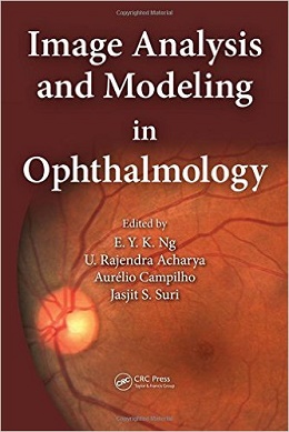
Image Analysis and Modeling in Ophthalmology, 1ed
Digital fundus images can effectively diagnose glaucoma and diabetes retinopathy, while infrared imaging can show changes in the vascular tissues. Likening the eye to the conventional camera, Image Analysis and Modeling in Ophthalmology explores the application of advanced image processing in ocular imaging. This book considers how images can be used to effectively diagnose ophthalmologic problems. It introduces multi-modality image processing algorithms as a means for analyzing subtle changes in the eye. It details eye imaging, textural imaging, and modeling, and highlights specific imaging and modeling techniques.
The book covers the detection of diabetes retinopathy, glaucoma, anterior segment eye abnormalities, instruments on detection of glaucoma, and development of human eye models using computational fluid dynamics and heat transfer principles to predict inner temperatures of the eye from its surface temperature. It presents an ultrasound biomicroscopy (UBM) system for anterior chamber angle imaging and proposes an automated anterior segment eye disease classification system that can be used for early disease diagnosis and treatment management. It focuses on the segmentation of the blood vessels in high-resolution retinal images and describes the integration of the image processing methodologies in a web-based framework aimed at retinal analysis.
The authors introduce the A-Levelset algorithm, explore the ARGALI system to calculate the cup-to-disc ratio (CDR), and describe the Singapore Eye Vessel Assessment (SIVA) system, a holistic tool which brings together various technologies from image processing and artificial intelligence to construct vascular models from retinal images. The text furnishes the working principles of mechanical and optical instruments for the diagnosis and healthcare administration of glaucoma, reviews state-of-the-art CDR calculation detail, and discusses the existing methods and databases.
Image Analysis and Modeling in Ophthalmology includes the latest research development in the field of eye modeling and the multi-modality image processing techniques in ocular imaging. It addresses the differences, performance measures, advantages and disadvantages of various approaches, and provides extensive reviews on related fields.
About the Author
E Y K Ng, received his PhD at Cambridge University. He is a faculty member at the Nanyang Technological University in the School of Mechanical and Aerospace Engineering. Ng is editor-in-chief for the J. of Mechanics in Medicine and Biology and J. Med. Imaging and Health Informatics; assoc. editor for Int. J. of Rotating Machinery, Computational Fluid Dynamics J., Int. J. of Breast Cancer, Chinese J. of Medicine, Open Medical Informatics J., Open Numerical Methods J., and J. of Healthcare Engineering; and strategy assoc. editor-in-chief for World J. of Clinical Oncology. Ng has published more than 255 SCI journal papers, and co-edited 11 books including Human Eye Imaging and Modeling by CRC (2011).
U. Rajendra Acharya, PhD, DEng is a visiting faculty in Ngee Ann Polytechnic, Singapore. He is also an adjunct professor at the University of Malaya, Malaysia; adjunct faculty in Singapore Institute of Technology-University of Glasgow, Singapore; associate faculty in SIM University, Singapore; and adjunct faculty in Manipal Institute of Technology, Manipal, India. He received his Ph.D. from the National Institute of Technology Karnataka, Surathkal, India and D Engg from Chiba University, Japan. He has published more than 275 papers in refereed international SCI-IF journals, international conference proceedings, textbook chapters, and books. His major interests are in biomedical signal processing, bio-imaging, data mining, visualization, and biophysics.
Aurélio Campilho, is a full professor in the Department of Electrical and Computer Engineering, Faculty of Engineering, University of Porto (FEUP), Portugal. He served as president of the Biomedical Engineering Institute, as director of the doctoral program in Electrical and Computer Engineering at FEUP, and as a member of the Scientific Committee of the Master Degree in Bioengineering at the University of Porto. His current research interests include the areas of medical image analysis, image processing, and computer vision. He served as associate editor of the journals IEEE Transactions on Biomedical Engineering and of the Machine Vision Applications journal. He authored one book, co-edited 12 books, and published more than 150 papers in journals and conferences.
Jasjit S. Suri, MS, PhD, MBA, Fellow AIMBE, has spent over 25 years in the field of biomedical engineering/sciences and its management. He received his masters from University of Illinois, Chicago, doctorate from University of Washington, Seattle, and executive management education from Weatherhead School of Management, Case Western Reserve University, Cleveland. Dr. Suri has written over 350 peer-reviewed publications, and is a committee member of several journals and companies. Dr. Suri was crowned with the President’s Gold medal in 1980 and the Fellow of American Institute of Medical and Biological Engineering (AIMBE), awarded by National Academy of Sciences, Washington DC in 2004.
Contents
۱. Ultrasound Biomicroscopic Imaging of the Anterior Chamber Angle
۲. Automated Glaucoma Identifcation Using Retinal Fundus Images: A Hybrid Texture Feature Extraction Paradigm
۳. Ensemble Classifcation Applied to Retinal Blood Vessel Segmentation: Theory and Implementation
۴. Computer Vision Algorithms Applied to Retinal Vessel Segmentation and Quantifcation of Vessel Caliber
۵. Segmentation of the Vascular Network of the Retina
۶. Automatic Analysis of the Microaneurysm Turnover in a Web-Based Framework for Retinal Analysis
۷. A-Levelset-Based Automatic Cup-to-Disc Ratio Measurement for Glaucoma Diagnosis from Fundus Image
۸. The Singapore Eye Vessel Assessment System
۹. Quantifcation of Diabetic Retinopathy Using Digital Fundus Images
۱۰. Diagnostic Instruments for Glaucoma Detection
۱۱. Automated Cup-to-Disc Ratio Estimation for Glaucoma Diagnosis in Retinal Fundus Images
۱۲. Arteriovenous Ratio Calculation Using Image-Processing Techniques
۱۳. Survey on Techniques Used in Iris Recognition Systems
۱۴. Formal Design and Development of an Anterior Segment Eye Disease Classifcation System
۱۵. Modeling of Laser-Induced Thermal Damage to the Retina and the Cornea
۱۶. Automatization of Dry Eye Syndrome Tests
۱۷. Thermal Modeling of the Ageing Eye
۱۸. A Perspective Review on the Use of In Vivo Confocal Microscopy in the Ocular Surface
۱۹. Computational Modeling of Thermal Damage Induced by Laser in a Choroidal Melanoma
لینک کوتاه : https://bookbaz.ir/?p=37604
نویسنده : Eddie Y. K. Ng , U. Rajendra Acharya
ناشر : CRC Press; 1 edition
سال انتشار : 2014
زبان کتاب : انگلیسی
نوع فایل : PDF
تعداد صفحات : 409
(ISBN) شابک : 1466559306
قیمت کتاب درآمازون : $107.91
حجم فایل : 45 MB
کتاب های مرتبط:
 دانلود کتاب اطلس ویدئویی بخیه چشم: اصول و تکنیک ها + ویدئو
Video Atlas of Ophthalmic Suturing: Fundamentals and Techniques, 1ed + Video
 دانلود کتاب بهینه سازی نتایج زیر بهینه حاصل از جراحی آب مروارید + ویدئو
Optimizing Suboptimal Results Following Cataract Surgery, 1ed + Video
 دانلود کتاب چشم پزشکی عصبی مصور + ویدئو
Neuro-Ophthalmology Illustrated, 3ed + Video
 دانلود کتاب پزشکی تورنتو نوت (نسخه ۲۰۱۵)
Toronto Notes 2015 (Essential Med Notes), 31ed
 دانلود کتاب راهنمای پایندگی چشم پزشکی عصبی
The Neuro-Ophthalmology Survival Guide, 2ed
 دانلود کتاب جراحی لیزر فمتوثانیه در چشم پزشکی
Femtosecond Laser Surgery in Ophthalmology, 1ed
 دانلود کتاب راهنمای چشم پزشکی بخش چشم و گوش بیمارستان ماساچوست
The Massachusetts Eye and Ear Infirmary Illustrated Manual of Ophthalmology, 5ed
 دانلود کتاب چشم پزشکی و استرابیسم کودکان تیلور و هویت
Taylor and Hoyt’s Pediatric Ophthalmology and Strabismus, 6ed
 دانلود کتاب اطلس آنژیوگرافی فوندوس
Atlas of Fundus Angiography, 1ed
 دانلود کتاب جراحی اکولوپلاستیک
 Oculoplastic Surgery, 2ed
دانلود کتاب بهینه سازی نتایج زیر بهینه حاصل از جراحی آب مرواریدOptimizing Suboptimal Results Following Cataract Surgery, 1ed
 دانلود کتاب مجموعه ویدئویی جراحی پلک و دور چشم + کتاب
Eyelid and Periorbital Surgery Video Collection, 2ed + PDF
 دانلود کتاب گلوکوم چندلر و گرانت
Chandler and Grant’s Glaucoma, 5ed
 دانلود کتاب اطلس چشم پزشکی بالینی
Atlas of Clinical Ophthalmology, 2ed
 دانلود کتاب اطلس رنگی جراحی پلاستیک چشم + ویدئو
Colour Atlas of Ophthalmic Plastic Surgery, 4ed + Video
 دانلود کتاب جراحی استرابیسم: رویکردهای نوآورانه و کلاسیک + ویدئو
Strabismus Surgery: Innovative and Classic Approaches, 1ed + Video
 دانلود کتاب اپیدمیولوژی چشم
Ophthalmic Epidemiology, 1ed
 دانلود کتاب چشم پزشکی یانوف و داکر
Yanoff & Duker Ophthalmology, 6ed
 دانلود کتاب موارد بالینی در یووئیت: تشخیص و مدیریت افتراقی
Clinical Cases in Uveitis: Differential Diagnosis and Management, 1ed
 دانلود کتاب خلاصه چشم پزشکی بالینی کانسکی
Kanski’s Synopsis of Clinical Ophthalmology, 4ed
 دانلود کتاب تکنیک های عملی در جراحی شبکیه چشم + ویدئو
Operative Techniques in Vitreoretinal Surgery, 1ed + Video
 دانلود کتاب چشم پزشکی بالینی: یک رویکرد سیستماتیک
Clinical Ophthalmology: A Systematic Approach 7ed
 دانلود کتاب چشم پزشکی آکسفورد آمریکا
Oxford American Handbook of Ophthalmology, 1ed
 دانلود کتاب تصویربرداری چشم پزشکی و کاربردها
Ophthalmological Imaging and Applications, 1ed
 دانلود کتاب چشم پزشکی عصبی لیو، والپ و گالتا: تشخیص و مدیریت
Liu, Volpe, and Galetta’s Neuro-Ophthalmology: Diagnosis and Management, 3ed
 دانلود کتاب درسی چشم پزشکی
Textbook of Ophthalmology, 1ed
 دانلود کتاب جراحی لنز داخل چشمی: انتخاب، عوارض و موارد پیچیده + ویدئو
Intraocular Lens Surgery: Selection, Complications, and Complex Cases, 1ed + Video
 دانلود کتاب قرنیه (۲ جلدی) + ویدئو
Cornea, 2-Volume Set, 4ed + Video
 دانلود کتاب مرور چشم پزشکی
 Review of Ophthalmology, 3ed
 دانلود کتاب مشاور بالینی فری ۲۰۱۶ (راه حل های پزشکی فری)
Ferri’s Clinical Advisor 2016: 5 Books in 1, 1ed
 دانلود کتاب بلفاروپلاستی آسیایی و چروک پلک + ویدئو
Asian Blepharoplasty and the Eyelid Crease, 3ed + Video
 دانلود کتاب راهنمای جراحی شبکیه چشم
Handbook of Vitreoretinal Surgery, 1ed
 دانلود کتاب جراحی انکساری چشم + ویدئو
Refractive Surgery, 3ed + Video
 دانلود کتاب جراحی آب مروارید کودکان: تکنیک، عوارض و مدیریت
Pediatric Cataract Surgery: Techniques, Complications and Management, 2ed
 دانلود کتاب راهنمای چشم ویلز: تشخیص و درمان بیماری های چشم
 The Wills Eye Manual: Office and Emergency Room Diagnosis and Treatment of Eye Disease, 6ed
 دانلود کتاب عمل لنز تماسی
Contact Lens Practice, 4ed
 دانلود کتاب اطلس رنگی چشم پزشکی
 Color Atlas of Ophthalmology, 2ed
دانلود کتاب راهنمای پایندگی چشم پزشکی عصبی + ویدئوThe Neuro-Ophthalmology Survival Guide, 2ed + Video
 دانلود کتاب جراحی های کم تهاجمی گلوکوم
Minimally Invasive Glaucoma Surgery, 1ed
 دانلود کتاب چشم: علوم پایه در عمل + ویدئو
The Eye: Basic Sciences in Practice, 5ed + Video
 دانلود کتاب معاینه چشم پزشکی دوک الدر
The Duke Elder Exam of Ophthalmology, 1ed
 دانلود کتاب فلج صورت: تکنیک های توانبخشی
Facial Paralysis: Rehabilitation Techniques, 1ed
 دانلود کتاب راهنمای چشم ویلز
The Wills Eye Manual, 7ed
 دانلود کتاب پاتولوژی چشمی
 Ocular Pathology, 8ed
 دانلود کتاب بیولوژی سلول بنیادی و پزشکی احیا در چشم پزشکی
Stem Cell Biology and Regenerative Medicine in Ophthalmology
 دانلود کتاب آب سیاه چشم (۲ جلدی)
Glaucoma: 2-Volume Set, 2ed
 دانلود کتاب جراحی پلاستیک چشمی + ویدئو
Oculoplastic Surgery, 3ed + Video
 دانلود کتاب جراحی و روشهای چشم پزشکی کودکان
Pediatric Ophthalmology Surgery and Procedures, 1ed
 دانلود کتاب رویه های بالینی در مراقبت های اولیه چشم + ویدئو
Clinical Procedures in Primary Eye Care, 5ed + Video
 دانلود کتاب بررسی موارد آسیب شناسی چشمی
 Ocular Pathology Case Reviews: Expert Consult, 1ed
 دانلود کتاب تشخیص و درمان چشم یانوف
Ophthalmic Diagnosis and Treatment, 3ed
 دانلود کتاب اطلس جراحی پلاستیک و ترمیمی چشم (ویرایش ۲۰۱۷)
Video Atlas of Oculofacial Plastic and Reconstructive Surgery, 2ed
 دانلود کتاب اورژانس چشم: راهنمای تمرین کننده
Eye Emergencies: A Practitioner’s Guide, 2ed
 دانلود کتاب اطلس شبکیه یانوزی (ویرایش ۲۰۱۷)
The Retinal Atlas, 2ed
 دانلود کتاب اسرار چشم پزشکی (ویرایش ۲۰۱۶)
Ophthalmology Secrets in Color, 4ed
 دانلود کتاب چشم پزشکی عمومی وگان و آزبری
Vaughan & Asbury’s General Ophthalmology, 19ed
 دانلود کتاب لنز های چشمی
Contact Lenses, 6ed
دانلود کتاب جراحی لیزر فمتوثانیه در چشم پزشکی + ویدئوFemtosecond Laser Surgery in Ophthalmology, 1ed + Video
 دانلود کتاب جراحی چشم: اصول و تمرین + ویدئو
Ophthalmic Surgery: Principles and Practice, 4ed + Video
دانلود کتاب راهنمای چشم پزشکی بخش چشم و گوش بیمارستان ماساچوست + ویدئوThe Massachusetts Eye and Ear Infirmary Illustrated Manual of Ophthalmology, 5ed + Video
 دانلود کتاب نورشناسی هندسی و بصری
Geometrical and Visual Optics, 3ed
 دانلود کتاب شبکیه چشم رایان (۳ جلدی) + ویدئو
Ryan’s Retina, 3-Vol, 7ed + Video
 دانلود کتاب تکنیک های لیزر در چشم پزشکی
Laser Techniques in Ophthalmology, 1ed
 دانلود کتاب جراحی اندوسکوپیک هیپوفیز: غدد و متابولیسم، چشمی-عصبی و مدیریت جراحی
Endoscopic Pituitary Surgery: Endocrine, Neuro-Ophthalmologic, and Surgical Management, 1ed
 دانلود کتاب یادداشت های درسی: چشم پزشکی
 Lecture Notes: Ophthalmology, 11ed
 دانلود کتاب اطلس چشم پزشکی لانگ
Ophthalmology A Pocket Textbook Atlas, 2ed
 دانلود کتاب اطلس بیماری ماکولا گس (۲ جلدی)
Gass’ Atlas of Macular Diseases: Expert Consult, 2-Volume Set, 5ed
 دانلود کتاب دژنراسیون ماکولا مرتبط با سن
Age-Related Macular Degeneration, 2ed
 دانلود کتاب روش های جراحی چشم
Ophthalmic Surgical Procedures, 2ed
 دانلود کتاب حقایق سریع: چشم پزشکی
 Fast Facts: Ophthalmology, 2ed
 دانلود کتاب آنژیوگرافی فلورسین عمقی سانکارا
The Sankara Nethralaya Atlas of Fundus Fluorescein Angiography, 2ed
 دانلود کتاب جراحی پلک و دور چشم (۲ جلدی)
Eyelid and Periorbital Surgery, 2-Vol, 2ed
دانلود کتاب قرنیه (۲ جلدی)Cornea, 2-Volume Set, 4ed
 دانلود کتاب اولتراسونوگرافی چشمی
Ophthalmic Ultrasonography, 1ed
 دانلود کتاب ملزومات OCT در بیماری های چشم + ویدئو
Essentials of OCT in Ocular Disease, 1ed + Video
 دانلود کتاب هنر جراحی ناخنک چشم
The Art of Pterygium Surgery, 1ed
دانلود کتاب چشم پزشکی عصبی لیو، والپ و گالتا: تشخیص و مدیریت + ویدئوLiu, Volpe, and Galetta’s Neuro-Ophthalmology: Diagnosis and Management, 3ed + Video
 دانلود کتاب قرنیه (۲ جلدی)
Cornea, 2-Volume Set, 5ed
 دانلود کتاب اسرار چشم پزشکی
 Ophthalmology Secrets, 5ed
دانلود کتاب جراحی انکساری چشمRefractive Surgery, 3ed
 دانلود کتاب مراقبت از چشم
Ophthalmic Care, 2ed
 دانلود کتاب ابزار جراحی چشم
Ophthalmic Surgical Instruments, 1ed
 دانلود کتاب چشم پزشکی بالینی کودکان
Pediatric Clinical Ophthalmology
 دانلود کتاب مفاهیم کنونی در ملانوم Uveal (تحولات در چشم پزشکی)
Current Concepts in Uveal Melanoma
 دانلود کتاب چشم پزشکی بالینی کانسکی: یک رویکرد سیستماتیک
Kanski’s Clinical Ophthalmology: A Systematic Approach, 8ed
 دانلود کتاب اطلس آناتومی بالینی و جراحی حدقه ای
 Atlas of Clinical and Surgical Orbital Anatomy, 2ed
 دانلود کتاب اطلس رنگی جراحی اکولوپلاستیک
 Color Atlas of Oculoplastic Surgery, 2ed
 دانلود کتاب عوارض ناشی از جراحی شیشه ای-شبکیه
Complications of Vitreo-Retinal Surgery, 1ed
 دانلود کتاب بیماری های سطح چشم: قرنیه، ملتحمه و پرده اشک آور
Ocular Surface Disease: Cornea, Conjunctiva and Tear Film, 1ed
 دانلود کتاب آموزش در چشم پزشکی آکسفورد
Training in Ophthalmology (Oxford Specialty Training), 2ed
 دانلود کتاب جراحی پلاستیک چشم: ضروریات
Oculoplastic Surgery: The Essentials, 1ed
 دانلود کتاب شبکیه چشم رایان (۳ جلدی، ویرایش ۲۰۱۸)
Ryan’s Retina: 3 Volume Set, 6ed
دانلود کتاب بلفاروپلاستی آسیایی و چروک پلکAsian Blepharoplasty and the Eyelid Crease, 3ed
دانلود کتاب ملزومات OCT در بیماری های چشمEssentials of OCT in Ocular Disease, 1ed
دانلود کتاب جراحی پلاستیک چشمیOculoplastic Surgery, 3ed
 دانلود کتاب دوره علوم پایه و بالینی آکادمی چشم پزشکی آمریکا ۲۰۲۰-۲۰۱۹ (۱۳ جلدی)
BCSC 2019-2020 (Basic and Clinical Science Course), 13-Vol, 1e
 دانلود کتاب تکنیک های اساسی جراحی چشم
 Basic Techniques of Ophthalmic Surgery, 3ed
 دانلود کتاب راهنمای OCT شبکیه چشم کودکان و ارتباط چشم و مغز
Handbook of Pediatric Retinal OCT and the Eye-Brain Connection, 1ed
 دانلود کتاب جراحی پیشرفته بخش قدامی چشم مصور
Illustrated Advanced Anterior Segment Surgery, 1ed
دانلود کتاب چشم: علوم پایه در عملThe Eye: Basic Sciences in Practice, 5ed
دانلود کتاب قرنیه (۲ جلدی) + ویدئوCornea, 2-Volume Set, 5ed + Video
دانلود کتاب شبکیه چشم رایان (۳ جلدی)Ryan’s Retina, 3-Vol, 7ed
 دانلود کتاب یوئیت ویتکاپ و نوسنبلات: اصول و عملکرد بالینی
Whitcup and Nussenblatt’s Uveitis: Fundamentals and Clinical Practice, 5ed
 دانلود کتاب رویکرد بالینی به اختلالات عصبی چشمی
 A Clinical Approach to Neuro-Ophthalmic Disorders, 1ed
 دانلود کتاب ۵۰ مطالعه ای که هر چشم پزشک باید بداند
۵۰Studies Every Ophthalmologist Should Know, 1ed
 دانلود کتاب چشم پزشکی جامعه
Textbook of Community Ophthalmology, 1ed
 دانلود کتاب اطلس بازسازی Oculofacial: اصول و فنون برای ترمیم نقص پریاکولار
Atlas of Oculofacial Reconstruction: Principles and Techniques for the Repair of Periocular Defects
 دانلود کتاب استرابیسم و تنبلی چشم کودکان
Handbook of Pediatric Strabismus and Amblyopia
 دانلود کتاب دایره المعارف چشم (۴ جلدی)
Encyclopedia of the Eye, Four-Volume Set, 1ed
 دانلود کتاب آسیب شناسی چشمی یانوف
Ocular Pathology, 7ed
 دانلود کتاب اطلس جراحی اکولوپلاستیک و مداری
Atlas of Oculoplastic and Orbital Surgery, 1ed
 دانلود کتاب راهنمای بررسی چشم پزشکی
 Ophthalmology Review Manual, 2ed
 دانلود کتاب تخصص های بالینی آکسفورد
Oxford Handbook of Clinical Specialties, 9ed
 دانلود کتاب ایمپلنت فیکوامولسیفیکیشن و لنز داخل چشمی: تسلط تکنیک ها و عوارض در عمل جراحی کاتاراکت + ویدئو
Phacoemulsification and Intraocular Lens Implantation: Mastering Techniques and Complications in Cataract Surgery, 2ed + Video
 دانلود کتاب پیوند قرنیه اندوتلیال: مهارت DSEK، DMEK و PDEK + ویدئو
Endothelial Keratoplasty: Mastering DSEK, DMEK, and PDEK, 1ed + Video
 دانلود کتاب روش های بالینی برای معاینه چشم
 Clinical Procedures for Ocular Examination, 4ed
 دانلود کتاب تمرین لنز تماسی افرون
Contact Lens Practice, 3ed
 دانلود کتاب مشکلات در جراحی اطراف حدقه: راهنمای ترمیمی + ویدئو
Problems in Periorbital Surgery: A Repair Manual, 1ed + Video
 دانلود کتاب مسیرهای بالینی در چشم پزشکی عصبی
Clinical Pathways in Neuro-Ophthalmology, 3ed
 دانلود کتاب مرور چشم پزشکی: رویکرد مطالعه موردی
Ophthalmology Review: A Case-Study Approach, 2ed
 دانلود کتاب سم بوتولینوم در جوان سازی صورت
Botulinum Toxin in Facial Rejuvenation, 2ed
 دانلود کتاب راهنمای آزمون آماده سازی برای دانشجویان چشم پزشکی
 Exam Preparatory Manual for Undergraduates Ophthalmology, 1ed
 دانلود کتاب چشم پزشکی بالینی کانسکی
Kanski’s Clinical Ophthalmology, 9ed
دانلود کتاب هنر جراحی ناخنک چشم + ویدئوThe Art of Pterygium Surgery, 1ed + Video
 دانلود کتاب اصول اساسی جراحی چشم
 Basic Principles of Ophthalmic Surgery, 4ed
 دانلود کتاب الکترونیستاگموگرافی و ویدئونیستاگموگرافی
 Electronystagmography/ Videonystagmography, 2ed
 دانلود کتاب جراحی آندوسکوپی حدقه چشم
Endoscopic Surgery of the Orbit, 1ed
دانلود کتاب جراحی آندوسکوپی حدقه چشم + ویدئوEndoscopic Surgery of the Orbit, 1ed + Video
 دانلود کتاب فلپ های موضعی در بازسازی صورت بِیکر
Local Flaps in Facial Reconstruction, 4ed
 دانلود کتاب دستیار چشم پزشکی
 The Ophthalmic Assistant, 11ed
دانلود کتاب فلپ های موضعی در بازسازی صورت بِیکر + ویدئوLocal Flaps in Facial Reconstruction, 4ed + Video
 دانلود کتاب عوارض لنز تماسی افرون
Contact Lens Complications, 4ed
 دانلود کتاب تکنیک های جراحی پلاستیک چشم + ویدئو
Techniques in Ophthalmic Plastic Surgery, 2ed + Video
 دانلود کتاب دوره علوم پایه و بالینی آکادمی چشم پزشکی آمریکا ۲۰۲۲-۲۰۲۱ (۱۳ جلدی)
BCSC 2021-2022 (Basic and Clinical Science Course), 13-Vol, 1ed
 دانلود کتاب اپتیک چشم انسان
Optics of the Human Eye, 2ed
 دانلود کتاب جراحی آب مروارید استاینرت
Steinert’s Cataract Surgery, 4ed
دانلود کتاب چشم پزشکی و استرابیسم کودکان تیلور و هویت + ویدئوTaylor and Hoyt’s Pediatric Ophthalmology and Strabismus, 6ed + Video
 دانلود کتاب قوز قرنیه: تشخیص و درمان
Keratoconus: Diagnosis and Management, 1ed
 دانلود کتاب مقدمه ای بر اپتیک بصری: رویکرد تابشی + ویدئو
Introduction to Visual Optics: A Light Approach, 1ed + Video
 دانلود کتاب ناهنجاری های بینایی دید دوچشمی پیکول
Pickwell’s Binocular Vision Anomalies, 6ed
 دانلود کتاب دید کم: اصول و مدیریت
Low Vision: Principles and Management, 1ed
 دانلود کتاب چشم پزشکی عصبی بالینی والش و هویت
Walsh & Hoyt’s Clinical Neuro-Ophthalmology, 4ed
 دانلود کتاب هنر جراحی آب مروارید انکساری: برای رزیدنت ها، همراهان و مبتدیان
The Art of Refractive Cataract Surgery: For Residents, Fellows, and Beginners, 1ed
 دانلود کتاب جراحی شبکیه Vitreo (ملزومات در چشم پزشکی)
Vitreo-retinal Surgery: Progress III
 دانلود کتاب چشم: علوم پایه در عمل
The Eye: Basic Sciences in Practice, 4ed
 دانلود کتاب دژنراسیون ماکولا
 Macular Degeneration: Science and Medicine in Practice
 دانلود کتاب یادگیری جراحی استرابیسم: یک رویکرد موردی
Learning Strabismus Surgery: A Case-Based Approach, 1ed
 دانلود کتاب مشاوره در آب سیاه چشم: ۴۹ سوال بالینی
Curbside Consultation in Glaucoma: 49 Clinical Questions, 2ed
 دانلود کتاب مبانی و عملکرد چشم پزشکی آلبرت و جاکوبیک (۴ جلدی)
Albert & Jakobiec’s Principles & Practice of Ophthalmology, 4-Vol, 3ed
 دانلود کتاب اطلس بالینی روش های جراحی چشمی و چشمی چهره ای
Clinical Atlas of Procedures in Ophthalmic and Oculofacial Surgery, 2ed