
دانلود کتاب تشخیص افتراقی در تصویربرداری عصبی: مغز و مننژ
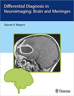
Differential Diagnosis in Neuroimaging: Brain and Meninges, 1ed
…an outstanding way to learn imaging anatomy and corresponding diseases….This [series] is a highly desirable set of books and would be a valuable addition to any neuroradiologist’s personal collection, and certainly to any departmental or sectional library. — AJNR Blog
Authored by renowned neuroradiologist Steven P. Meyers, Differential Diagnosis in Neuroimaging: Brain and Meninges is a stellar guide for identifying and diagnosing brain pathologies based on location and neuroimaging results. The succinct text reflects more than 25 years of hands-on experience gleaned from advanced training and educating residents and fellows in radiology, neurosurgery, and neurology. The high-quality MRI, CT, PET, PET/CT, conventional angiography, and X-ray images have been collected over Dr. Meyers’s lengthy career, presenting an unsurpassed visual learning tool.
The distinctive ‘three-column table plus images’ format is easy to incorporate into clinical practice, setting this book apart from larger, disease-oriented radiologic tomes. The layout enables readers to quickly recognize and compare abnormalities based on high-resolution images.
Key Highlights
- Tabular columns organized by anatomical abnormality include brain imaging findings and a summary of key clinical data that correlates to the images
- Comprehensive imaging of the brain, ventricles, meninges, and neurovascular system in both children and adults, including congenital/developmental anomalies and acquired disease
- More than 1,900 figures illustrate the radiological appearance of intracranial lesions, masses, neurodegenerative disorders, ischemia and infarction, and more
This visually rich resource is a must-have diagnostic tool for radiologists, neurosurgeons, and neurologists, and residents and fellows. The highly practical format makes it ideal for daily rounds, as well as a robust study guide for physicians preparing for board exams.
Contents
۱ Brain (Intra-Axial Lesions)
Table 1.1 Congenital and histogenic malformations of the brain
Table 1.2 Supratentorial solitary intra-axial mass lesions
Table 1.3 Solitary intra-axial lesions in the posterior cranial fossa (infratentorial)
Table 1.4 Multiple intra-axial lesions in the brain
Table 1.5 Multiple or diffuse lesions involving white matter in children
Table 1.6 Bilateral lesions involving the basal ganglia and/or thalami
Table 1.7 Neurodegenerative disorders
Table 1.8 Ischemia and infarction involving the brain and/or brainstem in adults
Table 1.9 Ischemia and infarction involving the brain and/or brainstem in children
Table 1.10 Intrasellar and juxtasellar lesions
Table 1.11 Lesions in the pineal region
۲ Ventricles and Cisterns
Table 2.1 Lateral ventricles— common masses
Table 2.2 Common third ventricular masses
Table 2.3 Fourth ventricular lesions
Table 2.4 Excessively small ventricles
Table 2.5 Dilated ventricles
Table 2.6 Abnormal or altered configuration of the ventricles
Table 2.7 Solitary intraventricular lesions in children
Table 2.8 Solitary intraventricular lesions in adults
Table 2.9 Contrast-enhancing ventricular margins
۳ Extra-Axial Lesions
Table 3.1 Solitary extra-axial mass lesions
Table 3.2 Multifocal extra-axial lesions
۴ Meninges
Table 4.1 Abnormalities involving the dura
Table 4.2 Multifocal and/or diffuse leptomeningeal abnormalities
۵ Vascular Abnormalities
Table 5.1 Congenital and developmental vascular anomalies/variants
Table 5.2 Acquired vascular disease
لینک کوتاه : https://bookbaz.ir/?p=55655
نویسنده : Steven P. Meyers
ناشر : Thieme; 1 edition
سال انتشار : 2017
زبان کتاب : انگلیسی
نوع فایل : PDF
تعداد صفحات : 664
(ISBN) شابک : 1604067004
قیمت کتاب درآمازون : $112.00
حجم فایل : 40 MB
کتاب های مرتبط:
 دانلود کتاب تشخیص افتراقی در تصویربرداری عصبی: ستون فقرات
Differential Diagnosis in Neuroimaging: Spine, 1ed
 دانلود کتاب SPECT و SPECT/CT: راهنمای بالینی
SPECT and SPECT/CT: A Clinical Guide, 1ed
 دانلود کتاب مشکلات رایج در بیماریهای مولتیپل اسکلروزیس و میلین زدا CNS
Common Pitfalls in Multiple Sclerosis and CNS Demyelinating Diseases, 1ed
 دانلود کتاب نورونولوژی و تصویربرداری عصبی سکته مغزی
Neurosonology and Neuroimaging of Stroke, 2ed
 دانلود کتاب MRI کمی مغز: اصول اندازه گیری فیزیکی
Quantitative MRI of the Brain: Principles of Physical Measurement, 2ed
 دانلود کتاب پردازش تصویربرداری پزشکی: تکنیک های پیشرفته تئوری فازی
Medical Image Processing: Advanced Fuzzy Set Theoretic Techniques, 1ed
 دانلود کتاب ملزومات رادیولوژی اسکلتی یاچم و رو (۲ جلدی) + CD
Yochum and Rowe’s Essentials of Skeletal Radiology, 2-Vol, 3ed + CD
 دانلود کتاب تصمیم گیری برای جراحی حداقل تهاجمی ستون فقرات
 Decision Making for Minimally Invasive Spine Surgery, 1ed
 دانلود کتاب راهنمای روش های رادیولوژی چپمن و ناکیِلنی
Chapman & Nakielny’s Guide to Radiological Procedures, 6ed
 دانلود کتاب مرور بورد پزشکی هسته ای: سوال و جواب های خود ارزیابی
Nuclear Medicine Board Review: Questions and Answers for Self-Assessment, 3ed
 دانلود کتاب مغز و اعصاب بالینی امینوف
Lange Clinical Neurology, 10ed
 دانلود کتاب اصول و تمرین جراحی سوراخ کلید مغز + ویدئو
Principles and Practice of Keyhole Brain Surgery, 1ed + Video
 دانلود کتاب تروما اعصاب و مراقبت بحرانی از ستون فقرات
 Neurotrauma and Critical Care of the Spine, 2ed
 دانلود کتاب چشم انداز محاسباتی و پردازش تصویری پزشکی
 Computational Vision and Medical Image Processing V: VipIMAGE 2015
 دانلود کتاب رادیوگرافی PREP (مرور برنامه و آمادگی آزمون)
Radiography PREP (Program Review and Exam Preparation), 8ed
 دانلود کتاب تصویربرداری پستان (سری تشخیص مستقیم در رادیولوژی)
Breast Imaging (Direct Diagnosis in Radiology: DX-Direct!), 1ed
 دانلود کتاب تصویربرداری قلبی (سری تشخیص مستقیم در رادیولوژی)
Cardiac Imaging (Direct Diagnosis in Radiology: DX-Direct!), 1ed
 دانلود کتاب پرسش و پاسخ تصویربرداری گوارشی رادکِیس
RadCases Plus Q&A Gastrointestinal Imaging, 2ed
 دانلود کتاب تصویربرداری از مغز با MRI و CT: رویکرد الگوی تصویری
Brain Imaging with MRI and CT: An Image Pattern Approach, 1ed
 دانلود کتاب اطلس توپوگرافی و پاتوتوپوگرافی آناتومی سر و گردن
Atlas of Topographical and Pathotopographical Anatomy of the Head and Neck, 1ed
 دانلود کتاب تصویربرداری قلب و عروق ESC
The ESC Textbook of Cardiovascular Imaging, 3ed
 دانلود کتاب دوز، فواید و خطر در تصویربرداری پزشکی
 Dose, Benefit, and Risk in Medical Imaging, 1ed
 دانلود کتاب پرتودرمانی عملی: فیزیک و تجهیزات
Practical Radiotherapy: Physics and Equipment, 3ed
 دانلود کتاب مغز و اعصاب کودکان
 Handbook of Pediatric Neurology, 1ed
 دانلود کتاب رادیولوژی ۱۰۱: مبانی و اصول تصویربرداری
Radiology 101: The Basics & Fundamentals of Imaging, 4ed
 دانلود کتاب پزشکی هسته ای: روش شناسی و کاربردهای بالینی
Nuclear Medicine Textbook: Methodology and Clinical Applications, 1ed
 دانلود کتاب طبقه بندی سیگنال مغزی EEG برای تشخیص اختلال تشنج صرع
 EEG Brain Signal Classification for Epileptic Seizure Disorder Detection, 1ed
 دانلود کتاب مقاله های برجسته در جراحی مغز و اعصاب
Landmark Papers in Neurosurgery, 2ed
 دانلود کتاب جراحی مغز و اعصاب یومانس و وین (۴ جلدی)
Youmans and Winn Neurological Surgery, 4-Vol, 7ed
 دانلود کتاب مبانی منطقی برای ترجمه بالینی در درمان سکته مغزی
 Rational Basis for Clinical Translation in Stroke Therapy, 1ed
 دانلود کتاب راهنمای اولیه برای رادیوگرافی دندان
 Basic Guide to Dental Radiography, 1ed
 دانلود کتاب سونوگرافی مراقبت اورژانسی
Emergency Point-of-Care Ultrasound, 2ed
 دانلود کتاب تصویربرداری تشخیصی: دهان و فک و صورت
Diagnostic Imaging: Oral and Maxillofacial, 2ed
 دانلود کتاب اختلاف نظر در جراحی عصبی: بیماری های عصبی-عروقی
Controversies in Neurological Surgery: Neurovascular Diseases, 1ed
 دانلود کتاب مراقبتهای ویژه جراحی مغز و اعصاب
 Neurosurgical Intensive Care, 2ed
 دانلود کتاب نوروآناتومی بالینی
Clinical Neuroanatomy, 29ed
 دانلود کتاب آناتومی آندوسکوپی ترانس نازال قاعده جمجمه و نواحی مجاور
Endoscopic Transnasal Anatomy of the Skull Base and Adjacent Areas, 1ed
 دانلود کتاب جراحی آندوسکوپی سینوس: آناتومی، بازسازی سه بعدی و روش جراحی + ویدئو
Endoscopic Sinus Surgery: Anatomy, Three-Dimensional Reconstruction, and Surgical Technique, 3ed + Videos
 دانلود کتاب راهنمای جراحی مغز و اعصاب گرینبرگ (ویرایش ۲۰۱۶)
Greenberg’s Handbook of Neurosurgery, 8ed
 دانلود کتاب مراقبت از بیمار در رادیوگرافی: همراه با معرفی تصویربرداری پزشکی
Patient Care in Radiography: With an Introduction to Medical Imaging, 9ed
 دانلود کتاب راهنمای جیبی رادیوگرافی مریل
Merrill’s Pocket Guide to Radiography, 14ed
 دانلود کتاب بیماری مویامویا: تشخیص و درمان
Moyamoya Disease: Diagnosis and Treatment, 1ed
 دانلود کتاب اولتراسوند تشخیصی عروقی
Diagnostic Ultrasound: Vascular, 1ed
 دانلود کتاب راهنمای OCT شبکیه: توموگرافی انسجام نوری
Handbook of Retinal OCT: Optical Coherence Tomography, 2ed
 دانلود کتاب تصویربرداری تشخیصی کودکان کافِی (۲ جلدی)
Caffey’s Pediatric Diagnostic Imaging, 2-Vol, 12ed
 دانلود کتاب مبانی جراحی مغز و اعصاب
 Neurosurgery Fundamentals, 1ed
 دانلود کتاب سونوگرافی در کمک به تولید مثل و اوایل بارداری
Ultrasound in Assisted Reproduction and Early Pregnancy, 1ed
 دانلود کتاب تصویربرداری قفسه سینه (سری تشخیص مستقیم در رادیولوژی)
Thoracic Imaging (Dx-Direct), 1ed
 دانلود کتاب سونوگرافی تشخیصی: شکم و لگن
 Diagnostic Ultrasound: Abdomen and Pelvis, 1ed
 دانلود کتاب رادیوسرجری استریوتاکتیک داخل جمجمه
Intracranial Stereotactic Radiosurgery, 2ed
 دانلود کتاب برنامه تحریک عمق مغز: مکانیسم، اصول و تمرین
Deep Brain Stimulation Programming: Mechanisms, Principles and Practice, 2ed
 دانلود کتاب مصور رنگی آناتومی اعصاب
 Neuroanatomy: An Illustrated Colour Text, 6ed
 دانلود کتاب مدیریت تطبیقی پاتولوژی ستون فقرات
 Comparative Management of Spine Pathology, 1ed
 دانلود کتاب صدمات اعصاب و عصب: درد، درمان، آسیب بیماری و دستورالعمل های آینده (جلد دوم)
Nerves and Nerve Injuries: Vol 2: Pain, Treatment, Injury, Disease and Future Directions
 دانلود کتاب آی سی یو اعصاب
The NeuroICU Book
 دانلود کتاب مغز و اعصاب بین المللی
International Neurology, 2ed
 دانلود کتاب اطلس آناتومی مقطعی، جلد ۳: ستون فقرات، پایانگاه، مفاصل: توموگرافی کامپیوتری و تصویربرداری رزونانس مغناطیسی
Pocket Atlas of Sectional Anatomy, Volume III: Spine, Extremities, Joints: Computed Tomography and Magnetic Resonance Imaging, 2ed
 دانلود کتاب جراحی ستون فقرات سینه ای: اصول و تکنیک ها
Surgery of the Thoracic Spine: Principles and Techniques, 1ed
 دانلود کتاب تفسیر تصاویر اسکلتی عضلانی: آناتومی، آسیب شناسی و گزارش اورژانس
Interpreting Musculoskeletal Images: Anatomy, Pathology and Emergency Reporting, 1ed
 دانلود کتاب اطلس رنگی آناتومی سونوگرافی بلاک
Color Atlas of Ultrasound Anatomy, 2ed
 دانلود کتاب راهنمای مطالعات اجرایی عصب بوشباچر
Buschbacher’s Manual of Nerve Conduction Studies, 3ed
 دانلود کتاب تصویربرداری تشخیصی کودکان
Diagnostic Imaging: Pediatrics, 4ed
 دانلود کتاب اصول جراحی مغز و اعصاب النبوجن
Principles of Neurological Surgery, 3ed
 دانلود کتاب اطلس آموزشی ماموگرافی
 Teaching Atlas of Mammography, 4ed
 دانلود کتاب سونوگرافی داپلر رنگی در زنان و زایمان
Color Doppler Sonography in Gynecology and Obstetrics, 1ed
 دانلود کتاب دیسپلازی استخوان: اطلس اختلالات ژنتیکی رشد اسکلت
Bone Dysplasias: An Atlas of Genetic Disorders of Skeletal Development, 4ed
 دانلود کتاب ۱۰۱ راه حل سی تی اسکن شکمی
 ۱۰۱Ct Abdomen Solutions, 1ed
 دانلود کتاب راهنمای سمیت عصبی
Handbook of Neurotoxicity, 2ed
 دانلود کتاب آرتروسکوپی پیشرفته AANA: شانه، آرنج و مچ دست، ران، زانو، پا و مچ پا (۵ جلدی)
AANA Advanced Arthroscopy: Shoulder, Elbow and Wrist, Hip, Knee, Foot and Ankle, 5-Vol, 1ed
 دانلود کتاب موارد تصویربرداری اسکلتی عضلانی تهران زاده
Musculoskeletal Imaging Cases (McGraw-Hill Radiology), 1ed
 دانلود کتاب متاستازهای مغز از تومورهای اولیه (۳ جلدی)
Brain Metastases from Primary Tumors, 3-Vol, 1ed
 دانلود کتاب اطلس ویدیویی پایش نوروفیزیولوژیک در جراحی تومورهای نفوذی مغز + ویدئو
Video Atlas of Neurophysiological Monitoring in Surgery of Infiltrating Brain Tumors, 1ed + Video
 دانلود کتاب معاینه بالینی عصبی
Neurological Clinical Examination, 4ed
 دانلود کتاب تشخیص افتراقی در تصویربرداری سونوگرافی
 Differential Diagnosis in Ultrasound Imaging, 2ed
 دانلود کتاب خون رسانی مجدد مغزی: تکنیک های جراحی بایپس در خارج به داخل جمجمه
Cerebral Revascularization: Techniques in Extracranial-to-Intracranial Bypass Surgery, 1ed
 دانلود کتاب چشم پزشکی عصبی مصور + ویدئو
Neuro-Ophthalmology Illustrated, 3ed + Video
 دانلود کتاب ۳ تفاوت برتر در رادیولوژی عروقی و مداخله ای
 Top 3 Differentials in Vascular and Interventional Radiology, 1ed
 دانلود کتاب راهنمای تصویربرداری عصبی نورو-انکولوژی
Handbook of Neuro-Oncology Neuroimaging, 2ed
 دانلود کتاب تصویربرداری سر و گردن (سری موارد رادیولوژی)
RadCases Head and Neck Imaging, 1ed
 دانلود کتاب راهنمای الکتروفیزیولوژی کلینیک مایو
Mayo Clinic Electrophysiology Manual, 1ed
 دانلود کتاب مرور بورد مراقبت های حاد و عصبی کلینیک مایو
Mayo Clinic Critical and Neurocritical Care Board Review, 1ed
 دانلود کتاب اصول سی تی اسکن بدن
Fundamentals of Body CT, 5ed
 دانلود کتاب الکترودیاگنوز در مغز و اعصاب بالینی امینف
Aminoff’s Electrodiagnosis in Clinical Neurology, 6ed
 دانلود کتاب بررسی موردی رادیولوژی: ستون فقرات
Radiology Case Review Series: Spine, 1ed
 دانلود کتاب بیومگنتیک ها: اصول و برنامه های برانگیختن بیومگنتیک و تصویربرداری
Biomagnetics: Principles and Applications of Biomagnetic Stimulation and Imaging, 1ed
 دانلود کتاب اولتراسوند دست و اندام فوقانی + ویدئو
Ultrasound of the Hand and Upper Extremity, 1ed + Video
 دانلود کتاب مشاهدات بحرانی در رادیولوژی برای دانشجویان پزشکی
Critical Observations in Radiology for Medical Students, 1ed
 دانلود کتاب تکنیک های اصلی در جراحی مغز و اعصاب عملی + ویدئو
Core Techniques in Operative Neurosurgery, 2ed + Video
 دانلود کتاب کتاب مبانی فیزیک تصویربرداری و رادیوبیولوژی سلمان
Selman’s The Fundamentals of Imaging Physics and Radiobiology, 10ed
 دانلود کتاب مراقبت سکته مغزی حاد
Acute Stroke Care, 3ed
 دانلود کتاب خود ارزیابی مغز و اعصاب: همگام با مغز و اعصاب برادلی
Neurology Self-Assessment: A Companion to Bradley’s Neurology in Clinical Practice, 1ed
 دانلود کتاب مرور پرسش و پاسخ اولتراسوند برای بورد
Ultrasound Q&A Review for the Boards, 1ed
 دانلود کتاب هدایت حین عمل MRI جراحی مغز و اعصاب
Intraoperative MRI-Guided Neurosurgery, 1ed
 دانلود کتاب تصویربرداری تشخیصی کودکان کافی (ویرایش ۲۰۱۹، ۲ جلدی)
Caffey’s Pediatric Diagnostic Imaging, 2-Volume Set, 13ed
 دانلود کتاب معرفی تصویربرداری نتر
Netter’s Introduction to Imaging, 1ed
 دانلود کتاب نوروبلاستوما: مداخلات مکانیسم های مولکولی و درمانی
Neuroblastoma: Molecular Mechanisms and Therapeutic Interventions, 1ed
 دانلود کتاب تصویربرداری در جراحی شکم
Imaging in Abdominal Surgery, 1ed
 دانلود کتاب تصویربرداری در گوارش
Imaging in Gastroenterology, 1ed
 دانلود کتاب تصویربرداری برای دانشجویان
Imaging for Students, 4ed
 دانلود کتاب تصویربرداری رزونانس مغناطیسی میکروسکوپی
Microscopic Magnetic Resonance Imaging, 1ed
 دانلود کتاب راهنمای پایندگی چشم پزشکی عصبی
The Neuro-Ophthalmology Survival Guide, 2ed
 دانلود کتاب کشیک مغز و اعصاب
 On Call Neurology, 4ed
 دانلود کتاب تصویربرداری تشخیصی: مغز
Diagnostic Imaging: Brain, 3ed
 دانلود کتاب تعادل ساژیتال ستون فقرات: از عادی به پاتولوژی
Sagittal Balance of the Spine: From Normal to Pathology, 1ed
 دانلود کتاب اصول و تمرین پزشکی خواب (۲ جلدی) + ویدئو
Principles and Practice of Sleep Medicine, 2-Vol, 7ed + Video
 دانلود کتاب پرینت سه بعدی برای رادیولوژیست
۳D Printing for the Radiologist, 1ed
 دانلود کتاب سری کارشناسی ستون فقرات AO: تغییر شکل ستون فقرات بزرگسالان (جلد ۴)
AOSpine Masters Series, Volume 4: Adult Spinal Deformities, 1ed
 دانلود کتاب اعصاب جمجمه: آناتومی، پاتولوژی، تصویربرداری
Cranial Nerves: Anatomy, Pathology, Imaging, 1ed
 دانلود کتاب نوروبیونیک ها: مهندسی پزشکی پروتز عصبی
Neurobionics: The Biomedical Engineering of Neural Prostheses, 1ed
 دانلود کتاب آناتومی تصویربرداری: قفسه سینه، شکم، لگن
Imaging Anatomy: Chest, Abdomen, Pelvis, 2ed
 دانلود کتاب رادیولوژی اعصاب تشخیصی و مداخله ای
Diagnostic and Interventional Neuroradiology, 1ed
 دانلود کتاب اولتراسوند: مرور کلی
Ultrasound: A Core Review, 1ed
 دانلود کتاب توانبخشی سکته مغزی: رویکرد مبتنی بر عملکرد
Stroke Rehabilitation: A Function-Based Approach, 4ed
 دانلود کتاب اطلس عمل جراحی مغز و اعصاب: جراحی مغز و اعصاب کاربردی
Neurosurgical Operative Atlas: Functional Neurosurgery, 3ed
 دانلود کتاب راهنمای سم شناسی عصبی رشد
Handbook of Developmental Neurotoxicology, 2ed
 دانلود کتاب موارد تصویربرداری پستان
Breast Imaging Cases
 دانلود کتاب تصویربرداری تشخیصی: پزشکی زایمان
Diagnostic Imaging: Obstetrics, 3ed
 دانلود کتاب ملزومات سونوگرافی شکمی و لگنی
Essentials of Abdomino-Pelvic Sonography, 1ed
 دانلود کتاب تفسیر رادیوگرافی قفسه سینه در بیماران قلبی کودک
Chest Radiographic Interpretation in Pediatric Cardiac Patients, 1ed
 دانلود کتاب اطلس بیهوشی منطقه ای هدایت اولتراسوند + ویدئو
Atlas of Ultrasound-Guided Regional Anesthesia, 3ed + Video
 دانلود کتاب آناتومی تصویربرداری: سر و گردن
Imaging Anatomy: Head and Neck, 1ed
 دانلود کتاب تصویربرداری تشخیصی دستگاه گوارش
Diagnostic Imaging: Gastrointestinal, 4ed
 دانلود کتاب تصویربرداری تشخیصی زایمان
Diagnostic Imaging: Obstetrics, 4ed
 دانلود کتاب تصویربرداری تشخیصی سر و گردن
Diagnostic Imaging: Head and Neck, 4ed
 دانلود کتاب تصویربرداری تشخیصی انکولوژی
Diagnostic Imaging: Oncology, 2ed
 دانلود کتاب راهنمای جراحی مغز و اعصاب کودکان
Handbook of Pediatric Neurosurgery, 1ed
 دانلود کتاب پرسش و پاسخ پزشکی هسته ای رادکِیس
RadCases Plus Q&A Nuclear Medicine, 2ed
 دانلود کتاب تصویربرداری رزونانس مغناطیسی (۳ جلدی)
Magnetic Resonance Imaging Handbook (MRI), 3-Vol, 1ed
 دانلود کتاب تصویربرداری تشخیصی نوزادان و کودکان ولز (جلد اول)
Diagnostic Imaging of Infants and Children, Vol-1, 1ed
 دانلود کتاب اطلس توموگرافی کامپیوتری پرتو مخروطی
Atlas of Cone Beam Computed Tomography, 1ed
 دانلود کتاب MRI در یک نگاه
MRI at a Glance, 2ed
 دانلود کتاب صدمات عصب کلاین و هادسون: صدمات عصب، انترپمنت و تومور
Kline and Hudson’s Nerve Injuries: Operative Results for Major Nerve Injuries, Entrapments and Tumors, 2ed
 دانلود کتاب سونوگرافی مداخله ای: راهنمای عملی و اطلس
Interventional Ultrasound: A Practical Guide and Atlas, 1ed
 دانلود کتاب فیزیک ضروری در تصویربرداری برای پرتونگار کلارک
Clark’s Essential Physics in Imaging for Radiographers, 1ed
 دانلود کتاب تصویربرداری ادراری تناسلی (سری موارد رادیولوژی)
Genitourinary Imaging (Radcase), 1ed
 دانلود کتاب اطلس درمان عصبی با بیحسی موضعی
 Atlas of Neural Therapy With Local Anesthetics, 3ed
 دانلود کتاب به دام افتادن اعصاب محیطی: تشخیص و مدیریت بالینی
Peripheral Nerve Entrapments: Clinical Diagnosis and Management, 1ed
 دانلود کتاب تصویربرداری تشخیصی: زنان و زایمان
Diagnostic Imaging: Gynecology, 2ed
 دانلود کتاب تصویربرداری زنان و زایمان: تشخیص و مراقبت از جنین
Obstetric Imaging: Fetal Diagnosis and Care, 2ed
 دانلود کتاب تصویربرداری اسکلتی عضلانی
Musculoskeletal Imaging, 2ed
 دانلود کتاب اندوسکوپی ترانس نازال قاعده جمجمه و جراحی مغز
Transnasal Endoscopic Skull Base and Brain Surgery, 1ed
 دانلود کتاب نوروپاتولوژی تکاملی
Developmental Neuropathology, 2ed
 دانلود کتاب موقعیت در رادیوگرافی کلارک
Clark’s Positioning in Radiography, 13ed
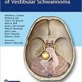 دانلود کتاب مدیریت جامع شوانوما وستیبولار
 Comprehensive Management of Vestibular Schwannoma, 1ed
 دانلود کتاب تصویربرداری تشخیصی قفسه سینه
Diagnostic Imaging: Chest, 3ed
 دانلود کتاب تصویربرداری تشخیصی زنان
Diagnostic Imaging: Gynecology, 3ed
 دانلود کتاب آنژیوگرافی مغزی کاربردی: آناتومی و پاتولوژی عروقی نرمال
Applied Cerebral Angiography: Normal Anatomy and Vascular Pathology, 1ed
 دانلود کتاب تعمیرات X-Ray: راهنمای جامع نصب و سرویس تجهیزات رادیوگرافی
X-Ray Repair: A Comprehensive Guide to the Installation and Servicing of Radiographic Equipment, 3ed
 دانلود کتاب جامع فیزیک پزشکی (۱۰ جلدی)
Comprehensive Biomedical Physics, 10-Vol, 1ed
 دانلود کتاب تصویربرداری سونوگرافی تشخیصی (مهندسی پزشکی)
Diagnostic Ultrasound Imaging: Inside Out, 2ed