
دانلود کتاب تصویربرداری رزونانس مغناطیسی: تصویربرداری از لگن، سیستم عضلانی و برنامه های کاربردی ویژه CAD
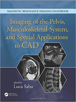
Magnetic Resonance Imaging Handbook: Imaging of the Pelvis, Musculoskeletal System, and Special Applications to CAD, 1ed
Magnetic resonance imaging (MRI) is a technique used in biomedical imaging and radiology to visualize internal structures of the body. Because MRI provides excellent contrast between different soft tissues, the technique is especially useful for diagnostic imaging of the brain, muscles, and heart.
In the past 20 years, MRI technology has improved significantly with the introduction of systems up to 7 Tesla (7 T) and with the development of numerous post-processing algorithms such as diffusion tensor imaging (DTI), functional MRI (fMRI), and spectroscopic imaging. From these developments, the diagnostic potentialities of MRI have improved impressively with an exceptional spatial resolution and the possibility of analyzing the morphology and function of several kinds of pathology.
Given these exciting developments, the Magnetic Resonance Imaging Handbook: Imaging of the Pelvis, Musculoskeletal System, and Special Applications to CAD is a timely addition to the growing body of literature in the field. Offering comprehensive coverage of cutting-edge imaging modalities, this book:
- Discusses MRI of the urinary system, pelvis, spine, soft tissues, lymphatics, and brain
- Explains how MRI can be used in fetal, pediatric, forensic, postmortem, and computer-aided diagnostic (CAD) applications
- Highlights each organ’s anatomy and pathological processes with high-quality images
- Examines the protocols and potentialities of advanced MRI scanners such as 7 T systems
- Includes extensive references at the end of each chapter to enhance further study
Thus, the Magnetic Resonance Imaging Handbook: Imaging of the Pelvis, Musculoskeletal System, and Special Applications to CAD provides radiologists and imaging specialists with a valuable, state-of-the-art reference on MRI.
About the Author
Luca Saba is full professor of Radiology in the University of Cagliari, Italy. Prof. Saba currently works at the Azienda Ospedaliero Universitaria of Cagliari. During his career, he has won 15 scientific and extracurricular awards, published more than 220 papers in high impact factor journals, presented more than 500 papers and posters at national and international congress events, spoken more than 45 times at national and international conferences, written 21 book chapters, edited 14 books, and reviewed more than 40 scientific journals. He is a member of the Italian Society of Medical Radiology, European Society of Radiology, Radiological Society of North America, American Roentgen Ray Society, and European Society of Neuroradiology.
Contents
۱: Magnetic Resonance Imaging of Kidneys and Ureters
۲: Carcinoma of the Bladder and Urethra
۳: Male Pelvis (Prostate, Seminal Vesicles, and Testes)
۴: Uterus and Vagina
۵: Benign and Malignant Conditions of the Ovaries and Peritoneum
۶: MRI of the Placenta and the Pregnant Patient
۷: MRI of Pelvic Floor Disorders
۸: Degenerative Disease of the Spine and Other Spondyloarthropathies
۹: Spine Infections
۱۰: Traumatic Disease of the Spine
۱۱: Neoplastic Disease of the Spine
۱۲: MR Pathology of Sacrum and Ilium
۱۳: Magnetic Resonance Imaging of Soft Tissues
۱۴: Temporomandibular Joints
۱۵: MR-Guided Interventional Radiology of the Musculoskeletal System
۱۶: MR of the Lymphatics
۱۷: Pediatric Applications
۱۸: Fetal MRI
۱۹: Postmortem and Forensic Magnetic Resonance Imaging
۲۰: Magnetic Resonance-Guided Focused Ultrasound
۲۱: PET/MRI: Concepts and Clinical Applications
۲۲: Computer-Aided Diagnosis with MR Images of the Brain
لینک کوتاه : https://bookbaz.ir/?p=32677
نویسنده : Luca Saba
ناشر : CRC Press; 1 edition
سال انتشار : 2016
زبان کتاب : انگلیسی
نوع فایل : PDF
تعداد صفحات : 562
(ISBN) شابک : 1482216213
قیمت کتاب درآمازون : $266.11
حجم فایل : 60 MB
کتاب های مرتبط:
 دانلود کتاب تصویربرداری زنان و زایمان کوپل: سری کارشناس رادیولوژی
Obstetric Imaging: Expert Radiology Series, 1ed
 دانلود کتاب تشخیص افتراقی در توموگرافی کامپیوتری
Differential Diagnosis in Computed Tomography, 2ed
 دانلود کتاب دوز، فواید و خطر در تصویربرداری پزشکی
 Dose, Benefit, and Risk in Medical Imaging, 1ed
 دانلود کتاب ۳ تفاوت برتر در رادیولوژی مغز و اعصاب
 Top 3 Differentials in Neuroradiology, 1ed
 دانلود کتاب MRI در یک نگاه
MRI at a Glance, 2ed
 دانلود کتاب تصویربرداری اسکلتی عضلانی
Musculoskeletal Imaging, 2ed
 دانلود کتاب تصویربرداری پزشکی آکسفورد
Oxford Handbook of Medical Imaging, 1ed
 دانلود کتاب MRI بافتها همراه با T2 یا T2*s کوتاه
MRI of Tissues with Short T2s or T2*s, 1ed
 دانلود کتاب راهنمای انکولوژی مداخله ای
 Manual of Interventional Oncology, 1ed
 دانلود کتاب ملزومات رادیوگرافی و رادیولوژی دندان
Essentials of Dental Radiography and Radiology, 5ed
 دانلود کتاب سری بررسی موردی رادیولوژی: تصویربرداری پستان
 Radiology Case Review Series: Breast Imaging, 1ed
 دانلود کتاب تعمیرات X-Ray: راهنمای جامع نصب و سرویس تجهیزات رادیوگرافی
X-Ray Repair: A Comprehensive Guide to the Installation and Servicing of Radiographic Equipment, 3ed
 دانلود کتاب موارد و مشکلات در تصویربرداری بدن: تصویربرداری قفسه سینه، شکم و زنان
Variants and Pitfalls in Body Imaging: Thoracic, Abdominal and Women’s Imaging, 2ed
 دانلود کتاب بیماری های آئورت: اطلس تصویربرداری تشخیصی بالینی
Aortic Diseases: Clinical Diagnostic Imaging Atlas, 1ed
 دانلود کتاب تصویربرداری قفسه سینه مولر
Muller’s Imaging of the Chest, 2ed
 دانلود کتاب پرسش و پاسخ رادیولوژی مداخله ای رادکِیس
RadCases Q&A Interventional Radiology, 2ed
 دانلود کتاب MRI در عمل
MRI in Practice, 5ed
 دانلود کتاب EXPERTddx: رادیولوژی شکم
 EXPERTddx: Abdomen: Published by Amirsys
 دانلود کتاب ExpertDDx: مغز و ستون فقرات
ExpertDDx: Brain and Spine, 2ed
 دانلود کتاب اصول اولتراسوند اسکلتی عضلانی
Fundamentals of Musculoskeletal Ultrasound, 3ed
 دانلود کتاب تصویربرداری در جراحی شکم
Imaging in Abdominal Surgery, 1ed
 دانلود کتاب تصویربرداری در گوارش
Imaging in Gastroenterology, 1ed
 دانلود کتاب اصول سی تی اسکن بدن
Fundamentals of Body CT, 5ed
 دانلود کتاب راهنمای تصویربرداری عصبی نورو-انکولوژی
Handbook of Neuro-Oncology Neuroimaging, 2ed
 دانلود کتاب رادیولوژی اضطراری
 ABC of Emergency Radiology, 3ed
 دانلود کتاب موارد تصویربرداری پستان
Breast Imaging Cases
 دانلود کتاب تصویربرداری تشخیصی: پزشکی زایمان
Diagnostic Imaging: Obstetrics, 3ed
 دانلود کتاب هدایت حین عمل MRI جراحی مغز و اعصاب
Intraoperative MRI-Guided Neurosurgery, 1ed
 دانلود کتاب ملزومات سونوگرافی شکمی و لگنی
Essentials of Abdomino-Pelvic Sonography, 1ed
 دانلود کتاب تفسیر رادیوگرافی قفسه سینه در بیماران قلبی کودک
Chest Radiographic Interpretation in Pediatric Cardiac Patients, 1ed
 دانلود کتاب تصویربرداری تشخیصی کودکان کافی (ویرایش ۲۰۱۹، ۲ جلدی)
Caffey’s Pediatric Diagnostic Imaging, 2-Volume Set, 13ed
 دانلود کتاب اطلس بیهوشی منطقه ای هدایت اولتراسوند + ویدئو
Atlas of Ultrasound-Guided Regional Anesthesia, 3ed + Video
 دانلود کتاب آناتومی تصویربرداری: سر و گردن
Imaging Anatomy: Head and Neck, 1ed
 دانلود کتاب تصویربرداری تشخیصی دستگاه گوارش
Diagnostic Imaging: Gastrointestinal, 4ed
 دانلود کتاب تصویربرداری تشخیصی زایمان
Diagnostic Imaging: Obstetrics, 4ed
 دانلود کتاب تصویربرداری تشخیصی سر و گردن
Diagnostic Imaging: Head and Neck, 4ed
 دانلود کتاب تصویربرداری تشخیصی انکولوژی
Diagnostic Imaging: Oncology, 2ed
 دانلود کتاب سونوگرافی مداخله ای: راهنمای عملی و اطلس
Interventional Ultrasound: A Practical Guide and Atlas, 1ed
 دانلود کتاب فیزیک ضروری در تصویربرداری برای پرتونگار کلارک
Clark’s Essential Physics in Imaging for Radiographers, 1ed
 دانلود کتاب تصویربرداری ادراری تناسلی (سری موارد رادیولوژی)
Genitourinary Imaging (Radcase), 1ed
 دانلود کتاب تشخیص افتراقی در تصویربرداری سونوگرافی
 Differential Diagnosis in Ultrasound Imaging, 2ed
 دانلود کتاب تصویربرداری تشخیصی: مغز
Diagnostic Imaging: Brain, 3ed
 دانلود کتاب تصویربرداری تشخیصی: زنان و زایمان
Diagnostic Imaging: Gynecology, 2ed
 دانلود کتاب تصویربرداری زنان و زایمان: تشخیص و مراقبت از جنین
Obstetric Imaging: Fetal Diagnosis and Care, 2ed
 دانلود کتاب موقعیت در رادیوگرافی کلارک
Clark’s Positioning in Radiography, 13ed
 دانلود کتاب تصویربرداری تشخیصی قفسه سینه
Diagnostic Imaging: Chest, 3ed
 دانلود کتاب تصویربرداری تشخیصی زنان
Diagnostic Imaging: Gynecology, 3ed
 دانلود کتاب تصویربرداری قفسه سینه و قلب و عروق
Chest and Cardiovascular Imaging, 3ed
 دانلود کتاب تصویربرداری از مغز با MRI و CT: رویکرد الگوی تصویری
Brain Imaging with MRI and CT: An Image Pattern Approach, 1ed
 دانلود کتاب آناتومی تصویربرداری: قفسه سینه، شکم، لگن
Imaging Anatomy: Chest, Abdomen, Pelvis, 2ed
 دانلود کتاب رادیولوژی اضطراری (سری موارد رادیولوژی)
Radcases Emergency Radiology, 1ed
 دانلود کتاب رادیولوژی اعصاب تشخیصی و مداخله ای
Diagnostic and Interventional Neuroradiology, 1ed
 دانلود کتاب اولتراسوند: مرور کلی
Ultrasound: A Core Review, 1ed
 دانلود کتاب پرسش و پاسخ تصویربرداری گوارشی رادکِیس
RadCases Plus Q&A Gastrointestinal Imaging, 2ed
 دانلود کتاب اندازه گیری و طبقه بندی در رادیولوژی اسکلتی عضلانی
Measurements and Classifications in Musculoskeletal Radiology, 1ed
 دانلود کتاب یادگیری رادیولوژی هِرینگ: شناخت مبانی
Learning Radiology: Recognizing the Basics, 3ed
 دانلود کتاب رادیولوژی ۱۰۱: مبانی و اصول تصویربرداری
Radiology 101: The Basics & Fundamentals of Imaging, 4ed
 دانلود کتاب تصویربرداری عمومی اسکلتی عضلانی
Basic Musculoskeletal Imaging, 1ed
 دانلود کتاب اندوسونوگرافی
 Endosonography, 4ed
 دانلود کتاب تعویض ترنس کاتتر دریچه آئورت
Transcatheter Aortic Valve Implantation, 1ed
 دانلود کتاب پرسش و پاسخ پزشکی هسته ای رادکِیس
RadCases Plus Q&A Nuclear Medicine, 2ed
 دانلود کتاب تصویربرداری بیماری های بینابینی ریه (سری رادیولوژی کلینیکو)
Clinico Radiological Series: Imaging of Interstitial Lung Diseases, 1ed
 دانلود کتاب اطلس آموزشی ماموگرافی
 Teaching Atlas of Mammography, 4ed
 دانلود کتاب توموگرافی پرتو مخروطی کامپیوتری در ارتودنسی: موارد مصرف، بینش و نوآوری
Cone Beam Computed Tomography in Orthodontics: Indications, Insights, and Innovations, 1ed
 دانلود کتاب سری مرور موردی رادیولوژی: تصویربرداری قفسه سینه
 Radiology Case Review Series: Thoracic Imaging, 1ed
 دانلود کتاب راهنمای رادیو مابولیزاسیون: فیزیک، بیولوژی، پزشکی هسته ای و تصویربرداری
Handbook of Radioembolization: Physics, Biology, Nuclear Medicine, and Imaging, 1ed
 دانلود کتاب اطلس جیبی آناتومی مقطعی: ستون فقرات، اندام، مفاصل
 Pocket Atlas of Sectional Anatomy: Spine Extremities Joints, 3ed
 دانلود کتاب چشم انداز محاسباتی و پردازش تصویری پزشکی
 Computational Vision and Medical Image Processing V: VipIMAGE 2015
 دانلود کتاب رادیوگرافی PREP (مرور برنامه و آمادگی آزمون)
Radiography PREP (Program Review and Exam Preparation), 8ed
 دانلود کتاب CT قلبی بالینی: آناتومی و عملکرد + ویدئو
Clinical Cardiac CT: Anatomy and Function, 2ed + Video
 دانلود کتاب سونوگرافی کودکان
 Pediatric Ultrasound, 1ed
 دانلود کتاب روشهای اسکلتی عضلانی هدایت شده با سونوگرافی در پزشکی ورزشی
Ultrasound Guided Musculoskeletal Procedures in Sports Medicine, 1ed
 دانلود کتاب تصویربرداری تومور قفسه سینه (سری رادیولوژی کلینیکو)
Clinico Radiological Series: Imaging Of Chest Tumors, 1ed
 دانلود کتاب مرور بالینی مغز و اعصاب مبتنی بر تصویر
Neurology Image-Based Clinical Review, 1ed
 دانلود کتاب توسعه دیجیتال ریه: از اولین سی تی ریه تا هوش مصنوعی بالینی
Developing the Digital Lung: From First Lung CT to Clinical AI, 1ed
 دانلود کتاب سونوگرافی مراقبت اورژانسی + ویدئو
Emergency Point-of-Care Ultrasound, 2ed + Video
 دانلود کتاب تصویربرداری از استخوان ها و مفاصل: یک روش مختصر و چند وجهی
Imaging of Bones and Joints: A Concise, Multimodality Approach, 1ed
 دانلود کتاب تصویربرداری و تجسم در اتاق عمل مدرن: راهنمای جامع برای پزشکان
Imaging and Visualization in The Modern Operating Room: A Comprehensive Guide for Physicians, 2015th
 دانلود کتاب آناتومی مقطعی برای متخصصان تصویربرداری
 Sectional Anatomy for Imaging Professionals, 3ed
 دانلود کتاب تصویربرداری پستان (سری تشخیص مستقیم در رادیولوژی)
Breast Imaging (Direct Diagnosis in Radiology: DX-Direct!), 1ed
 دانلود کتاب تصویربرداری قلبی (سری تشخیص مستقیم در رادیولوژی)
Cardiac Imaging (Direct Diagnosis in Radiology: DX-Direct!), 1ed
 دانلود کتاب مداخله انکولوژی (راهنمای عملی در مداخلات رادیولوژی)
Interventional Oncology (Practical Guides in Interventional Radiology), 1ed
 دانلود کتاب تصویربرداری تشخیصی: ادراری تناسلی
Diagnostic Imaging: Genitourinary, 3ed
 دانلود کتاب تصویربرداری تشخیصی مغز
 Diagnostic Imaging: Brain, 4ed
 دانلود کتاب قدرت فرا صوت: کاربرد فرا صوت شدت بالا
Power Ultrasonics: Applications of High-Intensity Ultrasound, 1ed
 دانلود کتاب MRI اسکلتی عضلانی آسیف سفیودین
Asif Saifuddin’s Musculoskeletal MRI, 2ed
 دانلود کتاب سونوگرافی تشخیصی کودکان
Diagnostic Pediatric Ultrasound, 1ed
 دانلود کتاب تصویربرداری رزونانس مغناطیسی میکروسکوپی
Microscopic Magnetic Resonance Imaging, 1ed
 دانلود کتاب مقدمه ای بر اولترا سونوگرافی عروقی زوئیبل
Zwiebel, Introduction of Vascular Ultrasonography, 6ed
 دانلود کتاب اصول تصویربرداری اسکلتی عضلانی مک کینز
Fundamentals of Musculoskeletal Imaging, 4ed
 دانلود کتاب تصویربرداری اشعه ایکس: اصول، تکنیک های صنعتی و کاربرد
X-Ray Imaging: Fundamentals, Industrial Techniques and Applications, 1ed
 دانلود کتاب مرور سریع رادیولوژی
 Rapid Review of Radiology, 1ed
 دانلود کتاب نورورادیولوژی: طیف و تکامل بیماری
Neuroradiology: Spectrum and Evolution of Disease, 1ed
 دانلود کتاب اطلس سونوگرافی سر و گردن
Atlas of Head and Neck Ultrasound, 1ed
 دانلود کتاب سونوگرافی اسکلتی عضلانی
Musculoskeletal Ultrasound, 1ed
 دانلود کتاب جریان خون مغزی و متابولیسم
 Cerebral Blood Flow and Metabolism, 1ed
 دانلود کتاب پاتولوژی رادیوگرافی جامع
Comprehensive Radiographic Pathology, 6ed
 دانلود کتاب رادیولوژی تشخیصی: تصویربرداری اطفال
Diagnostic Radiology: Paediatric Imaging, 3ed
 دانلود کتاب اولتراسونوگرافی مغز پیش از تولد
Ultrasonography of the Prenatal Brain, 3ed
 دانلود کتاب موقعیت ها و تکنیک های رادیوگرافی بانترِیگر
Bontrager’s Handbook of Radiographic Positioning and Techniques, 8ed
 دانلود کتاب سونوگرافی چندپارامتری برای ارزیابی بیماری کبد پراکنده: یک رویکرد عملی
Multiparametric Ultrasound for the Assessment of Diffuse Liver Disease: A Practical Approach, 1ed
 دانلود کتاب تصویربرداری دستگاه گوارش (سری تشخیص مستقیم در رادیولوژی)
Gastrointestinal Imaging (Direct Diagnosis in Radiology: DX-Direct!), 1ed
 دانلود کتاب آناتومی مقطعی انسان: اطلس بخش های بدن، تصاویر CT و MRI
Human Sectional Anatomy: Atlas of Body Sections, CT and MRI Images, 4ed
 دانلود کتاب پرتودرمانی بالینی سرطان: علائم، تکنیک ها و نتایج
Clinical Radiation Oncology: Indications, Techniques, and Results, 3ed
 دانلود کتاب بیماریهای شکم و لگن ۲۰۱۸-۲۰۲۱: تصویربرداری تشخیصی
Diseases of the Abdomen and Pelvis 2018-2021: Diagnostic Imaging, 1ed
 دانلود کتاب اطلس پاتولوژی آناتومیک با تصویربرداری
Atlas of Anatomic Pathology with Imaging
 دانلود کتاب موارد پیچیده در اکوکاردیوگرافی
 Complex Cases in Echocardiography, 1ed
 دانلود کتاب آزمون CORE – همگام با کیس
Crack the CORE Exam – Case Companion, 1ed
 دانلود کتاب آموختن فیزیک رادیولوژیک از طریق موارد
Radiologic Physics Taught Through Cases, 1ed
 دانلود کتاب CT و MRI کامل بدن (۲ جلدی)
CT and MRI of the Whole Body, 2-Vol, 5ed
 دانلود کتاب سونوگرافی ناهنجاری مادرزادی جنین: تشخیص افتراقی و شاخص پیش آگهی
Ultrasound of Congenital Fetal Anomalies: Differential Diagnosis and Prognostic Indicators, 2ed
 دانلود کتاب فیزیک تصویربرداری PET و SPECT
Physics of PET and SPECT Imaging, 1ed
 دانلود کتاب اطلس آموزشی تصویربرداری شکم
Teaching Atlas of Abdominal Imaging, 1ed
 دانلود کتاب تصویربرداری تشخیصی: ترومای اسکلتی عضلانی
Diagnostic Imaging: Musculoskeletal Trauma, 3ed
 دانلود کتاب پزشکی هسته ای (سری موارد رادیولوژی)
Nuclear Medicine (RadCases), 1ed
 دانلود کتاب جامع فیزیک پزشکی (۱۰ جلدی)
Comprehensive Biomedical Physics, 10-Vol, 1ed
 دانلود کتاب اطلس تصویربرداری عروقی ریوی
Atlas of Pulmonary Vascular Imaging, 1ed
 دانلود کتاب راهنمای بالینی IR: راهنمای مختصر برای رادیولوژی مداخله ای
 Pocketbook of Clinical IR: A Concise Guide to Interventional Radiology, 1ed
 دانلود کتاب رادیولوژی مغز و اعصاب پری ناتال: از جنین تا نوزاد
Perinatal Neuroradiology: From the Fetus to the Newborn, 1ed
 دانلود کتاب تشخیص افتراقی در تصویربرداری عصبی: ستون فقرات
Differential Diagnosis in Neuroimaging: Spine, 1ed
 دانلود کتاب سرطان پستان: تشخیص زودرس با ماموگرافی
Breast Cancer: Early Detection with Mammography: Casting Type Calcifications Sign of a Subtype with Deceptive Features, 1ed
 دانلود کتاب نورونولوژی و تصویربرداری عصبی سکته مغزی
Neurosonology and Neuroimaging of Stroke, 2ed
 دانلود کتاب موارد رادیولوژی دستگاه ادراری تناسلی
Genitourinary Radiology Cases
 دانلود کتاب رادیولوژی ضروری: معرفی بالینی تصویربرداری پاتوفیزیولوژی
 Essential Radiology: Clinical Presentation Pathophysiology Imaging, 3ed
 دانلود کتاب اصول CT ریه با وضوح بالا
Fundamentals of High-Resolution Lung CT, 1ed
 دانلود کتاب SPECT و SPECT/CT: راهنمای بالینی
SPECT and SPECT/CT: A Clinical Guide, 1ed
 دانلود کتاب انحراف جریان آنوریسم مغزی
Flow Diversion of Cerebral Aneurysms, 1ed
 دانلود کتاب EXPERTddx: رادیولوژی سر و گردن
 EXPERTddx: Head and Neck: Published by Amirsys
 دانلود کتاب تصویربرداری بالینی ایزنبرگ: اطلس تشخیص افتراقی
Eisenberg’s Clinical Imaging: An Atlas of Differential Diagnosis, 5ed
 دانلود کتاب الگوهای تصویربرداری مغز و ستون فقرات
 Brain and Spine Imaging Patterns, 1ed
 دانلود کتاب مبانی تصویربرداری کودکان
 Fundamentals of Pediatric Imaging, 3ed
 دانلود کتاب The Essential Guide to the New FRCR: Pt. 1 (MasterPass), 1ed
 دانلود کتاب رادیوتراپی کودکان
 Pediatric Radiation Oncology, 5ed
 دانلود کتاب انکولوژی تشعشع مبتنی بر شواهد
Handbook of Evidence-Based Radiation Oncology, 2ed
 دانلود کتاب حل مشکل در تصویربرداری شکم
 Problem Solving in Abdominal Imaging, 1ed
 دانلود کتاب پزشکی بالینی آکسفورد
Oxford Handbook of Clinical Medicine, 9ed
 دانلود کتاب پزشکی هسته ای: سری بررسی موردی
Nuclear Medicine: Case Review Series, 2ed
 دانلود کتاب راهنمای سرطان پستان و بیماری مربوط به پستان
Handbook of Breast Cancer and Related Breast Disease, 1ed
 دانلود کتاب آندوسکوپی دستگاه گوارش: فن آوری های جدید و تغییر پارادایم
Gastrointestinal Endoscopy: New Technologies and Changing Paradigms, 2015th
 دانلود کتاب اسکن سونوگرافی زنان و زایمان
Gynaecological Ultrasound Scanning, 1ed
 دانلود کتاب توموگرافی کامپیوتری قلب و عروق آکسفورد
Cardiovascular Computed Tomography, 2ed
 دانلود کتاب برنامه ریزی درمان در پرتو انکولوژی
Handbook of Treatment Planning in Radiation Oncology, 3ed
 دانلود کتاب تصویربرداری سر و گردن (سری موارد رادیولوژی)
RadCases Head and Neck Imaging, 1ed
 دانلود کتاب یادگیری رادیولوژی هِرینگ: شناخت مبانی + ویدئو
Learning Radiology: Recognizing the Basics, 3ed + Video
 دانلود کتاب راهنمای عملی روش های تحقیقی در رادیولوژی
 Research Methods in Radiology: A Practical Guide, 1ed
 دانلود کتاب مغز آزبورن: تصویربرداری، پاتولوژی و آناتومی
Osborn’s Brain: Imaging, Pathology, and Anatomy, 1ed
 دانلود کتاب تصویربرداری اورژانسی از بیماران در معرض خطر
Emergency Imaging of At-Risk Patients, 1ed
 دانلود کتاب CT ریه با وضوح بالا
High-Resolution CT of the Lung, 5ed
 دانلود کتاب تصویربرداری مولکولی هدفمند (تصویربرداری در تشخیص پزشکی و درمان)
Targeted Molecular Imaging (Imaging in Medical Diagnosis and Therapy), 1ed
 دانلود کتاب تصویربرداری تشخیصی پستان
Diagnostic Imaging: Breast, 3ed