
دانلود کتاب تصویربرداری چشم پزشکی و کاربردها
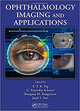
Ophthalmological Imaging and Applications, 1ed
Edited by and featuring contributions from world-class researchers, Ophthalmological Imaging and Applications offers a unified work of the latest human eye imaging and modeling techniques that have been proposed and applied to the diagnosis of ophthalmologic problems, including inflammation, cataracts, diabetic retinopathy, and glaucoma. With a focus on theory, basic principles, and results derived from research, the book:
- Explores various morphological, textural, higher-order spectral, and wavelet transformation techniques used to extract salient features from images of the human eye
- Examines 2D and 3D finite element and boundary element models of the human eye developed to simulate thermal steady-state conditions
- Addresses the difficult task of benchmarking the validity of human eye imaging techniques and computer-simulated results with experimental measurements
Intended to be a companion to Image Analysis and Modeling in Ophthalmology, this volume covers several aspects of multimodal ophthalmological imaging and applications, presenting information in an accessible manner to appeal to a wide audience of students, researchers, and practitioners. Ophthalmological Imaging and Applications considers promising simulations that pave the way for new possibilities in computational methods for eye health care.
About the Author
Contents
۱. Retinal Vascular Imaging in Clinical Research
۲. Detection, Modeling, and Analysis of the Major Temporal Arcade in Fundus Images of the Retina
۳. Application of Higher-Order Spectra Cumulants for Diabetic Retinopathy Detection Using Digital Fundus Images
۴. Quality Measures for Retinal Images
۵. Graph Search Retinal Vessel Tracking
۶. Fundus Autofluorescence Imaging: Fundamentals and Clinical Relevance
۷. Needs/Requirements and Design of Retinal Imaging and Image Processing Methods for Diabetic Retinopathy in the Indian Context
۸. Application of Ocular Fundus Photography and Angiography
۹. Optic Nerve Analysis and Imaging in Relation to Glaucoma
۱۰. Imaging of the Eye after Glaucoma Surgery
۱۱. Confocal Microscopy of Cornea
۱۲. Corneal Topography and Tomography: The Orbscan II
۱۳. Automatic Analysis of Scanning Laser Ophthalmoscope Sequences for Arteriovenous Passage Time Measurement
۱۴. Optical Coherence Tomography
۱۵. Role of Optical Coherence Tomography in Anterior Segment Imaging
۱۶. Anterior Segment Imaging with Anterior Segment Optical Coherence Tomography
۱۷. Cyst Detection in OCT Images for Pathology Characterization
۱۸. Scanning Laser Ophthalmoscope Fundus Perimetry: The Microperimetry
۱۹. In Vivo Confocal Microscopy: Imaging of the Ocular Surface
۲۰. Biomechanical Modeling of Blood Vessels for Interpretation of Tortuosity Estimates
۲۱. Hybrid Finite Element Simulation for Bioheat Transfer in the Human Eye
۲۲. Effects of Electromagnetic Fields on Specifc Absorption Rate and Heat Transfer in the Human Eye
۲۳. Dry Eye Characterization by Analyzing Tear Film Images
لینک کوتاه : https://bookbaz.ir/?p=53720
نویسنده : E. Y. K. Ng , U. Rajendra Acharya
ناشر : CRC Press; 1 edition
سال انتشار : 2014
زبان کتاب : انگلیسی
نوع فایل : PDF
تعداد صفحات : 519
(ISBN) شابک : 1466559136
قیمت کتاب درآمازون : $249.95
حجم فایل : 38 MB
کتاب های مرتبط:
 دانلود کتاب تعمیرات X-Ray: راهنمای جامع نصب و سرویس تجهیزات رادیوگرافی
X-Ray Repair: A Comprehensive Guide to the Installation and Servicing of Radiographic Equipment, 3ed
 دانلود کتاب تصویربرداری تشخیصی: زنان و زایمان
Diagnostic Imaging: Gynecology, 2ed
 دانلود کتاب رادیولوژی اعصاب تشخیصی و مداخله ای
Diagnostic and Interventional Neuroradiology, 1ed
 دانلود کتاب یادگیری رادیولوژی هِرینگ: شناخت مبانی
Learning Radiology: Recognizing the Basics, 3ed
 دانلود کتاب ۳ تفاوت برتر در رادیولوژی مغز و اعصاب
 Top 3 Differentials in Neuroradiology, 1ed
 دانلود کتاب مرور بالینی مغز و اعصاب مبتنی بر تصویر
Neurology Image-Based Clinical Review, 1ed
 دانلود کتاب تصویربرداری در جراحی شکم
Imaging in Abdominal Surgery, 1ed
 دانلود کتاب تصویربرداری در گوارش
Imaging in Gastroenterology, 1ed
 دانلود کتاب اصول سی تی اسکن بدن
Fundamentals of Body CT, 5ed
 دانلود کتاب رویه های بالینی در مراقبت های اولیه چشم + ویدئو
Clinical Procedures in Primary Eye Care, 5ed + Video
 دانلود کتاب موارد تصویربرداری پستان
Breast Imaging Cases
 دانلود کتاب تصویربرداری تشخیصی: پزشکی زایمان
Diagnostic Imaging: Obstetrics, 3ed
 دانلود کتاب هدایت حین عمل MRI جراحی مغز و اعصاب
Intraoperative MRI-Guided Neurosurgery, 1ed
 دانلود کتاب ملزومات سونوگرافی شکمی و لگنی
Essentials of Abdomino-Pelvic Sonography, 1ed
 دانلود کتاب تفسیر رادیوگرافی قفسه سینه در بیماران قلبی کودک
Chest Radiographic Interpretation in Pediatric Cardiac Patients, 1ed
 دانلود کتاب تصویربرداری تشخیصی کودکان کافی (ویرایش ۲۰۱۹، ۲ جلدی)
Caffey’s Pediatric Diagnostic Imaging, 2-Volume Set, 13ed
 دانلود کتاب اطلس بیهوشی منطقه ای هدایت اولتراسوند + ویدئو
Atlas of Ultrasound-Guided Regional Anesthesia, 3ed + Video
 دانلود کتاب آناتومی تصویربرداری: سر و گردن
Imaging Anatomy: Head and Neck, 1ed
 دانلود کتاب تصویربرداری تشخیصی دستگاه گوارش
Diagnostic Imaging: Gastrointestinal, 4ed
 دانلود کتاب تصویربرداری تشخیصی زایمان
Diagnostic Imaging: Obstetrics, 4ed
 دانلود کتاب تصویربرداری تشخیصی سر و گردن
Diagnostic Imaging: Head and Neck, 4ed
 دانلود کتاب تصویربرداری تشخیصی انکولوژی
Diagnostic Imaging: Oncology, 2ed
 دانلود کتاب MRI در یک نگاه
MRI at a Glance, 2ed
 دانلود کتاب سونوگرافی مداخله ای: راهنمای عملی و اطلس
Interventional Ultrasound: A Practical Guide and Atlas, 1ed
 دانلود کتاب فیزیک ضروری در تصویربرداری برای پرتونگار کلارک
Clark’s Essential Physics in Imaging for Radiographers, 1ed
 دانلود کتاب تصویربرداری ادراری تناسلی (سری موارد رادیولوژی)
Genitourinary Imaging (Radcase), 1ed
 دانلود کتاب تشخیص افتراقی در تصویربرداری سونوگرافی
 Differential Diagnosis in Ultrasound Imaging, 2ed
 دانلود کتاب تصویربرداری تشخیصی: مغز
Diagnostic Imaging: Brain, 3ed
 دانلود کتاب تصویربرداری زنان و زایمان: تشخیص و مراقبت از جنین
Obstetric Imaging: Fetal Diagnosis and Care, 2ed
 دانلود کتاب تصویربرداری اسکلتی عضلانی
Musculoskeletal Imaging, 2ed
 دانلود کتاب موقعیت در رادیوگرافی کلارک
Clark’s Positioning in Radiography, 13ed
 دانلود کتاب تصویربرداری تشخیصی قفسه سینه
Diagnostic Imaging: Chest, 3ed
 دانلود کتاب تصویربرداری تشخیصی زنان
Diagnostic Imaging: Gynecology, 3ed
 دانلود کتاب تصویربرداری قفسه سینه و قلب و عروق
Chest and Cardiovascular Imaging, 3ed
 دانلود کتاب تصویربرداری از مغز با MRI و CT: رویکرد الگوی تصویری
Brain Imaging with MRI and CT: An Image Pattern Approach, 1ed
 دانلود کتاب تشخیص افتراقی در توموگرافی کامپیوتری
Differential Diagnosis in Computed Tomography, 2ed
 دانلود کتاب آسیب شناسی چشمی یانوف
Ocular Pathology, 7ed
 دانلود کتاب آناتومی تصویربرداری: قفسه سینه، شکم، لگن
Imaging Anatomy: Chest, Abdomen, Pelvis, 2ed
 دانلود کتاب رادیولوژی اضطراری (سری موارد رادیولوژی)
Radcases Emergency Radiology, 1ed
 دانلود کتاب اولتراسوند: مرور کلی
Ultrasound: A Core Review, 1ed
 دانلود کتاب راهنمای آزمون آماده سازی برای دانشجویان چشم پزشکی
 Exam Preparatory Manual for Undergraduates Ophthalmology, 1ed
 دانلود کتاب پرسش و پاسخ تصویربرداری گوارشی رادکِیس
RadCases Plus Q&A Gastrointestinal Imaging, 2ed
 دانلود کتاب آموختن فیزیک رادیولوژیک از طریق موارد
Radiologic Physics Taught Through Cases, 1ed
 دانلود کتاب چشم: علوم پایه در عمل
The Eye: Basic Sciences in Practice, 4ed
 دانلود کتاب رادیولوژی ۱۰۱: مبانی و اصول تصویربرداری
Radiology 101: The Basics & Fundamentals of Imaging, 4ed
 دانلود کتاب تصویربرداری عمومی اسکلتی عضلانی
Basic Musculoskeletal Imaging, 1ed
 دانلود کتاب اندوسونوگرافی
 Endosonography, 4ed
 دانلود کتاب تعویض ترنس کاتتر دریچه آئورت
Transcatheter Aortic Valve Implantation, 1ed
 دانلود کتاب پرسش و پاسخ پزشکی هسته ای رادکِیس
RadCases Plus Q&A Nuclear Medicine, 2ed
 دانلود کتاب تصویربرداری بیماری های بینابینی ریه (سری رادیولوژی کلینیکو)
Clinico Radiological Series: Imaging of Interstitial Lung Diseases, 1ed
 دانلود کتاب توموگرافی پرتو مخروطی کامپیوتری در ارتودنسی: موارد مصرف، بینش و نوآوری
Cone Beam Computed Tomography in Orthodontics: Indications, Insights, and Innovations, 1ed
 دانلود کتاب تصویربرداری پزشکی آکسفورد
Oxford Handbook of Medical Imaging, 1ed
 دانلود کتاب سری مرور موردی رادیولوژی: تصویربرداری قفسه سینه
 Radiology Case Review Series: Thoracic Imaging, 1ed
 دانلود کتاب راهنمای رادیو مابولیزاسیون: فیزیک، بیولوژی، پزشکی هسته ای و تصویربرداری
Handbook of Radioembolization: Physics, Biology, Nuclear Medicine, and Imaging, 1ed
 دانلود کتاب دوز، فواید و خطر در تصویربرداری پزشکی
 Dose, Benefit, and Risk in Medical Imaging, 1ed
 دانلود کتاب اطلس جیبی آناتومی مقطعی: ستون فقرات، اندام، مفاصل
 Pocket Atlas of Sectional Anatomy: Spine Extremities Joints, 3ed
 دانلود کتاب چشم انداز محاسباتی و پردازش تصویری پزشکی
 Computational Vision and Medical Image Processing V: VipIMAGE 2015
 دانلود کتاب سری بررسی موردی رادیولوژی: تصویربرداری پستان
 Radiology Case Review Series: Breast Imaging, 1ed
 دانلود کتاب رادیوگرافی PREP (مرور برنامه و آمادگی آزمون)
Radiography PREP (Program Review and Exam Preparation), 8ed
 دانلود کتاب CT قلبی بالینی: آناتومی و عملکرد + ویدئو
Clinical Cardiac CT: Anatomy and Function, 2ed + Video
 دانلود کتاب سونوگرافی کودکان
 Pediatric Ultrasound, 1ed
 دانلود کتاب روشهای اسکلتی عضلانی هدایت شده با سونوگرافی در پزشکی ورزشی
Ultrasound Guided Musculoskeletal Procedures in Sports Medicine, 1ed
 دانلود کتاب تجزیه و تحلیل تصویر و مدل سازی در چشم پزشکی
 Image Analysis and Modeling in Ophthalmology, 1ed
 دانلود کتاب MRI بافتها همراه با T2 یا T2*s کوتاه
MRI of Tissues with Short T2s or T2*s, 1ed
 دانلود کتاب تصویربرداری تومور قفسه سینه (سری رادیولوژی کلینیکو)
Clinico Radiological Series: Imaging Of Chest Tumors, 1ed
 دانلود کتاب توسعه دیجیتال ریه: از اولین سی تی ریه تا هوش مصنوعی بالینی
Developing the Digital Lung: From First Lung CT to Clinical AI, 1ed
 دانلود کتاب سونوگرافی مراقبت اورژانسی + ویدئو
Emergency Point-of-Care Ultrasound, 2ed + Video
 دانلود کتاب تصویربرداری از استخوان ها و مفاصل: یک روش مختصر و چند وجهی
Imaging of Bones and Joints: A Concise, Multimodality Approach, 1ed
 دانلود کتاب تصویربرداری و تجسم در اتاق عمل مدرن: راهنمای جامع برای پزشکان
Imaging and Visualization in The Modern Operating Room: A Comprehensive Guide for Physicians, 2015th
 دانلود کتاب آناتومی مقطعی برای متخصصان تصویربرداری
 Sectional Anatomy for Imaging Professionals, 3ed
 دانلود کتاب تصویربرداری پستان (سری تشخیص مستقیم در رادیولوژی)
Breast Imaging (Direct Diagnosis in Radiology: DX-Direct!), 1ed
 دانلود کتاب تصویربرداری قلبی (سری تشخیص مستقیم در رادیولوژی)
Cardiac Imaging (Direct Diagnosis in Radiology: DX-Direct!), 1ed
 دانلود کتاب بیماری ویترئورتینال: تشخیص، مدیریت و موارد بالینی
Vitreoretinal Disease: Diagnosis, Management, and Clinical Pearls, 2ed
 دانلود کتاب موارد و مشکلات در تصویربرداری بدن: تصویربرداری قفسه سینه، شکم و زنان
Variants and Pitfalls in Body Imaging: Thoracic, Abdominal and Women’s Imaging, 2ed
 دانلود کتاب مداخله انکولوژی (راهنمای عملی در مداخلات رادیولوژی)
Interventional Oncology (Practical Guides in Interventional Radiology), 1ed
 دانلود کتاب تصویربرداری زنان و زایمان کوپل: سری کارشناس رادیولوژی
Obstetric Imaging: Expert Radiology Series, 1ed
 دانلود کتاب تصویربرداری اسکلتی عضلانی اکسفورد
Musculoskeletal Imaging (Oxford Specialist Handbooks in Radiology), 1ed
 دانلود کتاب درسی موقعیت و آناتومی مرتبط با رادیوگرافی
 Textbook of Radiographic Positioning and Related Anatomy, 8ed
 دانلود کتاب اطلس آنژیوگرافی فوندوس
Atlas of Fundus Angiography, 1ed
 دانلود کتاب اطلس پاتولوژی آناتومیک با تصویربرداری
Atlas of Anatomic Pathology with Imaging
 دانلود کتاب تصویربرداری تشخیصی: ترومای اسکلتی عضلانی
Diagnostic Imaging: Musculoskeletal Trauma, 3ed
 دانلود کتاب تکنیک های جراحی پلاستیک چشم + ویدئو
Techniques in Ophthalmic Plastic Surgery, 2ed + Video
 دانلود کتاب اطلس تصویربرداری عروقی ریوی
Atlas of Pulmonary Vascular Imaging, 1ed
 دانلود کتاب سونوگرافی تشخیصی هیگن-انسرت (۲ جلدی)
Textbook of Diagnostic Sonography, 2-Vol, 7ed
 دانلود کتاب تشخیص افتراقی در تصویربرداری عصبی: ستون فقرات
Differential Diagnosis in Neuroimaging: Spine, 1ed
 دانلود کتاب سرطان پستان: تشخیص زودرس با ماموگرافی
Breast Cancer: Early Detection with Mammography: Casting Type Calcifications Sign of a Subtype with Deceptive Features, 1ed
 دانلود کتاب اطلس جراحی پلاستیک و ترمیمی چشم + ویدئو
Video Atlas of Oculofacial Plastic and Reconstructive Surgery, 2ed + Video
 دانلود کتاب نورونولوژی و تصویربرداری عصبی سکته مغزی
Neurosonology and Neuroimaging of Stroke, 2ed
 دانلود کتاب SPECT و SPECT/CT: راهنمای بالینی
SPECT and SPECT/CT: A Clinical Guide, 1ed
 دانلود کتاب مجموعه ویدئویی جراحی پلک و دور چشم + کتاب
Eyelid and Periorbital Surgery Video Collection, 2ed + PDF
 دانلود کتاب رادیوتراپی کودکان
 Pediatric Radiation Oncology, 5ed
 دانلود کتاب رادیولوژی ضروری: معرفی بالینی تصویربرداری پاتوفیزیولوژی
 Essential Radiology: Clinical Presentation Pathophysiology Imaging, 3ed
 دانلود کتاب نورورادیولوژی: الزامات (ویرایش ۲۰۱۷)
Neuroradiology: The Requisites, 4ed
 دانلود کتاب گلوکوم چندلر و گرانت
Chandler and Grant’s Glaucoma, 5ed
 دانلود کتاب پزشکی تورنتو نوت (نسخه ۲۰۱۵)
Toronto Notes 2015 (Essential Med Notes), 31ed
 دانلود کتاب تشخیص افتراقی در سونوگرافی
 Differential Diagnosis in Ultrasound, 2ed
 دانلود کتاب اطلس چشم پزشکی بالینی
Atlas of Clinical Ophthalmology, 2ed
 دانلود کتاب اطلس رنگی جراحی پلاستیک چشم + ویدئو
Colour Atlas of Ophthalmic Plastic Surgery, 4ed + Video
 دانلود کتاب پرونده آموزشی تصویربرداری سر و گردن
 Head and Neck Imaging: A Teaching File, 2ed
 دانلود کتاب آسان سازی دسترسی وریدی
 Venous Access Made Easy, 1ed
 دانلود کتاب EXPERTddx: رادیولوژی سر و گردن
 EXPERTddx: Head and Neck: Published by Amirsys
 دانلود کتاب پوکی استخوان مارکوس و فلدمن (۲ جلدی)
Osteoporosis, 2-Volume Set, Fourth Edition
 دانلود کتاب تصویربرداری سه بعدی در اندودنتیکس
۳D Imaging in Endodontics, 1ed
 دانلود کتاب ضروریات انکولوژی رادیوتراپی بالینی
Essentials of Clinical Radiation Oncology, 1ed
 دانلود کتاب اطلس ویدئویی بخیه چشم: اصول و تکنیک ها + ویدئو
Video Atlas of Ophthalmic Suturing: Fundamentals and Techniques, 1ed + Video
 دانلود کتاب بهینه سازی نتایج زیر بهینه حاصل از جراحی آب مروارید + ویدئو
Optimizing Suboptimal Results Following Cataract Surgery, 1ed + Video
 دانلود کتاب چشم پزشکی عصبی مصور + ویدئو
Neuro-Ophthalmology Illustrated, 3ed + Video
 دانلود کتاب اکوکاردیوگرافی عملی نوزاد
Practical Neonatal Echocardiography, 1ed
 دانلود کتاب رادیولوژی دستگاه گوارش گور و لوین + ویدئو
Textbook of Gastrointestinal Radiology, 5ed + Video
 دانلود کتاب آسان سازی سی تی اسکن قلب
Cardiac CT Made Easy, 3ed
 دانلود کتاب جراحی استرابیسم: رویکردهای نوآورانه و کلاسیک + ویدئو
Strabismus Surgery: Innovative and Classic Approaches, 1ed + Video
 دانلود کتاب حل مسئله در تصویربرداری کودکان
Problem Solving in Pediatric Imaging, 1ed
 دانلود کتاب آزمون اصلی را بترکانید (۲ جلدی)
Crack The Core Exam, 2-Vol, 9ed
 دانلود کتاب آنژیوگرافی آبرامز: رادیولوژی مداخله ای
Abrams’ Angiography: Interventional Radiology, 3ed
 دانلود کتاب تصویربرداری CT آشکارساز چندگانه (۲ جلدی)
Multi-Detector CT Imaging Handbook, 2-Vol, 1ed
 دانلود کتاب ۳ تفاوت برتر در رادیولوژی: بررسی موردی
Top 3 Differentials in Radiology: A Case Review, 1ed
 دانلود کتاب آندوسکوپی تشخیصی (سری فیزیک پزشکی و مهندسی پزشکی)
Diagnostic Endoscopy (Series in Medical Physics and Biomedical Engineering), 1ed
 دانلود کتاب سونوگرافی شکم: گام به گام
Abdominal Ultrasound: Step by Step, 2ed
 دانلود کتاب تصویربرداری اسکلتی عضلانی: یک فایل آموزشی
Musculoskeletal Imaging: A Teaching File, 3ed
 دانلود کتاب جریان خون مغزی و متابولیسم
 Cerebral Blood Flow and Metabolism, 1ed
 دانلود کتاب استراتژی برای برنامه ریزی درمان رادیوتراپی
 Strategies for Radiation Therapy Treatment Planning, 1ed
 دانلود کتاب تصویربرداری اسکلتی عضلانی: موارد ضروری
Musculoskeletal Imaging: The Requisites, 4ed
 دانلود کتاب ExpertDDx: اسکلتی عضلانی
ExpertDDx: Musculoskeletal, 2ed
 دانلود کتاب اپیدمیولوژی چشم
Ophthalmic Epidemiology, 1ed
 دانلود کتاب چشم پزشکی یانوف و داکر
Yanoff & Duker Ophthalmology, 6ed
 دانلود کتاب اطلس تصویربرداری ترکیبی: آناتومی مقطعی برای PET/CT، PET/MRI و SPECT/CT (3 جلدی)
Atlas of Hybrid Imaging: Sectional Anatomy for PET/CT, PET/MRI and SPECT/CT, 1ed
 دانلود کتاب CT ریه با وضوح بالا
High-Resolution CT of the Lung, 5ed
 دانلود کتاب رادیولوژی تشخیصی: آخرین پیشرفت ها و فیزیک کاربردی در تصویربرداری
Diagnostic Radiology: Recent Advances and Applied Physics in Imaging, 2ed
 دانلود کتاب انکولوژی تابشی: تصویربرداری و درمان
Radiation Oncology: Imaging and Treatment
 دانلود کتاب یادداشت های درسی: رادیولوژی
 Lecture Notes: Radiology, 3ed
 دانلود کتاب یک رویکرد عملی به اکوکاردیوگرافی بالینی
 A Practical Approach to Clinical Echocardiography, 1ed
 دانلود کتاب تصویربرداری قلبی (رادیولوژی چرخشی)
Cardiac Imaging (Rotations in Radiology), 1ed
 دانلود کتاب موارد چالش برانگیز در تصویربرداری اسکلتی عضلانی
 Challenging Cases in Musculoskeletal Imaging, 1ed
 دانلود کتاب پزشکی فوتونیک وُدین (۳ جلدی)
Biomedical Photonics Handbook, 3-Vol, 2ed
 دانلود کتاب اندازه گیری در سونوگرافی
 Measurement in Ultrasound, 2ed
 دانلود کتاب سونوگرافی تشخیصی روماک (۲ جلدی)
Diagnostic Ultrasound, 2-Volume Set, 5ed
 دانلود کتاب تصویربرداری تشخیصی: دهان و فک و صورت
Diagnostic Imaging: Oral and Maxillofacial, 2ed
 دانلود کتاب اصول و ابزار سونوگرافی
 Sonography Principles and Instruments, 9ed
 دانلود کتاب سونوگرافی در زنان و زایمان کالن (ویرایش ۲۰۱۷) + ویدئو
Callen’s Ultrasonography in Obstetrics and Gynecology, 6ed + Video
 دانلود کتاب جراحی پلاستیک و بازسازی پستان
Plastic and Reconstructive Surgery of the Breast, 2ed
 دانلود کتاب موردی ماموگرافی
 Mammography Casebook, 1ed
 دانلود کتاب اولتراسوند دست و اندام فوقانی + ویدئو
Ultrasound of the Hand and Upper Extremity, 1ed + Video
 دانلود کتاب اثرات درمان رادیوتراپی
 Radiation Therapy Treatment Effects, 1ed
 دانلود کتاب رادیولوژی برای متخصص دندانی فرومر
Frommer’s Radiology for the Dental Professional, 10ed
 دانلود کتاب تمرین رادیولوژی مداخله ای + ویدئو
The Practice of Interventional Radiology, 1ed + Video
 دانلود کتاب تصویربرداری پزشکی برای متخصصین سلامت
Medical Imaging for Health Professionals, 1ed
 دانلود کتاب توموگرافی محاسباتی پرتوی مخروطی فک و صورت
Maxillofacial Cone Beam Computed Tomography, 1ed
 دانلود کتاب پرسش و پاسخ تصویربرداری عصبی رادکِیس
RadCases Plus Q&A Neuro Imaging, 2ed
 دانلود کتاب موارد بالینی در یووئیت: تشخیص و مدیریت افتراقی
Clinical Cases in Uveitis: Differential Diagnosis and Management, 1ed
 دانلود کتاب حل مسئله در تصویربرداری قفسه سینه
Problem Solving in Chest Imaging, 1ed