دانلود کتاب جراحی اسب + ویدئو
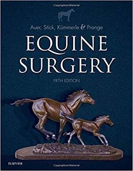
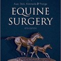
Equine Surgery, 5ed + Video
Equip yourself for success with the only book on the market that covers all aspects of equine surgery! Equine Surgery, 5th Edition prepares you to manage each surgical condition by understanding its pathophysiology and evaluating alternative surgical approaches. Explanations in the book describe how to avoid surgical infections, select and use instruments, and perfect fundamental surgical techniques including incisions, cautery, retractions, irrigation, surgical suction, wound closure, dressings, bandages, and casts. In addition to diagnostic imaging and orthopedic coverage, it includes in-depth information on anesthesia, the integumentary system (including wound management, reconstructive surgery, and skin grafting), the alimentary system, respiratory, and urogenital systems.
- Complete coverageof all the information needed to study for theAmerican and European College of Veterinary Surgeons Board Examinations makes this edition an excellent study tool.
- Section on anesthesiology and pain management prepares you to manage these critical aspects of any surgery.
- Extensive, up-to-date orthopedic coverage includes joint disorders and joint trauma.
- Section on integumentary system contains information on wound management, reconstructive surgery, and skin grafting.
- Section on the alimentary system covers postoperative care, complications and reoperation guidelines.
- New techniques in vascular surgery keep you up-to-date with best practices.
- NEW! Expert Consult site offering 40+ videos of surgeons performing techniques so that you can quickly access drug and equipment information.
- NEW! Expansion of minimally invasive surgical techniques includes laser ablation procedures, implantation of plates against bones in orthopedic procedures, and laparoscopic procedures for soft tissue injuries.
- NEW! World-renowned contributors, featuring two new associate editors include over 70 of the most experienced and expert equine specialist surgeons, each providing current and accurate information.
- NEW! Current advances in imaging detect musculoskeletal conditions in the sports horse.
Contents
I Surgical Biology
II Surgical Methods
III Recent Advances in Anesthesia
IV Integumentary System
V Alimentary System
VI Respiratory System
VII Nervous System
VIII Eye and Adnexa
IX Reproductive System
X Urinary System
XI Diagnostic Imaging
XII Musculoskeletal System
Video Contents
۱۳.۱ Video illustration of how a foggy image can be resolved.
۱۳.۲ Video explanation of how to perform an extracorporeal 4-S modifed Roeder slipknot.
۱۳.۳ Video demonstration of the technique for transvaginal access to the abdominal cavity under endoscopic guidance.
۱۳.۴ Video demonstration of the technique for trocar exchange.
۱۶.۱ One-handed knot typing technique.
۱۶.۲ Two-handed knot typing technique.
۱۶.۳ Hand-tied sliding knot 1.
۱۶.۴ Hand-tied sliding knot 2.
۱۶.۵ Instrument-tied simple knot.
۱۶.۶ Instrument-tied surgical knot.
۱۶.۷ Simple continuous suture pattern.
۱۶.۸ Cruciate suture pattern.
۱۶.۹ Vertical mattress suture pattern.
۱۶.۱۰ Horizontal mattress suture pattern.
۱۶.۱۱ Ford interlocking suture pattern.
۱۶.۱۲ Connell and Cushing suture pattern.
۱۶.۱۳ Lembert suture pattern.
۱۶.۱۴ Pursestring suture pattern.
۱۶.۱۵ Far-near-near-far suture pattern.
۱۶.۱۶ Three-loop pulley suture technique.
۱۶.۱۷ Three-loop pulley suture technique.
۱۶.۱۸ Technique for tying an Aberdeen knot.
۱۶.۱۹ Chinese fnger trap technique.
۱۶.۲۰ Use of the skin stapler to close a skin incision.
۳۲.۱ Demonstrating the pathway for laparoscopic approach to the epiploic foramen.
۳۲.۲ Demonstrating the ideal placement of the gastropancreatic fold against the caudal aspect of the caudate liver lobe using the Babcock forceps.
۳۲.۳ Tacking of the gastropancreatic fold.
۴۸.۱ Self-retaining tracheostomy tube insertion.
۵۱.۱ Vertebral canal endoscopy: cervical myeloscopy and lumbosacral epiduroscopy.
۵۵.۱ Left femoral nerve paralysis following prolonged anesthesia in dorsal recumbency with caudally extended hind limbs.
۸۵.۱ Typical motion of a horse affected with fbrotic myopathy of the semitendinosus muscle.
۸۵.۲ Typical motion of a horse with classical stringhalt: involuntary and exaggerated flexion mainly of the left hindlimb.
۸۵.۳ A horse with rupture of the left peroneus tertius.
۸۵.۴ Typical motion of a horse with a ruptured extensor caripi radialis tendon, frst at a walk and then at a trot.
۸۷.۱ A 4-month-old Arabian foal is shown walking toward the viewer next to its dam. During protraction of each fore limb the MCP region describes an outward arch.
لینک کوتاه : https://bookbaz.ir/?p=119007
نویسنده : Jorg A. Auer Dr Med Vet MS
ناشر : Saunders; 5 edition
سال انتشار : 2019
زبان کتاب : انگلیسی
نوع فایل : MP4 + PDF (کیفیت اصلی)
تعداد صفحات : 1898
(ISBN) شابک : 0323484204
قیمت کتاب درآمازون : $184.70
حجم فایل : 600 MB


