
دانلود کتاب روش چندگانه تصویربرداری پستان: اطلس لازم و ملزوم
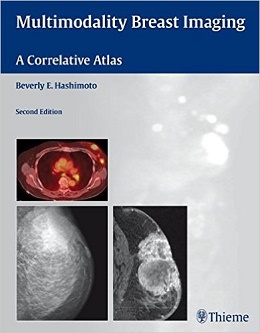
Multimodality Breast Imaging: A Correlative Atlas, 2ed
Praise for the previous edition:
“Well organised and beautifully illustrated…A good book for trainee breast radiologists and radiographers…[and] an extremely useful reference textbook for more experienced practitioners.”–RAD Magazine
The second edition of this generously illustrated case-based reference provides a systematic visual collection of pathologic entities and a detailed assessment of how to optimize sonographic technique as well as how to approach the integration of mammography, sonography, MRI and PET/CT in breast cancer diagnosis. The book begins with a focus on teaching practical methods to analyze and incorporate mammographic, sonographic, and magnetic resonance findings in the clinical setting. The closing chapters are devoted to illustrating the applications of PET as demonstrated by specific clinical cases.
Features of the second edition:
- Emphasis on the importance of high-resolution sonography
- Three new chapters on the use of MRI and PET in breast imaging
- Numerous new case studies — including helpful pearls and pitfalls — that focus on common and uncommon examples of metastatic and non-metastatic disease
- Charts and outlines that provide rapid reference for the clinical workup of a lesion
- More than 800 images that help identify both mammographic and sonographic abnormalities
This thorough reference is ideal for radiologists, mammographers, oncologists, gynecologists and all clinicians looking to broaden their visual sonographic experience. Its user-friendly format makes it a handy text for radiology residents in breast rotations.
Review
“[Five stars] Clear and concise…a good introduction for residents…the illustrations and annotations are uniformly excellent and easy to understand…very helpful.”–Doody’s Review
[Five stars] Clear and concise…a good introduction for residents…the illustrations and annotations are uniformly excellent and easy to understand…very helpful.–Doody’s Review
Contents
۱. Approach to Mammographic Analysis
۲. Sonographic Technique and Cross-Correlation with Mammography
۳. Approach to Magnetic Resonance Imaging
۴. PET-CT in Breast Cancer Diagnosis
۵. Circumscribed Masses: Fat-Containing Masses
۶. Circumscribed Masses: Medium- or High-Density Masses
۷. Irregular Masses: Benign Masses
۸. Irregular Masses: Malignant Masses
۹. Calcifications: Large Linear
۱۰. Calcifications: Milk of Calcium
۱۱. Calcifications: Large Round
۱۲. Calcifications: Round Lucent Center
۱۳. Calcifications: Eggshell/Rim
۱۴. Calcifications: Coarse/Popcorn-Fibroadenoma
۱۵. Calcifications: Dystrophic
۱۶. Calcifications: Small Round/Punctate
۱۷. Calcifications: Amorphous/Indistinct Microcalcifications
۱۸. Calcifications: Heterogeneous/Pleomorphic Microcalcifications.
۱۹. Calcifications: Fine Linear/Branching Microcalcifications
۲۰. Global Asymmetry
۲۱. Focal Asymmetry
۲۲. Peripheral Architectural Distortion
۲۳. Central Architectural Distortion
۲۴. Benign Male Breast
۲۵. Malignant Male Breast
۲۶. Augmentation Mammoplasty
۲۷. Reduction Mammoplasty
۲۸. After Diagnostic or Therapeutic Procedures for Neoplasm
۲۹. Patient Unable to Tolerate Mammogram
۳۰. Palpable Masses
۳۱. Mammogram Underestimates Tumor Size
۳۲. Mass in Unusual Locations
۳۳. Ductal Abnormalities
۳۴. Mammographically Occult MRI Lesions
۳۵. Staging
۳۶. Assessing Response to Therapy
۳۷. Identifications of Recurrent Disease
۳۸. Additional Malignancies
۳۹. Unknown Primary
۴۰. Pitfalls of PET
لینک کوتاه : https://bookbaz.ir/?p=38419
نویسنده : Beverly Hashimoto
ناشر : Thieme; 2 edition
سال انتشار : 2010
زبان کتاب : انگلیسی
نوع فایل : PDF
تعداد صفحات : 665
(ISBN) شابک : 1604061715
قیمت کتاب درآمازون : $179.99
حجم فایل : $179.99
کتاب های مرتبط:
 دانلود کتاب فیزیک ضروری در تصویربرداری برای پرتونگار کلارک
Clark’s Essential Physics in Imaging for Radiographers, 1ed
 دانلود کتاب تعویض ترنس کاتتر دریچه آئورت
Transcatheter Aortic Valve Implantation, 1ed
 دانلود کتاب توموگرافی پرتو مخروطی کامپیوتری در ارتودنسی: موارد مصرف، بینش و نوآوری
Cone Beam Computed Tomography in Orthodontics: Indications, Insights, and Innovations, 1ed
 دانلود کتاب سونوگرافی مراقبت اورژانسی + ویدئو
Emergency Point-of-Care Ultrasound, 2ed + Video
 دانلود کتاب مداخله انکولوژی (راهنمای عملی در مداخلات رادیولوژی)
Interventional Oncology (Practical Guides in Interventional Radiology), 1ed
 دانلود کتاب تصویربرداری زنان و زایمان کوپل: سری کارشناس رادیولوژی
Obstetric Imaging: Expert Radiology Series, 1ed
 دانلود کتاب سونوگرافی کودکان
 Pediatric Ultrasound, 1ed
 دانلود کتاب تصویربرداری تشخیصی پستان
Diagnostic Imaging: Breast, 3ed
 دانلود کتاب آناتومی تصویربرداری: سونوگرافی
Imaging Anatomy: Ultrasound, 2ed
 دانلود کتاب قفسه سینه ExpertDDx
ExpertDDx: Chest, 2ed
 دانلود کتاب توموسنتز پستان
 Breast Tomosynthesis, 1ed
 دانلود کتاب تصویربرداری رزونانس مغناطیسی مغز و ستون فقرات (دو جلدی)
Magnetic Resonance Imaging of the Brain and Spine, 2-Vol, 4ed
 دانلود کتاب سیگنال و سیستم های تصویربرداری پزشکی
 Medical Imaging Signals and Systems, 2ed
 دانلود کتاب سرطان پستان: تشخیص زودرس با ماموگرافی
Breast Cancer: Early Detection with Mammography: Crushed Stone-like Calcifications: The Most Frequent Malignant Type, 1ed
 دانلود کتاب اطلس آناتومی مقطعی، جلد ۳: ستون فقرات، پایانگاه، مفاصل: توموگرافی کامپیوتری و تصویربرداری رزونانس مغناطیسی
Pocket Atlas of Sectional Anatomy, Volume III: Spine, Extremities, Joints: Computed Tomography and Magnetic Resonance Imaging, 2ed
 دانلود کتاب تصویربرداری در جراحی شکم
Imaging in Abdominal Surgery, 1ed
 دانلود کتاب تصویربرداری در گوارش
Imaging in Gastroenterology, 1ed
 دانلود کتاب اصول سی تی اسکن بدن
Fundamentals of Body CT, 5ed
 دانلود کتاب سونوگرافی داپلر رنگی در زنان و زایمان
Color Doppler Sonography in Gynecology and Obstetrics, 1ed
 دانلود کتاب موارد تصویربرداری پستان
Breast Imaging Cases
 دانلود کتاب تصویربرداری تشخیصی: پزشکی زایمان
Diagnostic Imaging: Obstetrics, 3ed
 دانلود کتاب هدایت حین عمل MRI جراحی مغز و اعصاب
Intraoperative MRI-Guided Neurosurgery, 1ed
 دانلود کتاب ملزومات سونوگرافی شکمی و لگنی
Essentials of Abdomino-Pelvic Sonography, 1ed
 دانلود کتاب تفسیر رادیوگرافی قفسه سینه در بیماران قلبی کودک
Chest Radiographic Interpretation in Pediatric Cardiac Patients, 1ed
 دانلود کتاب تصویربرداری تشخیصی کودکان کافی (ویرایش ۲۰۱۹، ۲ جلدی)
Caffey’s Pediatric Diagnostic Imaging, 2-Volume Set, 13ed
 دانلود کتاب اطلس بیهوشی منطقه ای هدایت اولتراسوند + ویدئو
Atlas of Ultrasound-Guided Regional Anesthesia, 3ed + Video
 دانلود کتاب آناتومی تصویربرداری: سر و گردن
Imaging Anatomy: Head and Neck, 1ed
 دانلود کتاب تصویربرداری تشخیصی دستگاه گوارش
Diagnostic Imaging: Gastrointestinal, 4ed
 دانلود کتاب تصویربرداری تشخیصی زایمان
Diagnostic Imaging: Obstetrics, 4ed
 دانلود کتاب تصویربرداری تشخیصی سر و گردن
Diagnostic Imaging: Head and Neck, 4ed
 دانلود کتاب تصویربرداری تشخیصی انکولوژی
Diagnostic Imaging: Oncology, 2ed
 دانلود کتاب MRI در یک نگاه
MRI at a Glance, 2ed
 دانلود کتاب سونوگرافی مداخله ای: راهنمای عملی و اطلس
Interventional Ultrasound: A Practical Guide and Atlas, 1ed
 دانلود کتاب تصویربرداری ادراری تناسلی (سری موارد رادیولوژی)
Genitourinary Imaging (Radcase), 1ed
 دانلود کتاب تشخیص افتراقی در تصویربرداری سونوگرافی
 Differential Diagnosis in Ultrasound Imaging, 2ed
 دانلود کتاب تصویربرداری تشخیصی: مغز
Diagnostic Imaging: Brain, 3ed
 دانلود کتاب تصویربرداری تشخیصی: زنان و زایمان
Diagnostic Imaging: Gynecology, 2ed
 دانلود کتاب تصویربرداری زنان و زایمان: تشخیص و مراقبت از جنین
Obstetric Imaging: Fetal Diagnosis and Care, 2ed
 دانلود کتاب تصویربرداری اسکلتی عضلانی
Musculoskeletal Imaging, 2ed
 دانلود کتاب موقعیت در رادیوگرافی کلارک
Clark’s Positioning in Radiography, 13ed
 دانلود کتاب تصویربرداری تشخیصی قفسه سینه
Diagnostic Imaging: Chest, 3ed
 دانلود کتاب تصویربرداری تشخیصی زنان
Diagnostic Imaging: Gynecology, 3ed
 دانلود کتاب تصویربرداری قفسه سینه و قلب و عروق
Chest and Cardiovascular Imaging, 3ed
 دانلود کتاب تصویربرداری از مغز با MRI و CT: رویکرد الگوی تصویری
Brain Imaging with MRI and CT: An Image Pattern Approach, 1ed
 دانلود کتاب تشخیص افتراقی در توموگرافی کامپیوتری
Differential Diagnosis in Computed Tomography, 2ed
 دانلود کتاب آناتومی تصویربرداری: قفسه سینه، شکم، لگن
Imaging Anatomy: Chest, Abdomen, Pelvis, 2ed
 دانلود کتاب رادیولوژی اضطراری (سری موارد رادیولوژی)
Radcases Emergency Radiology, 1ed
 دانلود کتاب رادیولوژی اعصاب تشخیصی و مداخله ای
Diagnostic and Interventional Neuroradiology, 1ed
 دانلود کتاب اولتراسوند: مرور کلی
Ultrasound: A Core Review, 1ed
 دانلود کتاب پرسش و پاسخ تصویربرداری گوارشی رادکِیس
RadCases Plus Q&A Gastrointestinal Imaging, 2ed
 دانلود کتاب یادگیری رادیولوژی هِرینگ: شناخت مبانی
Learning Radiology: Recognizing the Basics, 3ed
 دانلود کتاب رادیولوژی ۱۰۱: مبانی و اصول تصویربرداری
Radiology 101: The Basics & Fundamentals of Imaging, 4ed
 دانلود کتاب تصویربرداری عمومی اسکلتی عضلانی
Basic Musculoskeletal Imaging, 1ed
 دانلود کتاب اندوسونوگرافی
 Endosonography, 4ed
 دانلود کتاب پرسش و پاسخ پزشکی هسته ای رادکِیس
RadCases Plus Q&A Nuclear Medicine, 2ed
 دانلود کتاب تصویربرداری بیماری های بینابینی ریه (سری رادیولوژی کلینیکو)
Clinico Radiological Series: Imaging of Interstitial Lung Diseases, 1ed
 دانلود کتاب تصویربرداری پستان: ضروریات در رادیولوژی
Breast Imaging: The Requisites (Requisites in Radiology), 2ed
 دانلود کتاب تصویربرداری پزشکی آکسفورد
Oxford Handbook of Medical Imaging, 1ed
 دانلود کتاب سری مرور موردی رادیولوژی: تصویربرداری قفسه سینه
 Radiology Case Review Series: Thoracic Imaging, 1ed
 دانلود کتاب راهنمای رادیو مابولیزاسیون: فیزیک، بیولوژی، پزشکی هسته ای و تصویربرداری
Handbook of Radioembolization: Physics, Biology, Nuclear Medicine, and Imaging, 1ed
 دانلود کتاب تعمیرات X-Ray: راهنمای جامع نصب و سرویس تجهیزات رادیوگرافی
X-Ray Repair: A Comprehensive Guide to the Installation and Servicing of Radiographic Equipment, 3ed
 دانلود کتاب دوز، فواید و خطر در تصویربرداری پزشکی
 Dose, Benefit, and Risk in Medical Imaging, 1ed
 دانلود کتاب اطلس جیبی آناتومی مقطعی: ستون فقرات، اندام، مفاصل
 Pocket Atlas of Sectional Anatomy: Spine Extremities Joints, 3ed
 دانلود کتاب چشم انداز محاسباتی و پردازش تصویری پزشکی
 Computational Vision and Medical Image Processing V: VipIMAGE 2015
 دانلود کتاب سری بررسی موردی رادیولوژی: تصویربرداری پستان
 Radiology Case Review Series: Breast Imaging, 1ed
 دانلود کتاب رادیوگرافی PREP (مرور برنامه و آمادگی آزمون)
Radiography PREP (Program Review and Exam Preparation), 8ed
 دانلود کتاب CT قلبی بالینی: آناتومی و عملکرد + ویدئو
Clinical Cardiac CT: Anatomy and Function, 2ed + Video
 دانلود کتاب روشهای اسکلتی عضلانی هدایت شده با سونوگرافی در پزشکی ورزشی
Ultrasound Guided Musculoskeletal Procedures in Sports Medicine, 1ed
 دانلود کتاب پوکی استخوان مارکوس و فلدمن (۲ جلدی)
Osteoporosis, 2-Volume Set, Fourth Edition
 دانلود کتاب اطلس آسیب شناسی بافت نرم و استخوان: با بافت شناسی، سیتولوژی و رادیولوژی
Atlas of Soft Tissue and Bone Pathology: With Histologic, Cytologic, and Radiologic Correlations, 1ed
 دانلود کتاب تکنیک های پیشرفته در انتقال تصویر جراحی مغز و ستون فقرات
 Advanced Techniques in Image-Guided Brain and Spine Surgery, 1ed
 دانلود کتاب اطلس تعیین سن اسکلت: MRI دست و مچ دست در کودکان
Text-Atlas of Skeletal Age Determination: MRI of the Hand and Wrist in Children, 1ed
 دانلود کتاب تصویربرداری تشخیصی نقایص مادرزادی قلب
Diagnostic Imaging of Congenital Heart Defects, 1ed
 دانلود کتاب MRI بافتها همراه با T2 یا T2*s کوتاه
MRI of Tissues with Short T2s or T2*s, 1ed
 دانلود کتاب تصویربرداری تومور قفسه سینه (سری رادیولوژی کلینیکو)
Clinico Radiological Series: Imaging Of Chest Tumors, 1ed
 دانلود کتاب راهنمای اکوکاردیوگرافی: همگام با کتاب اکوکاردیوگرافی بالینی اوتو
Echocardiography Review Guide: Companion to the Textbook of Clinical Echocardiography, 3ed
 دانلود کتاب MRI اسکلتی عضلانی آسیف سفیودین
Asif Saifuddin’s Musculoskeletal MRI, 2ed
 دانلود کتاب سرطان پستان: هنر و علم تشخیص زود هنگام با ماموگرافی
Breast Cancer: The Art and Science of Early Detection with Mammography, 1ed
 دانلود کتاب اصول تصویربرداری کودکان
 Fundamentals of Pediatric Imaging, 2ed
 دانلود کتاب نورورادیولوژی: تشخیص های افتراقی کلیدی و سوالات بالینی
Neuroradiology: Key Differential Diagnoses and Clinical Questions, 2ed
 دانلود کتاب ۳ تفاوت برتر در رادیولوژی مغز و اعصاب
 Top 3 Differentials in Neuroradiology, 1ed
 دانلود کتاب مرور بالینی مغز و اعصاب مبتنی بر تصویر
Neurology Image-Based Clinical Review, 1ed
 دانلود کتاب رادیولوژی حوادث و اورژانس: راهنمای بقا
Accident and Emergency Radiology: A Survival Guide, 3ed
 دانلود کتاب اندام فوقانی کودکان
 The Pediatric Upper Extremity, 2015th
 دانلود کتاب تصویربرداری سر و گردن مقدماتی
Introductory Head and Neck Imaging, 1ed
 دانلود کتاب سونوگرافی تشخیصی: سر و گردن
Diagnostic Ultrasound: Head and Neck, 1ed
 دانلود کتاب اطلس جیبی اکوکاردیوگرافی
 Pocket Atlas of Echocardiography, 2ed
 دانلود کتاب راهنمای گام به گام اولتراسوند شکمی
 Abdominal Ultrasound: Step by Step, 3ed
 دانلود کتاب روش های کلارک در تصویربرداری تشخیصی
 Clark’s Procedures in Diagnostic Imaging, 1ed
 دانلود کتاب اطلس آندوسکوپی تشخیصی
Atlas of Diagnostic Endoscopy, 3ed
 دانلود کتاب سونوگرافی تشخیصی روماک (۲ جلدی)
Diagnostic Ultrasound, 2-Volume Set 6th Edition
 دانلود کتاب توسعه دیجیتال ریه: از اولین سی تی ریه تا هوش مصنوعی بالینی
Developing the Digital Lung: From First Lung CT to Clinical AI, 1ed
 دانلود کتاب اصول رادیولوژی دهان و فک و صورت
Fundamentals of Oral and Maxillofacial Radiology, 1ed
 دانلود کتاب آنالیز تصویر رادیوگرافی
 Radiographic Image Analysis, 4ed
 دانلود کتاب اشعه ایکس اسکلتی عضلانی برای دانشجویان و کارآموزان پزشکی
Musculoskeletal X-Rays for Medical Students and Trainees, 1ed
 دانلود کتاب ۳ تفاوت برتر در پزشکی هسته ای
 Top 3 Differentials in Nuclear Medicine, 1ed
 دانلود کتاب تصویربرداری تشخیصی: بیماری اسکلتی عضلانی غیر تروماتیک + ویدئو
Diagnostic Imaging: Musculoskeletal Non-Traumatic Disease, 3ed + Video
 دانلود کتاب تصویربرداری از استخوان ها و مفاصل: یک روش مختصر و چند وجهی
Imaging of Bones and Joints: A Concise, Multimodality Approach, 1ed
 دانلود کتاب ملزومات عملی شدت مدوله پرتو درمانی
Practical Essentials of Intensity Modulated Radiation Therapy, 3ed
 دانلود کتاب سونوگرافی عروقی عملی: راهنمای مصور
Practical Vascular Ultrasound: An Illustrated Guide, 1ed
 دانلود کتاب خط مرزی و عادی اولیه پاتولوژی فین فریشمیت کوهلر/زیمر
Freyschmidt’s ,Koehler/Zimmer, Borderlands of Normal and Early Pathological Fin, 5ed
 دانلود کتاب تصویربرداری شکم: الزامات اصلی
Abdominal Imaging: The Core Requisites, 1ed
 دانلود کتاب تصویربرداری در گوش و حلق و بینی
Imaging in Otolaryngology, 1ed
 دانلود کتاب تصویربرداری انکولوژیک: یک رویکرد چند رشته ای
Oncologic Imaging: A Multidisciplinary Approach, 2ed
 دانلود کتاب تصویربرداری و تجسم در اتاق عمل مدرن: راهنمای جامع برای پزشکان
Imaging and Visualization in The Modern Operating Room: A Comprehensive Guide for Physicians, 2015th
 دانلود کتاب آناتومی مقطعی برای متخصصان تصویربرداری
 Sectional Anatomy for Imaging Professionals, 3ed
 دانلود کتاب تصویربرداری پستان (سری تشخیص مستقیم در رادیولوژی)
Breast Imaging (Direct Diagnosis in Radiology: DX-Direct!), 1ed
 دانلود کتاب تصویربرداری قلبی (سری تشخیص مستقیم در رادیولوژی)
Cardiac Imaging (Direct Diagnosis in Radiology: DX-Direct!), 1ed
 دانلود کتاب اطلس رادیولوژی اورژانس: سیستم عروقی، قفسه سینه، شکم و لگن و سیستم تولید مثل
Atlas of Emergency Radiology: Vascular System, Chest, Abdomen and Pelvis, and Reproductive System 2015th
 دانلود کتاب آندوسونوگرافی + ویدئو
Endosonography, 3ed + Video
 دانلود کتاب تصویربرداری قفسه سینه (سری تشخیص مستقیم در رادیولوژی)
Thoracic Imaging (Dx-Direct), 1ed
 دانلود کتاب سونوگرافی تشخیصی: راهنما و اطلس مقدماتی
Endoscopic Ultrasound: An Introductory Manual and Atlas, 2ed
 دانلود کتاب ساختار سر و گردن ویکر
Structures of the Head and Neck, 1ed
 دانلود کتاب مداخلات در پزشکی ریه
 Interventions in Pulmonary Medicine, 2ed
دانلود کتاب اطلس بیهوشی منطقه ای هدایت اولتراسوندAtlas of Ultrasound-Guided Regional Anesthesia, 3ed
 دانلود کتاب فناوری اولتراسوند برای پزشکان بالینی
Ultrasound Technology for Clinical Practitioners, 1ed
 دانلود کتاب اطلس آموزشی تصویربرداری ارولوژی
 Teaching Atlas of Urologic Imaging, 1ed
 دانلود کتاب رادیولوژی پا و مچ پا
Foot and Ankle Radiology, 2ed
 دانلود کتاب تصویربرداری دستگاه گوارش (سری تشخیص مستقیم در رادیولوژی)
Gastrointestinal Imaging (Direct Diagnosis in Radiology: DX-Direct!), 1ed
 دانلود کتاب تصویربرداری رزونانس مغناطیسی: اصول فیزیکی و زیست شناختی
Magnetic Resonance Imaging: Physical and Biological Principles, 4ed
 دانلود کتاب اطلس جیبی آناتومی مقطعی: سر و گردن
Pocket Atlas of Sectional Anatomy: Head and Neck, 3ed
 دانلود کتاب اطلس آموزشی تصویربرداری شکم
Teaching Atlas of Abdominal Imaging, 1ed
 دانلود کتاب موارد رادیولوژی مغز و اعصاب
Neuroradiology Cases
 دانلود کتاب موارد اکوکاردیوگرافی عملی بالینی
Practical Bedside Echocardiography Cases, 1ed
 دانلود کتاب ۱۰۱ راه حل MRI مغز
 ۱۰۱MRI Brain Solutions, 1ed
 دانلود کتاب تصویربرداری تشخیصی: بیماری غیر تروماتیک اسکلتی عضلانی
Diagnostic Imaging: Musculoskeletal Non-Traumatic Disease, 2ed
 دانلود کتاب اولتراسوند تشخیصی اسکلتی عضلانی و راهنمای تزریق
Diagnostic Musculoskeletal Ultrasound and Guided Injection, 1ed
 دانلود کتاب پرتو درمانی والتر و میلر: فیزیک پرتو، درمان و انکولوژی
Walter and Miller’s Textbook of Radiotherapy: Radiation Physics, Therapy and Oncology, 7ed
 دانلود کتاب تصویربرداری استخوان گیجگاهی
 Imaging of the Temporal Bone, 4ed
 دانلود کتاب علم رادیولوژیک برای تکنولوژیست ها: فیزیک، بیولوژی و حفاظت
Radiologic Science for Technologists: Physics, Biology, and Protection, 11ed
 دانلود کتاب Vascular Ultrasound: B-Mode, Color Doppler and Duplex Ultrasound, Contrast-Enhanced Ultrasound, 1ed + Video
 دانلود کتاب بورد و بخش برای USMLE مراحل ۲ و ۳
Boards & Wards for USMLE Steps 2 & 3, 5ed
 دانلود کتاب بیماری های آئورت: اطلس تصویربرداری تشخیصی بالینی
Aortic Diseases: Clinical Diagnostic Imaging Atlas, 1ed
 دانلود کتاب تشخیص افتراقی در تصویربرداری عصبی: ستون فقرات
Differential Diagnosis in Neuroimaging: Spine, 1ed
 دانلود کتاب سرطان پستان: تشخیص زودرس با ماموگرافی
Breast Cancer: Early Detection with Mammography: Casting Type Calcifications Sign of a Subtype with Deceptive Features, 1ed
 دانلود کتاب نورونولوژی و تصویربرداری عصبی سکته مغزی
Neurosonology and Neuroimaging of Stroke, 2ed
 دانلود کتاب SPECT و SPECT/CT: راهنمای بالینی
SPECT and SPECT/CT: A Clinical Guide, 1ed
 دانلود کتاب CT ریه با وضوح بالا
High-Resolution CT of the Lung, 5ed
 دانلود کتاب سونوگرافی تشخیصی بالینی: شکم و ساختار ظاهری + کتاب کار
Diagnostic Medical Sonography: Abdomen and Superficial Structures + Workbook, 3ed
 دانلود کتاب راهنمای جیبی برای رادیوگراف های کلارک
Clark’s Pocket Handbook for Radiographers, 2ed
 دانلود کتاب سونوگرافی تشخیصی هیگن-انسرت (۲ جلدی)
Textbook of Diagnostic Sonography, 2-Vol, 7ed
دانلود کتاب نورونولوژی و تصویربرداری عصبی سکته مغزی + ویدئوNeurosonology and Neuroimaging of Stroke, 2ed + Video
 دانلود کتاب اولتراسوند مراقبت بحرانی
 Critical Care Ultrasound, 1ed
 دانلود کتاب تشخیص افتراقی در تصویربرداری کودکان
Differential Diagnosis in Pediatric Imaging, 1ed
 دانلود کتاب تصویربرداری هیبرید در پزشکی قلب و عروق
Hybrid Imaging in Cardiovascular Medicine, 1ed
 دانلود کتاب رادیوسرجری استریوتاکتیک و پرتو درمانی استریوتاکتیک بدن
Stereotactic Radiosurgery and Stereotactic Body Radiation Therapy, 1ed
 دانلود کتاب دانشنامه فیزیک پزشکی (۲ جلدی)
Encyclopaedia of Medical Physics, 2-Vol, 2ed
 دانلود کتاب راهنمای سی تی قلب و عروق EACVI
EACVI Handbook of Cardiovascular CT, 1ed
 دانلود کتاب کار برای آناتومی مقطعی برای متخصصان تصویربرداری
 Workbook for Sectional Anatomy for Imaging Professionals, 3ed
 دانلود کتاب رادیولوژی سر و گردن مَنکوسو (۲ جلدی)
Head and Neck Radiology, 2-Vol, 1ed