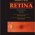
دانلود کتاب شبکیه چشم رایان + ویدئو
Ryan’s Retina: 3 Volume Set, 6ed + Video
The undisputed gold standard text in the field, Ryan’s Retina is your award-winning choice for the most current, authoritative information on new technologies, surgical approaches, scientific advances and diagnostic and therapeutic options for retinal diseases and disorders. Packed with timely updates throughout, new illustrations, and a dedicated team of editors who extend Dr. Ryan’s legacy in retina, this outstanding 6th Edition is a must-have reference for retinal specialists, ophthalmologists, and fellows in training.
- Offers the most comprehensive content available on retina, balancing the latest scientific research and clinical correlations, covering everything you need to know on retinal diagnosis, treatment, development, structure, function, and pathophysiology.
- Provides a truly global perspective from five highly esteemed section editors and more than 350 other world authorities from across Europe, Asia, Australasia, and the Americas.
-
- Bullets Includes new chapters on widefield imaging, intraoperative OCT imaging, medical management of diabetes mellitus and age-related macular degeneration, and senile retinoschisis.
- Includes more than 1,150 brand-new illustrations, scans, and photographs throughout.
- Covers the explosion of new imaging options across optical coherence tomography (OCT), fundus imaging, and autofluorescence imaging, including a greatly expanded OCT imaging chapter that features crucial information on OCT-Angiography (OCT-A).
- Presents new pharmacotherapy data and the latest approaches in anti-VEGF therapy for age-related macular degeneration, diabetic retinopathy, and venous occlusive disease.
- Features an expanded online video library highlighting the latest surgical techniques and new coverage of complications of vitreoretinal surgery.
- Contains thorough content updates in every area of retina, including advanced imaging technologies, gene therapy, inflammation and immune responses, white dot syndromes, epigenetic mechanisms, transplantation frontiers to improve retinal function, macular hole, myopic eye disease, ocular trauma, drug delivery to the posterior segment, advances in macular surgery, vitrectomy and complex retinal detachment, tumors, and retinal genetics and biology.
- Expert Consult™ eBook version included with purchase. This enhanced eBook experience allows you to search all of the text, figures, Q&As, and references from the book on a variety of devices.
Contents
۱. Fluorescein Angiography: Basic Principles and Interpretation
۲. Clinical Applications of Diagnostic Indocyanine Green Angiography
۳. Optical Coherence Tomography
۴. Autofluorescence Imaging
۵. Widefield Imaging
۶. Intraoperative OCT Imaging
۷. Advanced Imaging Technologies
۸. Advanced Image Analysis
۹. Electrogenesis of the Electroretinogram
۱۰. Clinical Electrophysiology
۱۱. Diagnostic Ophthalmic Ultrasound
۱۲. Color Vision and Night Vision
۱۳. Visual Acuity and Contrast Sensitivity
۱۴. Visual Fields in Retinal Disease
۱۵. The Development of the Retina
۱۶. Structure and Function of Rod and Cone Photoreceptors
۱۷. Function and Anatomy of the Mammalian Retina
۱۸. Cell Biology of the Retinal Pigment Epithelium
۱۹. Cell Biology of Retinal Glia
۲۰. Retinal and Choroidal Vasculature: Retinal Oxygenation
۲۱. Mechanisms of Normal Retinal Adhesion
۲۲. Structure, Function, and Pathology of Bruch’s Membrane
۲۳. Vitreous and Vitreoretinal Interface
۲۴. Mechanisms of Oxidative Stress in Retinal Injury
۲۵. Mechanisms of Endoplasmic Reticulum Stress in Retinal Disease
۲۶. Cell Death, Apoptosis, and Autophagy in Retinal Injury
۲۷. Inflammation and Immune Responses In Retinal Health and Disease
۲۸. Basic Mechanisms of Pathological Retinal and Choroidal Angiogenesis
۲۹. Blood-Retinal Barrier, Immune Privilege, and Autoimmunity
۳۰. Mechanisms of Macular Edema and Therapeutic Approaches
۳۱. Cellular Effects of Detachment and Reattachment on the Neural Retina and the Retinal Pigment Epithelium
۳۲. Pathogenesis of Serous Detachment of the Retina and Pigment Epithelium
۳۳. Genetic Mechanisms of Retinal Disease
۳۴. Mitochondrial Genetics of Retinal Disease
۳۵. Epigenetic Mechanisms of Retinal Disease
۳۶. Gene Therapy for Retinal Disease
۳۷. Stem Cells and Cellular Therapy
۳۸. Nanomedicine in Ophthalmology
۳۹. Neuroprotection
۴۰. Drug Delivery
More…
۱۳۵. Tuberous Sclerosis and the Eye
۱۳۶. Phakomatoses
۱۳۷. Retinal Metastases
۱۳۸. Remote Effects of Cancer on the Retina
۱۳۹. Melanocytoma of the Optic Disc
۱۴۰. Congenital Hypertrophy of the Retinal Pigment Epithelium
۱۴۱. Combined Hamartoma of the Retinal Pigment Epithelium and Retina
۱۴۲. Choroidal Nevi
۱۴۳. Epidemiology of Posterior Uveal Melanoma
۱۴۴. Prognosis of Posterior Uveal Melanoma
۱۴۵. Molecular Genetics of Choroidal Melanoma
۱۴۶. Pathology of Choroidal Melanoma
۱۴۷. Overview of Management of Posterior Uveal Melanoma
۱۴۸. Enucleation for Choroidal Melanomas
۱۴۹. Brachytherapy for Choroidal Melanoma
۱۵۰. Charged-Particle Irradiation of Uveal Melanoma
۱۵۱. Surgical Resection of Choroidal Melanoma
۱۵۲. Laser Treatment of Choroidal Melanoma
۱۵۳. Systemic Evaluation and Management of Patients with Metastatic Uveal Melanoma
۱۵۴. Collaborative Ocular Melanoma Study
۱۵۵. Choroidal Metastases
۱۵۶. Choroidal Osteoma
۱۵۷. Circumscribed Choroidal Hemangioma
۱۵۸. Miscellaneous Uveal Tumors
۱۵۹. Leukemias and Lymphomas
۱۶۰. Primary Vitreoretinal Lymphoma
Video Contents
Video 1: 6.1 Comparing Handheld and MicroscopeMounted Optical Coherence Tomography Imaging Systems
Video 2: 6.2 Three-Dimensional Reconstruction of a FullThickness Macular Hole with Associated Vitreomacular Traction Utilizing a Microscope Integrated Swept Source Optical Coherence Tomography System
Video 3: 6.3 Real-Time Volumetric Peeling with Forceps Visualized with a Swept Source Optical Coherence Tomography System
Video 4: 6.4 Real-Time Volumetric Peeling with Diamond-Dusted Membrane Scraper Visualized with a Swept Source Optical Coherence Tomography System
Video 5: 6.5 Intraoperative Optical Coherence Tomography Feedback during Macular Hole Surgery
Video 6: 36.1 Per?uouron Placement and Subretinal Injection Procedure
Video 7: 104.1 Techniques of Scleral Buckling
Video 8: 106.1 A Case of Primary Vitrectomy for Treating Rhegmatogenous Retinal Detachment
Video 9: 106.2 Primary Vitrectomy for Rhegmatogenous Retinal Detachment
Video 10: 109.1 Optimal Procedures for Retinal Detachment Repair
Video 11: 111.1 Relaxing Retinotomy and Retinectomy for Scarred Contracted Retina and Recurrent Inferior Retinal Detachment due to Proliferative Vitreoretinopathy
Video 12: 121.1 Posterior Hyaloid Detachment
Video 13: 121.2 Epiretinal Membrane Peeling
Video 14: 121.3 Internal Limiting Membrane Peeling
Video 15: 131.1 Subluxated Intraocular Lens without Haptics
Video 16: 131.2 Posterior Synechiae and Small Pupil during Vitrectomy
Video 17: 131.3 Induction of Posterior Vitreous Detachment in Retinal Detachment
Video 18: 131.4 Induction of Posterior Vitreous Detachment in High Myopia
Video 19: 131.5 Iatrogenic Retinal Breaks During Peeling
Video 20: 131.6 Internal Limiting Membrane Peeling
Video 21: 131.7 Subretinal Injection of Brilliant Blue
Video 22: 131.8 Reopening of Peripheral Retinal Breaks during Surgery for Submacular Hemorrhage
Video 23: 131.9 Surgical Management of Hypotony Maculopathy
Video 24: 131.10 Intraoperative Choroidal Detachment
Video 25: 131.11 Massive Suprachoroidal Hemorrhage (SCH)
Video 26: 131.12 Suprachoroidal Hemorrhage during Cataract Surgery
Video 27: 131.13 Subretinal Per?uorocarbon Bubble
Video 28: 131.14 Per?uorocarbon-Induced Macular Hole
Video 29: 131.15 Giant Retinal Tear with Slippage on
Video 30: 131.16 Proliferative Vitreoretinopathy and Subretinal Membrane
Video 31: 131.17 A Problem during 27G Vitrectomy
Video 32: 131.18 Vitreous Incarceration in Sclerotomies
Video 33: 131.19 Argus II Array Implantation
Video 34: 131.20 Subretinal SF6 Gas after Retinal Detachment Surgery
Video 35: 131.21 Removal of a Large Glass Intraocular Foreign Body
Video 36: 151.1 Surgical Resection of Choroidal Melanoma
جهت مشاهده نسخه همراه با ویدئوهای کامل ویرایش ۲۰۲۳ این کتاب کلیک کنید
لینک کوتاه : https://bookbaz.ir/?p=77035
نویسنده : Andrew P. Schachat MD , Charles P. Wilkinson MD
ناشر : Elsevier; 6 edition
سال انتشار : 2018
زبان کتاب : انگلیسی
نوع فایل : MP4 + PDF (کیفیت اصلی)
تعداد صفحات : 2756
(ISBN) شابک : 032340197X
قیمت کتاب درآمازون : $445.00
حجم فایل : 1000 MB


