
دانلود کتاب طبقه بندی ویروس: گزارش نهم کمیته بین المللی در طبقه بندی ویروس ها
Virus Taxonomy: Ninth Report of the International Committee on Taxonomy of Viruses, 1ed
The practical need to partition the world of viruses into distinguishable, universally agreed upon entities is the ultimate justification for developing a virus classification system. Since 1971, the International Committee on Taxonomy of Viruses (ICTV) operating on behalf of the world community of virologists has taken on the task of developing a single, universal taxonomic scheme for all viruses infecting animals (vertebrate, invertebrates, and protozoa), plants (higher plants and algae), fungi, bacteria, and archaea. The current report builds on the accumulated taxonomic construction of the eight previous reports dating back to 1971 and records the proceedings of the Committee since publication of the last report in 2005. Representing the work of more than 500 virologists worldwide, this report is the authoritative reference for virus organization, distinction, and structure.
Amazon.com Review
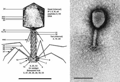
Figure 1: (Left) Diagram of Enterobacteria phage T4 (T4) showing detailed location of structural proteins. Head vertices consist of cleaved gp24. Gp20 is located at the head–tail connector. Collar and whiskers appear to be made of the same protein, gpwac. Sheath subunits (gp18) fit into holes in the base plate and short tail proteins (gp12) are shown in the quiescent state. The complex base is assembled from a central plug and six wedges. Tail fibers consist of three proteins. (From Eiserling, F.A. (1983). In: Bacteriophage T4 (C.K. Mathews, E.M. Kutter, G. Mosig and P.B. Berget, Eds.), American Society for Microbiology, Washington, DC; with permission.) (Right) Negative contrast electron micrograph of T4 particle stained with uranyl acetate. The bars represent 100 nm.
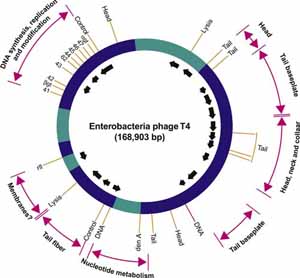
Figure 2: Simplified genetic map of Enterobacteria phage T4 (T4) showing clustering of genes with related functions, location of essential genes (solid bars) and direction and origin of transcripts (arrows). (From Freifelder, D. (Ed.) (1983)
Table 3: Cryo-EM-based reconstruction of bacteriophage phiKZ. Spiralling tail sheath vertices are highlighted. (Reconstruction courtesy of Andrei Fokine.)
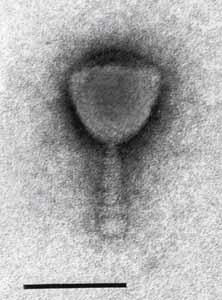
Table 4: Electron micrographs of Mycobacterium phage I3. Phage particles were positively stained with uranyl acetate. The bar represents 100 nm. (Image courtesy of H. Ackermann.)
لینک کوتاه : https://bookbaz.ir/?p=17615
نویسنده : Andrew MQ King , Elliot Lefkowitz
ناشر : Elsevier; 1 edition
سال انتشار : 2012
زبان کتاب : انگلیسی
نوع فایل : PDF
تعداد صفحات : 1338
(ISBN) شابک : 0123846846
قیمت کتاب درآمازون : $139.96
حجم فایل : 66 MB
کتاب های مرتبط:
 دانلود کتاب آزمایشات میکروبیولوژی: دیدگاه علم سلامت
Microbiology Experiments: A Health Science Perspective, 10ed
 دانلود کتاب باکتری: جهان بزرگ میکروب های واقعا کوچک
The Bacteria Book: The Big World of Really Tiny Microbes, 1ed
 دانلود کتاب راهنمای عفونت کودک: کتاب آبی
Manual of Childhood Infection: The Blue Book, 4ed
 دانلود کتاب ویروس شناسی بالینی
Clinical Virology, 4ed
 دانلود کتاب راهنمای ویروس شناسی بالینی
Clinical Virology Manual, 5ed
 دانلود کتاب پیشگیری و کنترل عفونت در یک نگاه
Infection Prevention and Control at a Glance, 1ed
 دانلود کتاب مدلسازی محاسباتی و تصویربرداری برای SARS-CoV-2 و COVID-19
Computational Modelling and Imaging for SARS-CoV-2 and COVID-19, 1ed
 دانلود کتاب زمینه ویروس شناسی نایپ (۲ جلدی)
Knipe’ Fields Virology, 2-Vol, 6ed
 دانلود کتاب میکروب شناسی پزشکی عملی برای پزشکان
Practical Medical Microbiology for Clinicians, 1ed
 دانلود کتاب پزشکی نظامی در عراق و افغانستان
Military Medicine in Iraq and Afghanistan, 1ed
 دانلود کتاب درمان ضد قارچ
 Antifungal Therapy, 2ed
 دانلود کتاب میکروبیولوژی کوان: رویکرد سیستمی
Cowan’s Microbiology: A Systems Approach, 7ed
 دانلود کتاب بیماری های عفونی کودکان فیگین و چری (۲جلدی)
Feigin and Cherry’s Textbook of Pediatric Infectious Diseases, 2-Vol, 8ed
 دانلود کتاب بیولوژی مولکولی و سلولی ویروس ها
Molecular and Cellular Biology of Viruses, 1ed
 دانلود کتاب بیماری های عفونی پیوند و انکولوژی کودکان
Pediatric Transplant and Oncology Infectious Diseases, 1ed
 دانلود کتاب بیماری عفونی بالینی شلوسبرگ
Schlossberg’s Clinical Infectious Disease, 3ed
 دانلود کتاب ویروس هپاتیت C و پیوند کبد
Hepatitis C Virus and Liver Transplantation, 2014th
 دانلود کتاب بیماری های مقاربتی مسری هولمز
Holmes’s Sexually Transmitted Diseases, 4ed
 دانلود کتاب راهنمای گرافیکی بیماری عفونی
 Graphic Guide to Infectious Disease, 1ed
 دانلود کتاب راهنمای پزشکی صحرایی و طبیعت آکسفورد
Oxford Handbook of Expedition and Wilderness Medicine, 2ed
 دانلود کتاب معضلات بالینی در بیماری کبد ویروسی
Clinical Dilemmas in Viral Liver Disease, 2ed
 دانلود کتاب بیماری های عفونی کودکان: موارد ضروری برای تمرین
Pediatric Infectious Diseases: Essentials for Practice, 2ed
 دانلود کتاب اصول و عملکرد بیماری های عفونی کودکان
 Principles and Practice of Pediatric Infectious Diseases, 6ed
 دانلود کتاب ایمونولوژی پایه: عملکردها و اختلالات سیستم ایمنی
Basic Immunology: Functions and Disorders of the Immune System, 7ed
 دانلود کتاب میکروبیولوژی بالینی به طرز مضحکی ساده شده
Clinical Microbiology Made Ridiculously Simple, 9ed
 دانلود کتاب درمان ضد میکروبی کودکان نلسون ۲۰۲۳
۲۰۲۳Nelson’s Pediatric Antimicrobial Therapy, 29ed
 دانلود کتاب ویروس: راهنمای مصور ۱۰۱ میکروب باور نکردنی
Virus: An Illustrated Guide to 101 Incredible Microbes, 3ed
 دانلود کتاب مبانی در میکروبیولوژی تالارو
Foundations in Microbiology, 10ed
 دانلود کتاب اصول و عملکرد بیماریهای عفونی مندل، داگلاس و بنت
Mandell, Douglas, and Bennett’s Principles and Practice of Infectious Diseases, 9ed
 دانلود کتاب کاربردهای آزمایشگاهی در میکروبیولوژی: رویکرد مطالعه موردی
Laboratory Applications in Microbiology: A Case Study Approach, 4ed
 دانلود کتاب دایره المعارف ویروس شناسی (۵ جلدی)
Encyclopedia of Virology, 5-Vol, 4ed
 دانلود کتاب ویروس ها: از درک تا بررسی
Viruses: From Understanding to Investigation, 2ed
 دانلود کتاب هپاتیت ویروسی
Viral Hepatitis, 4ed
 دانلود کتاب اصول ویروس شناسی فلینت (۲ جلدی)
Principles of Virology, 2-Vol, 4ed
 دانلود کتاب میکروب شناسی پزشکی موری
Medical Microbiology, 8ed
 دانلود کتاب مقدمه ای بر ویروس شناسی مدرن
Introduction to Modern Virology, 7ed
 دانلود کتاب ویروس شناسی ضروری انسان لوتن
Essential Human Virology, 1ed
 دانلود کتاب مکانیسم سرایت پاتوژن های باکتریایی
Virulence Mechanisms of Bacterial Pathogens, 5ed
 دانلود کتاب ویروس شناسی مولکولی ویروس های پاتوژنیک انسان
Molecular Virology of Human Pathogenic Viruses, 1ed
 دانلود کتاب پزشکی گرمسیری و بیماری های عفونی نوظهور هانتر
Hunter’s Tropical Medicine and Emerging Infectious Diseases, 10ed
 دانلود کتاب ویروس پاپیلومای انسانی
 Human Papillomavirus, 1ed
 دانلود کتاب رویکرد مطالعه موردی بیماریهای عفونی
 Infectious Diseases Case Study Approach, 1ed
 دانلود کتاب سل بالینی
 Clinical Tuberculosis, 6ed
 دانلود کتاب پاتوژنز باکتریایی: یک رویکرد مولکولی
Bacterial Pathogenesis: A Molecular Approach, 4ed
 دانلود کتاب مبانی میکروبیولوژی
 Foundations in Microbiology, 9ed
 دانلود کتاب ویروس شناسی
 Virology: An Illustrated Colour Text, 1ed
 دانلود کتاب ویروس شناسی مولکولی و کنترل فلاوی ویروس ها
Molecular Virology and Control of Flaviviruses, 1ed
 دانلود کتاب شاخص ویروس های اسپرینگر
The Springer Index of Viruses, 2ed
 دانلود کتاب مبانی میکروبیولوژی پومرویل
Pommerville’s Fundamentals of Microbiology, 11ed
 دانلود کتاب میکروبیولوژی عفونت های سیستم تنفسی
The Microbiology of Respiratory System Infections, 1ed
 دانلود کتاب نورو ویروس شناسی بالینی
Clinical Neurovirology, 2ed
 دانلود کتاب مدیریت بیماری های عفونی در مراقبت از کودکان و مدارس
Managing Infectious Diseases in Child Care and Schools, 6ed
 دانلود کتاب ویروس های انسانی در رسوبات لجن و خاک
Human Viruses In Sediments Sludges & Soils, 1ed
 دانلود کتاب بیماری های گرمسیری منسون
Manson’s Tropical Diseases, 24ed
 دانلود کتاب دانشنامه ویروس شناسی انسانی و پزشکی
Desk Encyclopedia of Human and Medical Virology, 1ed
 دانلود کتاب تاریخچه مختصری از باکتری ها
A Brief History of Bacteria, 1ed
 دانلود کتاب موارد بالینی در پزشکی گرمسیری
Clinical Cases in Tropical Medicine, 2ed
 دانلود کتاب بیماری ویروسی کرونا ۲۰۱۹ (کووید-۱۹): راهنمای بالینی
Coronavirus Disease 2019 (Covid-19): A Clinical Guide, 1ed
 دانلود کتاب هندبوک درمانی آنتی بادی (۴ جلدی)
Handbook of Therapeutic Antibodies, 2ed
 دانلود کتاب میکروبیولوژی مولکولی: اصول و تمرین تشخیصی
Molecular Microbiology: Diagnostic Principles and Practice, 3ed
 دانلود کتاب تب مغزی: چگونه واکسن ها از مننژیت و سایر بیماری های کشنده جلوگیری می کنند
Brain Fever: How Vaccines Prevent Meningitis And Other Killer Diseases, 1ed
 دانلود کتاب مبانی در میکروبیولوژی تالارو
Talaro’s Foundations in Microbiology, 12ed
 دانلود کتاب اصول و تمرین ویروس شناسی بالینی
Principles and Practice of Clinical Virology, 6ed
 دانلود کتاب بیماری های عفونی بزرگسالان: بیش از ۲۰۰ مورد مطالعاتی
Adult Infectious Diseases Over 200 Case Studies, 1ed
 دانلود کتاب راهنما و کتاب کار آزمایشگاهی در میکروبیولوژی
 Lab Manual and Workbook in Microbiology, 12ed
 دانلود کتاب تشخیص پاتوژنومیک عفونت های منتقله از راه جنسی
 Diagnostics to Pathogenomics of Sexually Transmitted Infections, 1ed
 دانلود کتاب واکسن های پلوتکین
Plotkin’s Vaccines, 8ed
 دانلود کتاب Textbook of SARS-CoV-2 and COVID-19: Epidemiology, Etiopathogenesis, Immunology, Clinical Manifestations, Treatment, Complications, and Preventive Measures, 1ed
 دانلود کتاب قرمز اطلس بیماری های عفونی کودکان
Red Book Atlas of Pediatric Infectious Diseases, 5ed
 دانلود کتاب دانشنامه ویروس شناسی (۵ جلدی)
Encyclopedia of Virology, 5-Volume Set, 3ed
 دانلود کتاب واکسن های پلوتکین
Plotkin’s Vaccines, 7ed
 دانلود کتاب راهنمای پزشکی ادراری تناسلی، اچ آی وی و سلامت جنسی آکسفورد
Oxford Handbook of Genitourinary Medicine, HIV, and S3xual Health, 3ed
 دانلود کتاب MKSAP 17 : بیماری عفونی
 MKSAP (R) 17 Infectious Disease, 17ed
 دانلود کتاب ویروس پاپیلومای انسانی: پژوهش در یک چشم انداز جهانی
Human Papillomavirus: Research in a Global Perspective, 1ed
 دانلود کتاب بیماری ریزاندامگان همزیست و مزمن انسان: ناترازشدن همزیستی به عنوان علت آسیب شناسی انسانی
The Human Microbiota and Chronic Disease: Dysbiosis as a Cause of Human Pathology, 1ed
 دانلود کتاب مبانی در میکروبیولوژی تالارو: اصول پایه
Talaro’s Foundations in Microbiology: Basic Principles, 12ed
Stress and Environmental Regulation of Gene Expression and Adaptation in Bacteria, 2-Vol, 1ed
 دانلود کتاب اصول ویروس شناسی فلینت (۲ جلدی)
Principles of Virology, 2-Vol, 5ed
 دانلود کتاب میکروبیولوژی با بیماری ها بر اساس طبقه بندی
Microbiology with Diseases by Taxonomy, 6ed
 دانلود کتاب اطلس پزشکی گرمسیری و انگل شناسی پیترز
Peters’ Atlas of Tropical Medicine and Parasitology, 7ed
 دانلود کتاب اطلس سرخ بیماریهای عفونی کودکان
 Red Book Atlas of Pediatric Infectious Diseases, 4ed
 دانلود کتاب تظاهرات مکوکوبتی بیماریهای ویروسی
Mucocutaneous Manifestations of Viral Diseases, 2ed
 دانلود کتاب عفونت های ویروسی تنفسی انسانی
Human Respiratory Viral Infections, 1ed
 دانلود کتاب میکروبیولوژی پرسکات
Prescott’s Microbiology, 11ed
 دانلود مجموعه ویدئویی بیماری عفونی Boards and Beyond 2021: Infectious Disease + Slides