
دانلود کتاب قرنیه (۲ جلدی) + ویدئو
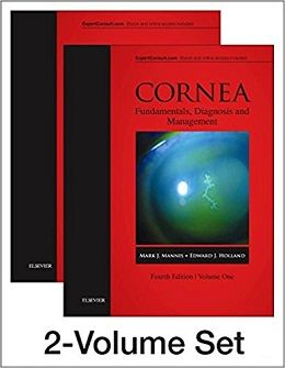
Cornea, 2-Volume Set, 4ed + Video
Highly praised in its first three editions, Cornea has become a market-leading cornerstone text and the immediate go-to resource for anyone working in this hugely popular and evolving sub-specialty. Offered over two volumes and featuring the knowledge of over 200 experts worldwide, it presents state-of-the-art coverage of the expanding range of contemporary corneal surgery, new diagnostic technology, and medical management of corneal and external disease as well as ocular surface disease. This updated edition includes ۲۰ brand-new chapters and ۶۰ video clips, while an enhanced focus on images provides key visual guidance in this challenging field.
- Exceptionally clear illustrations, diagnostic images, and step-by-step surgical photographs offer superb visual guidance.
- Expert Consult eBook version included with purchase. This enhanced eBook experience allows you to search all of the text, figures, images, videos, and references from the book on a variety of devices.
- ۲۰ brand-new chapters cover the latest advances in the field, such as DMEK, Ultra-Thin DSEK and DSAEK techniques; endothelial cell transplantation; keratoplasty and prosthokeratoplasty techniques; collagen cross-linking; and new refractive surgical techniques (presbyopic implants and SMILE surgery).
- ۶۰ video clips on Expert Consult show new footage of the latest corneal surgery techniques, including Boston Keratoprosthesis, corneal inlay surgery, and lenticule extraction.
- Boasts over 170 chapters with unique, cutting-edge content, as well as ۲,۳۰۰clear illustrations – ۶۷۰ of which are new to this edition.
- Presents a detailed exposition of the growing number of techniques forlamellar keratoplasty, including outcomes.
- Includes new sections on the latest developments in the management of ocular surface disease.
- Key point overviews in each chapter offer easier access to crucial information.
Review
Contents
۱ Cornea and Sclera
۲ The Conjunctiva
۳ Tear Film
۴ Eyelids and the Corneal Surface
۵ A Matrix of Pathologic Responses in the Cornea
۶ Examination of the Lids
۷ Slit Lamp Examination and Photography
۸ Tear Film Evaluation
۹ Corneal Diagnostic Techniques
۱۰ Practical Ophthalmic Microbiology for the Detection of Corneal Pathogens
۱۱ Molecular Genetics of Corneal Disease
۱۲ Keratometry and Topography
۱۳ Corneal Shape Analysis
۱۴ Specular Microscopy
۱۵ Confocal Microscopy
۱۶ High Resolution Ultrasound
۱۷ Anterior Segment Optical Coherence Tomography
۱۸ Congenital Corneal Opacities
۱۹ Peripheral Corneal Disease
۲۰ The Corneal Ulcer
۲۱ Corneal Edema
۲۲ Corneal Deposits
۲۳ The Red Eye
۲۴ Minimal Visual Loss
۲۵ Eye Banking
۲۶ Medical Standards for Eye Banking
۲۷ Malposition of the Eyelids
۲۸ Benign Lid Tumors
۲۹ Malignant Eyelid Tumors
۳۰ Blepharitis
۳۱ Meibomian Gland Dysfunction and Seborrhea
۳۲ Eyelid Infections
۳۳ Dry Eye
۳۴ Dacryoadenitisr Dacryocystitisr and Canaliculitis
۳۵ Epiphora
۳۶ Epithelial Tumors of the Conjunctiva
۳۷ Medical and Surgical Management of Ocular Surface Squamous Neoplasia
۳۸ Melanocytic Neoplasms of the Conjunctiva
More…
۱۳۸ Therapeutic Keratoplasty
۱۳۹ Surgical Management of Superficial Corneal and Conjunctival Disease
۱۴۰ Excimer Laser Phototherapeutic Keratectomy
۱۴۱ Management of Pterygium
۱۴۲ Conjunctival Flaps
۱۴۳ Indications for and Uses of Amniotic Membrane
۱۴۴ Surgical Management and Rehabilitation of Anterior Segment Trauma
۱۴۵ Iris Reconstruction Surgery
۱۴۶ Management of Scleral Perforation
۱۴۷ Collagen Crosslinking for Keratoconus
۱۴۸ Collagen Crosslinking for Post-Refractive Ectasia
۱۴۹ Collagen Crosslinking for Infectious Keratitis
۱۵۰ Indications for Keratoprosthesis
۱۵۱ Boston Keratoprosthesis Type 1 Surgical Technique
۱۵۲ Postoperative Management of Boston Keratoprosthesis Type 1
۱۵۳ Complications of Boston Keratoprosthesis Type 1
۱۵۴ Outcomes of Boston Keratoprosthesis Type 1
۱۵۵ Boston Keratoprosthesis Type 2 Surgical Techniques, Complicationsr and Outcomes
۱۵۶ OOKP
۱۵۷ Classification and Staging of Ocular Surface Disease
۱۵۸ Surgical Techniques for Ocular Surface Reconstruction
۱۵۹ Postoperative Management of Ocular Surface Reconstruction
۱۶۰ Corneal Transplantation in Ocular Surface Disease
۱۶۱ Decision Making in Refractive Surgery
۱۶۲ Patient Evaluation and Selection in Refractive Surgery
۱۶۳ Topographic Analysis in Keratorefractive Surgery
۱۶۴ Excimer Laser Surface Treatment
۱۶۵ Surface Ablation
۱۶۶ LASIK Technique
۱۶۷ LASIK for Myopia
۱۶۸ LASIK for Hyperopia
۱۶۹ LASIK Complications
۱۷۰ Corneal Ectasia
۱۷۱ Small Incision Lenticule Extraction
۱۷۲ Intrastromal Corneal Ring Segments
۱۷۳ Incisional Keratotomy
۱۷۴ Phakic Intraocular Lenses
۱۷۵ Corneal Inlay Surgery for Presbyopia
Video Contents
۵۰.۱ The Application of Cryopreserved Amniotic Membrane in the Treatment of Acute Stevens–Johnson Syndrome: Part 1
۵۰.۲ The Application of Cryopreserved Amniotic Membrane in the Treatment of Acute Stevens–Johnson Syndrome: Part 2
۵۰.۳ The Application of Cryopreserved Amniotic Membrane in the Treatment of Acute Stevens–Johnson Syndrome: Part 3
۱۱۰.۱ Penetrating Keratoplasty Using the Barron Trephine and Interrupted Sutures
۱۱۰.۲ Penetrating Keratoplasty Using the Hanna Trephine and Interrupted Sutures
۱۱۰.۳ Penetrating Keratoplasty Using the Slipknot Technique for Suturing
۱۱۰.۴ Penetrating Keratoplasty Using a Running Suture
۱۱۱.۱ An Intraoperative Suprachoroidal Hemorrhage During Penetrating Keratoplasty, which was Successfully Managed with the Assistance of a Cobo Temporary Keratoprosthesis
۱۱۲.۱ Donor Preparation
۱۱۲.۲ Dissection and Suturing
۱۱۲.۳ Laser Incision
۱۱۲.۴ Femto DALK
۱۱۷.۱ Big Bubble DALK: A Video Presentation
۱۱۸.۱ Techniques of Anterior Lamellar Keratoplasty: Stromal Delamination
۱۱۸.۲ Techniques of Anterior Lamellar Keratoplasty: ALTK 1
۱۱۸.۳ Techniques of Anterior Lamellar Keratoplasty: ALTK 2
۱۱۸.۴ Techniques of Anterior Lamellar Keratoplasty: Big Bubble
۱۱۸.۵ Techniques of Anterior Lamellar Keratoplasty: Big Bubble Using a Cannula
۱۱۸.۶ Techniques of Anterior Lamellar Keratoplasty: Donor Preparation
۱۱۹.۱ Rupture of DM in a Keratoconus Patient Shigeto Shimmura
۱۲۰.۱ Cannula Big-Bubble Technique, Bubble Test, and the New Opening of the Bubble
۱۲۰.۲ Air-Viscobubble Technique (AVB)
۱۲۰.۳ Layer-by-Layer Manual Dissection
۱۲۰.۴ dDALK Rupture Management
۱۲۰.۵ Subtotal Full Thickness Circular Cut of the Recipient Bed
۱۲۰.۶ Total Full Thickness Circular Cut of the Recipient Bed
۱۲۰.۷ Excessive Trephination and Perforation
۱۲۰.۸ Traumatic Postoperative DM Disinsertion
۱۲۶.۱ Eye Bank Preparation of DMEK Graft Tissue Using a Modifed SCUBA Technique
۱۳۰.۱ Ultrathin DSAEK Massimo Busin
۱۳۱.۱ DMEK Injectors
۱۳۱.۲ Peripheral Iridotomy
۱۳۲.۱ Torn Donor Graft During DMEK Graft Preparation Using SCUBA Technique
۱۳۴.۱ DSEK Under Top Hat PKP with Laplace Phenomenon
۱۳۴.۲ Suture Pull Through and Fixation
۱۳۶.۱ RHCIII Implantation Surgery May Griffth
۱۴۱.۱ Primary Pterygium Excision and Conjunctival Autograft with Fibrin Glue
۱۴۱.۲ Recurrent Pterygium Excision and Extensive Tenon’s Excision with Conjunctival Autograft Using Fibrin Glue
۱۴۳.۱ How to Know the Orientation of the Amniotic Membrane
۱۴۳.۲ Surgical Videos of Several Cases where Amniotic Membrane Transplantation was Performed
۱۴۳.۳ The Modifed Gundersen Approach Consists of Four Main Steps
۱۴۳.۴ Two-Step Approach to Treat Unilateral Total Limbal Stem Cell Defciency
۱۴۵.۱ Iris Oversew ,
۱۴۵.۲ Taco Down Fold of PCIOL for Insertion Behind 50-Series Iris Prostheses
۱۴۵.۳ CustomFlex Iris Device Injection and Capsular Bag Implantation With Overfolding
۱۵۱.۱ KPro Assembly and Surgery Jose de la Cruz
۱۵۵.۱ Boston Keratoprosthesis Type 2 Surgical Techniques Duna Raoof,
۱۵۶.۱ Stage 1 OOKP
۱۵۶.۲ Stage 2 OOKP
۱۵۸.۱ LR-CLAU
۱۵۸.۲ KLAL
۱۵۸.۳ CLAU-KLAL Modifed Cincinnati Procedure
۱۶۰.۱ Combined LR-CLAL/KLAL (Cincinnati Procedure) Followed by Penetrating Keratoplasty
۱۶۵.۱ LASEK Azar Flap Technique
۱۶۶.۱ Technique for Custom Femtosecond LASIK
۱۶۷.۱ Femtosecond LASIK
۱۶۹.۱ Anterior Chamber Gas Bubbles Blocking the Tracking of the Pupil
۱۷۱.۱ Small Incision Lenticule Extraction
۱۷۲.۱ Post LASIK Ectasia – Intrastromal CXL and Ring
۱۷۳.۱ Femtosecond Laser Astigmatic Keratotomy
۱۷۴.۱ Implantation of an Artisan Lens
۱۷۴.۲ Implantation of a Toric Artisan Lens
۱۷۴.۳ Implantation of an Artiflex Lens
۱۷۴.۴ Implantation of an ICL
۱۷۴.۵ Performing LASIK in a Patient with an Iris-Fixated Lens
۱۷۵.۱ Implanting the Kamra Small-Aperture Inlay
جهت مشاهده نسخه همراه با ویدئوهای کامل ویرایش ۲۰۲۲ این کتاب کلیک کنید
لینک کوتاه : https://bookbaz.ir/?p=89264
نویسنده : Mark J Mannis MD FACS , Edward J Holland MD
ناشر : Elsevier; 4 edition
سال انتشار : 2017
زبان کتاب : انگلیسی
نوع فایل : MP4 + PDF (کیفیت اصلی)
تعداد صفحات : 2054
(ISBN) شابک : 0323357571
قیمت کتاب درآمازون : $407.42
حجم فایل : 1800 MB
کتاب های مرتبط:
 دانلود کتاب قرنیه (۲ جلدی)
Cornea, 2-Volume Set, 5ed
 دانلود کتاب دایره المعارف چشم (۴ جلدی)
Encyclopedia of the Eye, Four-Volume Set, 1ed
 دانلود کتاب شبکیه چشم رایان (۳ جلدی، ویرایش ۲۰۱۸)
Ryan’s Retina: 3 Volume Set, 6ed
 دانلود کتاب مجموعه ویدئویی جراحی پلک و دور چشم + کتاب
Eyelid and Periorbital Surgery Video Collection, 2ed + PDF
 دانلود کتاب شبکیه چشم رایان (۳ جلدی)
Ryan’s Retina, 3-Vol, 7ed
 دانلود کتاب پیوند قرنیه اندوتلیال: مهارت DSEK، DMEK و PDEK
Endothelial Keratoplasty: Mastering DSEK, DMEK, and PDEK, 1ed
 دانلود کتاب فلپ های موضعی در بازسازی صورت بِیکر + ویدئو
Local Flaps in Facial Reconstruction, 4ed + Video
 دانلود کتاب راهنمای جراحی شبکیه چشم
Handbook of Vitreoretinal Surgery, 1ed
 دانلود کتاب بیماری های سطح چشم: قرنیه، ملتحمه و پرده اشک آور + ویدئو
Ocular Surface Disease: Cornea, Conjunctiva and Tear Film, 1ed + Video
 دانلود کتاب چشم پزشکی بالینی کانسکی: یک رویکرد سیستماتیک
Kanski’s Clinical Ophthalmology: A Systematic Approach, 8ed
 دانلود کتاب قرنیه (۲ جلدی)
Cornea, 2-Volume Set, 4ed
 دانلود کتاب تکنیک های مهارت در جراحی چشم
 Master Techniques in Ophthalmic Surgery, 2ed
 دانلود کتاب جراحی پلاستیک زیبایی چشم پوترمن + ویدئو
Putterman’s Cosmetic Oculoplastic Surgery, 4ed + Video
 دانلود کتاب دانشنامه پزشکی گیل (۹ جلدی)
Gale Encyclopedia of Medicine, 9-Vol, 2015th
 دانلود کتاب لنز های چشمی
Contact Lenses, 6ed
 دانلود کتاب اولتراسونوگرافی چشمی
Ophthalmic Ultrasonography, 1ed
 دانلود کتاب هنر جراحی ناخنک چشم
The Art of Pterygium Surgery, 1ed
 دانلود کتاب جراحی انکساری چشم
Refractive Surgery, 3ed
 دانلود کتاب ملزومات OCT در بیماری های چشم
Essentials of OCT in Ocular Disease, 1ed
 دانلود کتاب جراحی پلاستیک چشمی
Oculoplastic Surgery, 3ed
 دانلود کتاب چشم: علوم پایه در عمل
The Eye: Basic Sciences in Practice, 5ed
 دانلود کتاب روش های بالینی برای معاینه چشم
 Clinical Procedures for Ocular Examination, 4ed
 دانلود کتاب تمرین لنز تماسی افرون
Contact Lens Practice, 3ed
 دانلود کتاب جراحی آندوسکوپی حدقه چشم
Endoscopic Surgery of the Orbit, 1ed
 دانلود کتاب جراحی آب مروارید استاینرت
Steinert’s Cataract Surgery, 4ed
 دانلود کتاب هنر جراحی آب مروارید انکساری: برای رزیدنت ها، همراهان و مبتدیان
The Art of Refractive Cataract Surgery: For Residents, Fellows, and Beginners, 1ed
 دانلود کتاب چشم پزشکی یانوف و داکر + ویدئو
Yanoff & Duker Ophthalmology, 5ed + Video
 دانلود کتاب تکنیک های جراحی پلاستیک چشم
 Techniques in Ophthalmic Plastic Surgery, 2ed
 دانلود کتاب اطلس شبکیه یانوزی
The Retinal Atlas: Expert Consult, 1ed
 دانلود کتاب چشم پزشکی و انحراف چشم کودکان تیلور و هویت + ویدئو
Taylor and Hoyt’s Pediatric Ophthalmology and Strabismus, 5ed + Video
دانلود کتاب جراحی پلاستیک زیبایی چشم پوترمنPutterman’s Cosmetic Oculoplastic Surgery, 4ed
 دانلود کتاب راهنمای پایندگی چشم پزشکی عصبی
The Neuro-Ophthalmology Survival Guide, 2ed
 دانلود کتاب جراحی لیزر فمتوثانیه در چشم پزشکی
Femtosecond Laser Surgery in Ophthalmology, 1ed
 دانلود کتاب رویه های بالینی در مراقبت های اولیه چشم
 Clinical Procedures in Primary Eye Care, 5ed
 دانلود کتاب تکنیک های عملی در جراحی شبکیه چشم
 Operative Techniques in Vitreoretinal Surgery, 1ed
 دانلود کتاب دوره علوم پایه و بالینی آکادمی چشم پزشکی آمریکا ۲۰۲۳-۲۰۲۲ (۱۳ جلدی)
BCSC 2022-2023 (Basic and Clinical Science Course), 13-Vol, 1ed
 دانلود کتاب اطلس OCT شبکیه: توموگرافی انسجام نوری
Atlas of Retinal OCT: Optical Coherence Tomography, 2ed
 دانلود کتاب اطلس رنگی جراحی پلاستیک چشم
Colour Atlas of Ophthalmic Plastic Surgery, 4ed
 دانلود کتاب جراحی پلاستیک چشم: ترفندهای حرفه ای
Ophthalmic Plastic Surgery: Tricks of the Trade, 1ed
 دانلود کتاب راهنمای چشم پزشکی بخش چشم و گوش بیمارستان ماساچوست
The Massachusetts Eye and Ear Infirmary Illustrated Manual of Ophthalmology, 5ed
 دانلود کتاب یادداشت های پزشکی: چشم پزشکی
 Lecture Notes Ophthalmology, 12ed
 دانلود کتاب اختلالات کوروئید
 Choroidal Disorders, 1ed
 دانلود کتاب جراحی پلاستیک چشم قسمت فوقانی صورت
Ophthalmic Plastic Surgery of the Upper Face, 1ed
 دانلود کتاب جراحی استرابیسم: رویکردهای نوآورانه و کلاسیک
Strabismus Surgery: Innovative and Classic Approaches, 1ed
 دانلود کتاب چشم پزشکی و استرابیسم کودکان تیلور و هویت
Taylor and Hoyt’s Pediatric Ophthalmology and Strabismus, 6ed
 دانلود کتاب مقدمه ای بر اپتیک بصری: رویکرد تابشی
Introduction to Visual Optics: A Light Approach, 1ed
 دانلود کتاب چشم پزشکی یانوف و داکر + ویدئو
Yanoff & Duker Ophthalmology, 4ed + Video
 دانلود کتاب مشکلات در جراحی اطراف حدقه: راهنمای ترمیمی
Problems in Periorbital Surgery: A Repair Manual, 1ed
 دانلود کتاب اطلس مدیریت چند رشته ای بیماری های حدقه ای
Interdisciplinary Management of Orbital Diseases: Textbook and Atlas, 1ed
دانلود کتاب جراحی پلاستیک چشم: ترفندهای حرفه ای + ویدئوOphthalmic Plastic Surgery: Tricks of the Trade, 1ed + Video
 دانلود کتاب شبکیه چشم رایان (۳ جلدی)
Ryan’s Retina: Expert Consult Premium Edition Enhanced, 3-Vol, 5ed
 دانلود کتاب اصول و عملکرد چشم پزشکی آلبرت و جاکوبیک
Albert and Jakobiec’s Principles and Practice of Ophthalmology, 4ed
 دانلود کتاب اصول و عملکرد قرنیه کوپلند و افشاری
Copeland and Afshari’s Principles and Practice of Cornea, 1ed
 دانلود کتاب بهینه سازی نتایج زیر بهینه حاصل از جراحی آب مروارید
Optimizing Suboptimal Results Following Cataract Surgery, 1ed
 دانلود کتاب چشم پزشکی یانوف و داکر + ویدئو
Yanoff & Duker Ophthalmology, 6ed + Video
 دانلود کتاب بورد USMLE مرحله ۲ چشم پزشکی مدکوئست + ویدئو
MedQuest USMLE Step 2 High–Yield Ophthalmology, 6ed + Video
 دانلود کتاب ایمپلنت فیکوامولسیفیکیشن و لنز داخل چشمی: تسلط تکنیک ها و عوارض در عمل جراحی کاتاراکت
Phacoemulsification and Intraocular Lens Implantation: Mastering Techniques and Complications in Cataract Surgery, 2ed
دانلود کتاب اولتراسونوگرافی چشمی + ویدئوOphthalmic Ultrasonography, 1ed + Video
دانلود کتاب قرنیه (۲ جلدی) + ویدئوCornea, 2-Volume Set, 5ed + Video
 دانلود کتاب قوز قرنیه: تشخیص و درمان
Keratoconus: Diagnosis and Management, 1ed
 دانلود کتاب عفونت و التهاب قرنیه
 Corneal Infection and Inflammation, 1ed
 دانلود کتاب اطلس قرنیه
 Cornea Atlas, 3ed
 دانلود کتاب جراحی های کم تهاجمی گلوکوم
Minimally Invasive Glaucoma Surgery, 1ed
دانلود کتاب چشم: علوم پایه در عمل + ویدئوThe Eye: Basic Sciences in Practice, 5ed + Video
 دانلود کتاب معاینه چشم پزشکی دوک الدر
The Duke Elder Exam of Ophthalmology, 1ed
 دانلود کتاب فلج صورت: تکنیک های توانبخشی
Facial Paralysis: Rehabilitation Techniques, 1ed
 دانلود کتاب راهنمای چشم ویلز
The Wills Eye Manual, 7ed
 دانلود کتاب پاتولوژی چشمی
 Ocular Pathology, 8ed
 دانلود کتاب بیولوژی سلول بنیادی و پزشکی احیا در چشم پزشکی
Stem Cell Biology and Regenerative Medicine in Ophthalmology
 دانلود کتاب آب سیاه چشم (۲ جلدی)
Glaucoma: 2-Volume Set, 2ed
 دانلود کتاب گلوکوم چندلر و گرانت
Chandler and Grant’s Glaucoma, 5ed
دانلود کتاب جراحی پلاستیک چشمی + ویدئوOculoplastic Surgery, 3ed + Video
 دانلود کتاب جراحی و روشهای چشم پزشکی کودکان
Pediatric Ophthalmology Surgery and Procedures, 1ed
دانلود کتاب رویه های بالینی در مراقبت های اولیه چشم + ویدئوClinical Procedures in Primary Eye Care, 5ed + Video
 دانلود کتاب بررسی موارد آسیب شناسی چشمی
 Ocular Pathology Case Reviews: Expert Consult, 1ed
 دانلود کتاب تشخیص و درمان چشم یانوف
Ophthalmic Diagnosis and Treatment, 3ed
 دانلود کتاب اطلس چشم پزشکی بالینی
Atlas of Clinical Ophthalmology, 2ed
 دانلود کتاب اطلس جراحی پلاستیک و ترمیمی چشم (ویرایش ۲۰۱۷)
Video Atlas of Oculofacial Plastic and Reconstructive Surgery, 2ed
 دانلود کتاب اورژانس چشم: راهنمای تمرین کننده
Eye Emergencies: A Practitioner’s Guide, 2ed
 دانلود کتاب اطلس شبکیه یانوزی (ویرایش ۲۰۱۷)
The Retinal Atlas, 2ed
 دانلود کتاب اسرار چشم پزشکی (ویرایش ۲۰۱۶)
Ophthalmology Secrets in Color, 4ed
 دانلود کتاب چشم پزشکی عمومی وگان و آزبری
Vaughan & Asbury’s General Ophthalmology, 19ed
دانلود کتاب اطلس رنگی جراحی پلاستیک چشم + ویدئوColour Atlas of Ophthalmic Plastic Surgery, 4ed + Video
دانلود کتاب جراحی لیزر فمتوثانیه در چشم پزشکی + ویدئوFemtosecond Laser Surgery in Ophthalmology, 1ed + Video
 دانلود کتاب جراحی چشم: اصول و تمرین + ویدئو
Ophthalmic Surgery: Principles and Practice, 4ed + Video
دانلود کتاب راهنمای چشم پزشکی بخش چشم و گوش بیمارستان ماساچوست + ویدئوThe Massachusetts Eye and Ear Infirmary Illustrated Manual of Ophthalmology, 5ed + Video
 دانلود کتاب نورشناسی هندسی و بصری
Geometrical and Visual Optics, 3ed
دانلود کتاب شبکیه چشم رایان (۳ جلدی) + ویدئوRyan’s Retina, 3-Vol, 7ed + Video
 دانلود کتاب تکنیک های لیزر در چشم پزشکی
Laser Techniques in Ophthalmology, 1ed
 دانلود کتاب جراحی اندوسکوپیک هیپوفیز: غدد و متابولیسم، چشمی-عصبی و مدیریت جراحی
Endoscopic Pituitary Surgery: Endocrine, Neuro-Ophthalmologic, and Surgical Management, 1ed
 دانلود کتاب یادداشت های درسی: چشم پزشکی
 Lecture Notes: Ophthalmology, 11ed
 دانلود کتاب اطلس چشم پزشکی لانگ
Ophthalmology A Pocket Textbook Atlas, 2ed
 دانلود کتاب اطلس بیماری ماکولا گس (۲ جلدی)
Gass’ Atlas of Macular Diseases: Expert Consult, 2-Volume Set, 5ed
 دانلود کتاب دژنراسیون ماکولا مرتبط با سن
Age-Related Macular Degeneration, 2ed
 دانلود کتاب روش های جراحی چشم
Ophthalmic Surgical Procedures, 2ed
 دانلود کتاب حقایق سریع: چشم پزشکی
 Fast Facts: Ophthalmology, 2ed
 دانلود کتاب آنژیوگرافی فلورسین عمقی سانکارا
The Sankara Nethralaya Atlas of Fundus Fluorescein Angiography, 2ed
 دانلود کتاب جراحی پلک و دور چشم (۲ جلدی)
Eyelid and Periorbital Surgery, 2-Vol, 2ed
 دانلود کتاب اطلس ویدئویی بخیه چشم: اصول و تکنیک ها + ویدئو
Video Atlas of Ophthalmic Suturing: Fundamentals and Techniques, 1ed + Video
دانلود کتاب بهینه سازی نتایج زیر بهینه حاصل از جراحی آب مروارید + ویدئوOptimizing Suboptimal Results Following Cataract Surgery, 1ed + Video
دانلود کتاب ملزومات OCT در بیماری های چشم + ویدئوEssentials of OCT in Ocular Disease, 1ed + Video
 دانلود کتاب چشم پزشکی عصبی مصور + ویدئو
Neuro-Ophthalmology Illustrated, 3ed + Video
 دانلود کتاب چشم پزشکی عصبی لیو، والپ و گالتا: تشخیص و مدیریت + ویدئو
Liu, Volpe, and Galetta’s Neuro-Ophthalmology: Diagnosis and Management, 3ed + Video
دانلود کتاب جراحی استرابیسم: رویکردهای نوآورانه و کلاسیک + ویدئوStrabismus Surgery: Innovative and Classic Approaches, 1ed + Video
 دانلود کتاب اسرار چشم پزشکی
 Ophthalmology Secrets, 5ed
 دانلود کتاب مراقبت از چشم
Ophthalmic Care, 2ed
 دانلود کتاب ابزار جراحی چشم
Ophthalmic Surgical Instruments, 1ed
 دانلود کتاب چشم پزشکی بالینی کودکان
Pediatric Clinical Ophthalmology
 دانلود کتاب مفاهیم کنونی در ملانوم Uveal (تحولات در چشم پزشکی)
Current Concepts in Uveal Melanoma
 دانلود کتاب پزشکی تورنتو نوت (نسخه ۲۰۱۵)
Toronto Notes 2015 (Essential Med Notes), 31ed
 دانلود کتاب اطلس آناتومی بالینی و جراحی حدقه ای
 Atlas of Clinical and Surgical Orbital Anatomy, 2ed
 دانلود کتاب اطلس رنگی جراحی اکولوپلاستیک
 Color Atlas of Oculoplastic Surgery, 2ed
 دانلود کتاب عوارض ناشی از جراحی شیشه ای-شبکیه
Complications of Vitreo-Retinal Surgery, 1ed
 دانلود کتاب بیماری های سطح چشم: قرنیه، ملتحمه و پرده اشک آور
Ocular Surface Disease: Cornea, Conjunctiva and Tear Film, 1ed
 دانلود کتاب آموزش در چشم پزشکی آکسفورد
Training in Ophthalmology (Oxford Specialty Training), 2ed
 دانلود کتاب جراحی پلاستیک چشم: ضروریات
Oculoplastic Surgery: The Essentials, 1ed
 دانلود کتاب بلفاروپلاستی آسیایی و چروک پلک
Asian Blepharoplasty and the Eyelid Crease, 3ed
 دانلود کتاب دوره علوم پایه و بالینی آکادمی چشم پزشکی آمریکا ۲۰۲۰-۲۰۱۹ (۱۳ جلدی)
BCSC 2019-2020 (Basic and Clinical Science Course), 13-Vol, 1e
 دانلود کتاب تکنیک های اساسی جراحی چشم
 Basic Techniques of Ophthalmic Surgery, 3ed
 دانلود کتاب راهنمای OCT شبکیه چشم کودکان و ارتباط چشم و مغز
Handbook of Pediatric Retinal OCT and the Eye-Brain Connection, 1ed
 دانلود کتاب جراحی پیشرفته بخش قدامی چشم مصور
Illustrated Advanced Anterior Segment Surgery, 1ed
 دانلود کتاب اپیدمیولوژی چشم
Ophthalmic Epidemiology, 1ed
دانلود کتاب چشم پزشکی یانوف و داکرYanoff & Duker Ophthalmology, 6ed
 دانلود کتاب یوئیت ویتکاپ و نوسنبلات: اصول و عملکرد بالینی
Whitcup and Nussenblatt’s Uveitis: Fundamentals and Clinical Practice, 5ed
 دانلود کتاب رویکرد بالینی به اختلالات عصبی چشمی
 A Clinical Approach to Neuro-Ophthalmic Disorders, 1ed
 دانلود کتاب ۵۰ مطالعه ای که هر چشم پزشک باید بداند
۵۰Studies Every Ophthalmologist Should Know, 1ed
 دانلود کتاب چشم پزشکی جامعه
Textbook of Community Ophthalmology, 1ed
 دانلود کتاب اطلس بازسازی Oculofacial: اصول و فنون برای ترمیم نقص پریاکولار
Atlas of Oculofacial Reconstruction: Principles and Techniques for the Repair of Periocular Defects
 دانلود کتاب استرابیسم و تنبلی چشم کودکان
Handbook of Pediatric Strabismus and Amblyopia
 دانلود کتاب آسیب شناسی چشمی یانوف
Ocular Pathology, 7ed
 دانلود کتاب اطلس جراحی اکولوپلاستیک و مداری
Atlas of Oculoplastic and Orbital Surgery, 1ed
 دانلود کتاب راهنمای بررسی چشم پزشکی
 Ophthalmology Review Manual, 2ed
 دانلود کتاب تخصص های بالینی آکسفورد
Oxford Handbook of Clinical Specialties, 9ed
دانلود کتاب ایمپلنت فیکوامولسیفیکیشن و لنز داخل چشمی: تسلط تکنیک ها و عوارض در عمل جراحی کاتاراکت + ویدئوPhacoemulsification and Intraocular Lens Implantation: Mastering Techniques and Complications in Cataract Surgery, 2ed + Video
دانلود کتاب پیوند قرنیه اندوتلیال: مهارت DSEK، DMEK و PDEK + ویدئوEndothelial Keratoplasty: Mastering DSEK, DMEK, and PDEK, 1ed + Video
دانلود کتاب مشکلات در جراحی اطراف حدقه: راهنمای ترمیمی + ویدئوProblems in Periorbital Surgery: A Repair Manual, 1ed + Video
 دانلود کتاب مسیرهای بالینی در چشم پزشکی عصبی
Clinical Pathways in Neuro-Ophthalmology, 3ed
 دانلود کتاب مرور چشم پزشکی: رویکرد مطالعه موردی
Ophthalmology Review: A Case-Study Approach, 2ed
 دانلود کتاب سم بوتولینوم در جوان سازی صورت
Botulinum Toxin in Facial Rejuvenation, 2ed
 دانلود کتاب راهنمای آزمون آماده سازی برای دانشجویان چشم پزشکی
 Exam Preparatory Manual for Undergraduates Ophthalmology, 1ed
 دانلود کتاب چشم پزشکی بالینی کانسکی
Kanski’s Clinical Ophthalmology, 9ed
دانلود کتاب هنر جراحی ناخنک چشم + ویدئوThe Art of Pterygium Surgery, 1ed + Video
 دانلود کتاب اصول اساسی جراحی چشم
 Basic Principles of Ophthalmic Surgery, 4ed
 دانلود کتاب الکترونیستاگموگرافی و ویدئونیستاگموگرافی
 Electronystagmography/ Videonystagmography, 2ed
دانلود کتاب جراحی آندوسکوپی حدقه چشم + ویدئوEndoscopic Surgery of the Orbit, 1ed + Video
دانلود کتاب فلپ های موضعی در بازسازی صورت بِیکرLocal Flaps in Facial Reconstruction, 4ed
 دانلود کتاب دستیار چشم پزشکی
 The Ophthalmic Assistant, 11ed
 دانلود کتاب عوارض لنز تماسی افرون
Contact Lens Complications, 4ed
دانلود کتاب تکنیک های جراحی پلاستیک چشم + ویدئوTechniques in Ophthalmic Plastic Surgery, 2ed + Video
 دانلود کتاب موارد بالینی در یووئیت: تشخیص و مدیریت افتراقی
Clinical Cases in Uveitis: Differential Diagnosis and Management, 1ed