
دانلود کتاب پاتولوژی دهان: پاتولوژی بالینی مرتبط
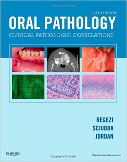
Oral Pathology: Clinical Pathologic Correlations, 6ed
Covering pathologic conditions by clinical appearance, Oral Pathology: Clinical Pathologic Correlations, 6th Edition uses an atlas-style format to help you identify, diagnose, and plan treatment for oral disease presentations. Two-page spreads include clinical photos of common conditions on one side while the facing page lists the central features, causes, and significance of each specific disease. Each chapter is organized by clinical appearance, such as white lesions, red-blue lesions, and cysts of the jaws and neck, and includes full-color photomicrographs and clinical photos to help you identify pathologic elements. This edition adds new coverage of oral cancer and new cone beam CT, regular CT, and MRI images. Expert authors Joseph Regezi, James Sciubba, and Richard Jordan provide a quick reference thatÕs ideal for the lab, NBDE review, or chairside use!
- An atlas-style Clinical Overview section makes it easy to find key information, with one page showing clinical photos of common conditions and the facing page listing symptoms, causes, and significance.
- Organization by clinical appearance — such as including all red-blue lesions in one chapter along with possible causes — matches what you would expect to see upon a patient’s presentation, and provides a practical tool for developing differential diagnoses and planning treatment.
- Hundreds of full-color clinical photographs and radiographs depict specific conditions for easier identification in real clinical scenarios.
- Full-color photomicrographs help you identify pathologic elements and provide correct diagnoses and treatment plans.
- Boxes and tables offer clear, at-a-glance information on the clinical features, diagnosis, and treatment for many conditions.
- Student resources on a companion Evolve website include 30 case studies, a 150-question practice exam, and case-based learning to prepare you for the NBDE and for clinical practice.
- Advanced imaging techniques include images such as CBCTs (cone beam computed tomography), regular CTs, and MRIs, reflecting the transition from a 2-dimensional to a 3-dimensional approach in acquiring data and reconstructing images, and helping you identify and assess disease entities — especially tumors of the salivary glands, jaws, and soft tissues.
- Expanded sections on etiology, treatment, and diagnosis of oral cancer (HPV) describe how subtypes can play an important role in the development and natural history of some head and neck cancers, most notably those arising in the oropharynx.
- Expanded section on bisphosphonate-related osteonecrosis of the jaws helps you effectively manage patients taking bisphosphonates to prevent bone loss, including information on oral complications, risk factors, and benefits.
Contents
۱. Vesiculobullous Diseases
۲. Ulcerative Conditions
۳. White Lesions
۴. Red-Blue Lesions
۵. Pigmented Lesions
۶. Verrucal-Papillary Lesions
۷. Connective Tissue Lesions
۸. Salivary Gland Diseases
۹. Lymphoid Lesions
۱۰. Cysts of the Jaws and Neck
۱۱. Odontogenic Tumors
۱۲. Benign Nonodontogenic Tumors
۱۳. Inflammatory Jaw Lesions
۱۴. Malignancies of the Jaws
۱۵. Metabolic and Genetic Diseases
۱۶. Abnormalities of Teeth
لینک کوتاه : https://bookbaz.ir/?p=14401
نویسنده : Joseph A. Regezi DDS MS , James J. Sciubba DMD PhD
ناشر : Saunders; 6 edition
سال انتشار : 2012
زبان کتاب : انگلیسی
نوع فایل : PDF
تعداد صفحات : 480
(ISBN) شابک : 1455702625
قیمت کتاب درآمازون : $98.45
حجم فایل : 90 MB
کتاب های مرتبط:
 دانلود کتاب اصول پاتوفیزیولوژی بولاک
Principles of Pathophysiology, 2ed
 دانلود کتاب عفونت و التهاب قرنیه
 Corneal Infection and Inflammation, 1ed
 دانلود کتاب پاتولوژی آندروود: یک رویکرد بالینی
Underwood’s Pathology: A Clinical Approach, 7ed
 دانلود کتاب پاتولوژی پستان دابس
Breast Pathology, 3ed
 دانلود کتاب جراحی بینی ساختاری: درس های آموخته شده در ۳۰ سال (۳ جلدی)
Structure Rhinoplasty: Lessons Learned in 30 Years, 1ed
 دانلود کتاب درمان ارتودنسی دندان های نهفته + ویدئو
Orthodontic Treatment of Impacted Teeth, 4ed + Video
 دانلود کتاب تشخیص افتراقی در پاتولوژی جراحی: سیستم گوارشی
 Differential Diagnoses in Surgical Pathology: Gastrointestinal System, 1ed
 دانلود کتاب جراحی فک و صورت بِرنان (۲ جلدی، ویرایش ۲۰۱۸)
Maxillofacial Surgery: 2-Volume Set, 3ed
 دانلود کتاب اصول اندودنتیکس
 The Principles of Endodontics, 3ed
 دانلود کتاب بیماری مادرزادی قلب: آنالیز بالینی، پاتولوژیک، جنینی و سگمنتال
Congenital Heart Disease: A Clinical, Pathological, Embryological, and Segmental Analysis, 1ed
 دانلود کتاب حل مشکلات بالینی در پریودنتولوژی و ایمپلنتولوژی
 Clinical Problem Solving in Periodontology and Implantology, 1ed
 دانلود کتاب پریودانتیکس
 Textbook of Periodontics, 1ed
 دانلود کتاب ملزومات پاتولوژی پوست ویدون
Weedon’s Skin Pathology Essentials: Expert Consult, 1ed
 دانلود کتاب مباحث کلی آسیب شناسی دهان بالینی
Clinical Outline of Oral Pathology, 4ed
 دانلود کتاب اصول پاتولوژی کلیه
Fundamentals of Renal Pathology, 2ed
 دانلود کتاب میکروبیولوژی دندانپزشکی اساسی
Essential Microbiology for Dentistry, 5ed
 دانلود کتاب تشخیص افتراقی در پاتولوژی جراحی گتوزو
Gattuso’s Differential Diagnosis in Surgical Pathology, 4ed
 دانلود کتاب مشکلات پزشکی در دندانپزشکی اسکالی
Scully’s Medical Problems in Dentistry, 7ed
 دانلود کتاب پاتولوژی تشخیصی: انکولوژی مولکولی
Diagnostic Pathology: Molecular Oncology, 1ed
 دانلود کتاب پاتولوژی تشخیصی: قفسه سینه
Diagnostic Pathology: Thoracic, 2ed
 دانلود کتاب پاتولوژی تشخیصی: استخوان
Diagnostic Pathology: Bone, 2ed
 دانلود کتاب چسبندگی در زمینه های دارویی، بیومدیکال و دندانپزشکی
Adhesion in Pharmaceutical, Biomedical, and Dental Fields, 1ed
 دانلود کتاب بیولوژی پالپ عاج دندان در دندانپزشکی ترمیمی
Pulp-Dentin Biology in Restorative Dentistry, 1ed
 دانلود کتاب سیتولوژی: اصول تشخیصی و همبستگی بالینی + ویدئو
Cytology: Diagnostic Principles and Clinical Correlates, 5ed + Video
 دانلود کتاب پاتولوژی تشخیصی: درماتوپاتولوژی غیرنئوپلاستیک
Diagnostic Pathology: Nonneoplastic Dermatopathology, 2ed
 دانلود کتاب پاتولوژی تشخیصی: پستان
Diagnostic Pathology: Breast, 2ed
 دانلود کتاب هماتوپاتولوژی
 Hematopathology, 2ed
 دانلود کتاب جراحی پلاستیک غیوران (۲ جلدی)
Plastic Surgery: Indications and Practice, 2-Volume Set, 1ed
 دانلود کتاب پاتولوژی تشخیصی: نوروپاتولوژی
 Diagnostic Pathology: Neuropathology, 2ed
 دانلود کتاب پاتولوژی تشخیصی: کالبد گشایی بیمارستانی
Diagnostic Pathology: Hospital Autopsy, 1ed
 دانلود کتاب انکولوژی عملی سر و گردن
Practical Head and Neck Oncology, 1ed
 دانلود کتاب درماپاتولوژی الستون
Dermatopathology, 3ed
 دانلود کتاب پاتولوژی استخوان و بافت نرم
Bone and Soft Tissue Pathology, 2ed
 دانلود کتاب پاتولوژی و ژنتیک تومورهای غدد اندام درون ریز
Pathology and Genetics of Tumours of Endocrine Organs
 دانلود کتاب پاتولوژی تشخیصی: سر و گردن
Diagnostic Pathology: Head and Neck, 2ed
 دانلود کتاب تشخیص های دشوار در پاتولوژی پستان
 Difficult Diagnoses in Breast Pathology, 1ed
 دانلود کتاب آسیب شناسی مولکولی و تشخیص سرطان (رشد سرطان و پیشرفت)
Molecular Pathology and Diagnostics of Cancer
 دانلود کتاب هماتوپاتولوژی: مبانی در آسیب شناسی تشخیصی
Hematopathology: Foundations in Diagnostic Pathology, 2ed
 دانلود کتاب آسیب شناسی پوست ویدون
Weedon’s Skin Pathology: Expert Consult, 3ed
 دانلود کتاب پیشرفت در پاتولوژی جراحی: سرطان آندومتر
 Advances in Surgical Pathology: Endometrial Carcinoma, 1ed
 دانلود کتاب نوروپاتولوژی جراحی عملی: یک رویکرد تشخیصی
Practical Surgical Neuropathology: A Diagnostic Approach, 2ed
 دانلود کتاب پیوند استخوان سینوس
 The Sinus Bone Graft, 3ed
 دانلود کتاب جراحی پیزوالکتریک استخوان
Piezoelectric Bone Surgery, 1ed
 دانلود کتاب پاتولوژی تشخیصی استخوان
Diagnostic Pathology: Bone, 3ed
 دانلود کتاب رینوپلاستی غیر جراحی
Non-Surgical Rhinoplasty, 1ed
 دانلود کتاب تشخیص افتراقی در پاتولوژی جراحی پستان
Differential Diagnoses in Surgical Pathology: Breast, 1ed
 دانلود کتاب پاتولوژی پایه رابینز و کومار
Robbins & Kumar Basic Pathology, 11ed
 دانلود کتاب جراحی دهان و فک و صورت فونسکا (۳ جلدی)
Fonseca Oral and Maxillofacial Surgery, 3-Volume Set, 2ed
 دانلود کتاب آناتومی بالینی: یک رویکرد مطالعه موردی
Clinical Anatomy: A Case Study Approach, 1ed
 دانلود کتاب جراحی سر، گردن و تیروئید
 Head, Neck and Thyroid Surgery, 1ed
 دانلود کتاب بورد بررسی تخصصی پاتولوژی تشریحی مک هیل
McGraw-Hill Specialty Board Review Anatomic Pathology, 1ed
 دانلود کتاب ملزومات عکاسی دندانپزشکی
 Essentials of Dental Photography, 1ed
 دانلود کتاب آزمایشات بالینی: چشم انداز روش شناختی
Clinical Trials: A Methodologic Perspective, 3ed
 دانلود کتاب راهنمای بالینی و آزمایشگاهی ایمپلنت اباتمنت دندانی
Clinical and Laboratory Manual of Dental Implant Abutments
 دانلود کتاب آناتومی سر و گردن برای دندانپزشکی نتر
Netter’s Head and Neck Anatomy for Dentistry, 3ed
 دانلود کتاب بررسی مورد جراحی پلاستیک: راهنمای مطالعه بورد دهان و دندان
Plastic Surgery Case Review: Oral Board Study Guide, 1ed
 دانلود کتاب راهنمای عمومی پیشگیری و کنترل عفونت در دندانپزشکی
Basic Guide to Infection Prevention and Control in Dentistry, 2ed
 دانلود کتاب معیارهای بالینی مواد دندانی
 Clinical Aspects of Dental Materials, 5ed
 دانلود کتاب پزشکی و رادیولوژی دهان
Oral Medicine and Radiology, 1ed
 دانلود کتاب اطلس پاتولوژی سر و گردن ونیگ
Atlas of Head and Neck Pathology, 4ed
 دانلود کتاب اطلس پزشکی پاتولوژی کلیه
Atlas of Medical Renal Pathology
 دانلود کتاب پاتولوژی پایه بیماری رابینز و کوتران
Robbins & Cotran Pathologic Basis of Disease, 9ed
 دانلود کتاب آسیب شناسی دستگاه گوارش لوین، وینستن و ریدل (۲ جلدی)
Lewin, Weinstein and Riddell’s Gastrointestinal Pathology and its Clinical Implications 2-Vol, 2ed
 دانلود کتاب روش های جراحی اسکلت صورت الیس و زیده
Surgical Approaches to the Facial Skeleton, 2ed
 دانلود کتاب جراحی دهان و فک و صورت پیشرفته (۳ جلدی)
A Textbook of Advanced Oral and Maxillofacial Surgery, 3-Vol, 1ed
 دانلود کتاب مدیریت دندانپزشکی بیمار باردار
Dental Management of the Pregnant Patient, 1ed
 دانلود کتاب روال جراحی اسکلت صورت
 Surgical Approaches to the Facial Skeleton, 3ed
 دانلود کتاب هیستولوژی دهانی تن کیت: توسعه، ساختار و عملکرد
Ten Cate’s Oral Histology: Development, Structure, and Function, 9ed
 دانلود کتاب پاتولوژی تشخیصی آندومتر
Diagnostic Endometrial Pathology, 2ed
 دانلود کتاب اطلس تشخیصی نئوپلازی مزانشیمی پوستی
Diagnostic Atlas of Cutaneous Mesenchymal Neoplasia, 1ed
 دانلود کتاب راهنمای مرجع روزانه دندانپزشکی بالینی
Clinical Dentistry Daily Reference Guide, 1ed
 دانلود کتاب آسیب شناسی دهان و فک و صورت نویل
Neville, Oral and Maxillofacial Pathology, 3ed
 دانلود کتاب آسیب شناسی عملی بافت نرم: روش تشخیصی
Practical Soft Tissue Pathology: A Diagnostic Approach, 1ed
 دانلود کتاب راهنمای عملی اختلالات مصرف مواد و تجویز ایمن آدا
The ADA Practical Guide to Substance Use Disorders and Safe Prescribing
 دانلود کتاب آناتومی سر و گردن برای دندانپزشکی نتر
Netter’s Head and Neck Anatomy for Dentistry, 2ed
 دانلود کتاب دندانپزشکی زیبایی معاصر فریدمن
Contemporary Esthetic Dentistry, 1ed
 دانلود کتاب آسیب شناسی پوست مک کی (۲ جلدی)
McKee’s Pathology of the Skin, 2-Vol, 4ed
 دانلود کتاب اساس پاتولوژیک بیماری رابینز و کوتران، نسخه حرفه ای
Robbins and Cotran Pathologic Basis of Disease, Professional Edition, 9ed
 دانلود کتاب زیبایی صورت: مفاهیم و تشخیص بالینی
Facial Aesthetics: Concepts and Clinical Diagnosis, 1ed
 دانلود کتاب اطلس رنگی تشخیص افتراقی در سیتوپاتولوژی اکسفولیاتیو و آسپیراسیون
 Color Atlas of Differential Diagnosis in Exfoliative and Aspiration Cytopathology, 2ed
 دانلود کتاب راهنمای جراحی دهان و فک و صورت
 Textbook of Oral and Maxillofacial Surgery, 4ed
 دانلود کتاب ایمپلنت های دندانی و پیوند استخوان
Dental Implants and Bone Grafts, 1ed
 دانلود کتاب جراحی پیشرفته دهان و فک و صورت
A Textbook of Advanced Oral and Maxillofacial Surgery
 دانلود کتاب جراحی و انکولوژی سر و گردن جاتین شاه
Jatin Shah’s Head and Neck Surgery and Oncology, 4ed
 دانلود کتاب آسیب شناسی آندروود: یک رویکرد بالینی
Underwood’s Pathology: a Clinical Approach, 6ed
 دانلود مجموعه کتاب های سبز پزشکی تصویری نتر (۹ جلدی)
The Netter Collection of Medical Illustrations (Netter Green Book Collection), 9-Vol
 دانلود کتاب میمیک نئوپلاستیک در درماتوپاتولوژی
Neoplastic Mimics in Dermatopathology, 1ed
 دانلود کتاب نوسازی دهانی: رویکرد مبتنی بر مورد
Oral Rehabilitation: A Case-Based Approach, 1ed
 دانلود کتاب دندانپزشکی جراحی
Textbook of Operative Dentistry, 3ed
 دانلود کتاب نانوساختارها برای پزشکی دهان
 Nanostructures for Oral Medicine, 1ed
 دانلود کتاب پاتولوژی عملی غشا سروزی
Practical Pathology of Serous Membranes, 1ed
 دانلود کتاب پاتولوژی برای مشاغل سلامت
Pathology for the Health Professions, 5ed
 دانلود کتاب دندانپزشکی میکرو تهاجمی: راهبردها و ابزارهای بالینی
Microinvasive Dentistry: Clinical Strategies and Tools, 1ed
 دانلود کتاب راهنمای نوروپاتولوژی عمومی اسکرول و پوریِر
Escourolle & Poirier’s Manual of Basic Neuropathology, 5ed
 دانلود کتاب اطلس رنگی پاتولوژی: اصول پاتولوژی، بیماری های مرتبط، عوارض
Color Atlas of Pathology: Pathologic Principles, Associated Diseases, Sequela, 1ed
 دانلود کتاب راهنمای دندانپزشکی سنتی پیش بالینی
Textbook of Preclinical Conservative Dentistry, 2ed
 دانلود کتاب پزشکی زیبایی: هنر و تکنیک ها
Aesthetic Medicine: Art and Techniques, 2012th Edition
 دانلود کتاب ترمیم ایمپلنت: راهنمای مرحله به مرحله
Implant Restorations: A Step-by-Step Guide, 4ed
 دانلود کتاب کشیک: اصول و پروتکل ها
On Call: Principles and Protocols, 6ed
 دانلود کتاب مولر سوم نهفته
 Impacted Third Molars, 1ed
 دانلود کتاب جراحی اندوسکوپیک گوش
Endoscopic Ear Surgery
 دانلود کتاب اسرار دندانپزشکی
 Dental Secrets, 4ed
 دانلود کتاب اصول و تمرین جراحی و انکولوژی سر و گردن
Principles and Practice of Head and Neck Surgery and Oncology, 2ed
 دانلود کتاب مورد دشوار در جراحی سرطان سر و گردن
The Difficult Case in Head and Neck Cancer Surgery, 1ed
 دانلود کتاب استفاده از منطقه خنثی در پروتز دندانی + ویدئو
Application of the Neutral Zone in Prosthodontics, 1ed + Video
 دانلود کتاب مدیریت شکاف لب و کام دهان: اطلس جامع
Cleft Lip and Palate Management: A Comprehensive Atlas, 1ed
 دانلود کتاب طبقه بندی تومورهای هماتوپویسیس و بافت لنفوئید WHO
WHO Classification of Tumours of Haematopoietic and Lymphoid Tissues, 4ed
 دانلود کتاب اولین کمک برای USMLE مرحله ۳
First Aid for the USMLE Step 3, 5ed
 دانلود کتاب پاتولوژی عصبی عضلانی آسان
Neuromuscular Pathology Made Easy, 1ed
 دانلود کتاب نقد و بررسی سریع آسیب شناسی
Rapid Review Pathology, 4ed
 دانلود کتاب اطلس سیتوپاتولوژی پانکراس به همراه هیستوپاتولوژیک
Atlas of Pancreatic Cytopathology with Histopathologic Correlations, 1ed
 دانلود کتاب ضایعات عروقی سر و گردن: تشخیص و مدیریت
Vascular Lesions of the Head and Neck: Diagnosis and Management, 1ed
 دانلود کتاب پاتولوژی تشخیصی گرینسون: دستگاه گوارش
Diagnostic Pathology: Gastrointestinal, 2ed
 دانلود کتاب پاتولوژی مثانه
 Bladder Pathology, 1ed
 دانلود کتاب اصول پاتولوژی پاتوما ۲۰۱۷ + ویدئو
Fundamentals of Pathology Pathoma 2017 + Video
 دانلود کتاب برنامه درمان سه بعدی مجازی جراحی فک
 ۳D Virtual Treatment Planning of Orthognathic Surgery, 1ed
 دانلود کتاب پریودنتولوژی بالینی نیومن و کارانزا
Newman and Carranza’s Clinical Periodontology, 13ed
 دانلود کتاب تکنیک های بالینی در دندانپزشکی کودکان
 Handbook of Clinical Techniques in Pediatric Dentistry, 1ed
 دانلود کتاب آناتومی، فیزیولوژی، بافت شناسی دهان و مورفولوژی دندان
Textbook of Oral Anatomy, Physiology, Histology and Tooth Morphology, 2ed
 دانلود کتاب لاواژ برونکوآلوئولار در تحقیقات پایه و پزشکی بالینی
Bronchoalveolar Lavage in Basic Research and Clinical Medicine, 1ed
 دانلود کتاب پاتوفیزیولوژی ریوی وست (ویرایش ۲۰۱۷)
West’s Pulmonary Pathophysiology, 9ed
 دانلود کتاب وکسینگ برای دانشجویان دندانپزشکی
Waxing for Dental Students, 1ed
 دانلود کتاب پزشکی در یک نگاه
Medicine at a Glance, 4ed
 دانلود کتاب مبانی برنامه ریزی درمان دندانی
 Fundamentals of Treatment Planning, 1ed
 دانلود کتاب پاتولوژی قلبی عروقی
Cardiovascular Pathology, 5ed
 دانلود کتاب اطلس تشخیص افتراقی در هماتوپاتولوژی نئوپلاستیک
Atlas of Differential Diagnosis in Neoplastic Hematopathology, 3ed
 دانلود کتاب آسیب شناسی: MCQs و مرور سریع
Pathology: Quick Review and MCQs, 4ed
 دانلود کتاب پریودنتولوژی و ایمپلنتولوژی بالینی نیومن و کارانزا
Newman and Carranza’s Clinical Periodontology and Implantology, 14ed
 دانلود کتاب پاتولوژی تشخیصی: ادراری تناسلی
Diagnostic Pathology: Genitourinary, 2ed
 دانلود کتاب فیزیولوژی ضروری برای دانشجویان دندانپزشکی
 Essentials Of Physiology For Dental Students, 2ed
 دانلود کتاب تکنیک ردیفگر دندان شفاف
Clear Aligner Technique, 1ed
 دانلود کتاب پاتولوژی تشخیصی تومورهای بافت نرم
Diagnostic Pathology: Soft Tissue Tumors, 3ed
 دانلود کتاب اطلس جراحی آندوسکوپی سینوس و قاعده جمجمه + ویدئو
Atlas of Endoscopic Sinus and Skull Base Surgery, 2ed + Video
 دانلود کتاب روشهای یکپارچه در جراحی زیبایی صورت + ویدئو
Integrated Procedures in Facial Cosmetic Surgery, 1ed + Video
 دانلود کتاب اطلس پاتولوژی جراحی زنان
 Atlas of Gynecologic Surgical Pathology, 4ed
 دانلود کتاب پاتولوژی پوست: تشخیص با اولین برداشت
Dermatopathology: Diagnosis by First Impression, 4ed
 دانلود کتاب مسیرهای پالپ دندانی کوهن
Cohen’s Pathways of the Pulp Expert Consult, 10ed
 دانلود کتاب نقد و بررسی تمام موضوعات دندانپزشکی
 Review of All Dental Subjects (ROADS), 1ed
 دانلود کتاب مرجع تست تشخیصی و آزمایشگاهی موزبی
Mosby’s Diagnostic and Laboratory Test Reference, 15ed
 دانلود کتاب جراحی ارتوگناتیک: اصول، برنامه ریزی و تمرین
Orthognathic Surgery: Principles, Planning and Practice, 1ed
 دانلود کتاب دندانپزشکی کم تهاجمی Supra-Gingival: رویکرد سالمتر برای ترمیم زیبایی
Supra-Gingival Minimally Invasive Dentistry: A Healthier Approach to Esthetic Restorations, 1ed
 دانلود کتاب برنامه درمانی ایمپلنت و پروتکل های بالینی مبتنی بر شواهد
Evidence-based Implant Treatment Planning and Clinical Protocols, 1ed
 دانلود کتاب اطلس رنگی بیماریهای دهان و فک و صورت نویل
Color Atlas of Oral and Maxillofacial Diseases, 1ed
 دانلود کتاب مهارت های دندانپزشکی پیش بالینی در یک نگاه
Pre-Clinical Dental Skills at a Glance, 1ed
 دانلود کتاب راهنمای پاتولوژی دهان و پزشکی دهان
Handbook of Oral Pathology and Oral Medicine, 1ed
دانلود کتاب پریودنتولوژی و ایمپلنتولوژی بالینی نیومن و کارانزا + ویدئوNewman and Carranza’s Clinical Periodontology and Implantology, 14ed + Video
 دانلود کتاب مبانی جراحی فک و صورت فرارو
Ferraro’s Fundamentals of Maxillofacial Surgery, 2ed
 دانلود کتاب آناتومی اجمالی مصور لیپینکات: سر و گردن
Lippincott Concise Illustrated Anatomy: Head & Neck, 3ed
 دانلود کتاب اصلاح نامرتبی دندان: درمان و استحکام
Open-Bite Malocclusion: Treatment and Stability, 1ed
 دانلود کتاب مهارت در جراحی زیبایی بینی: اطلس تکنیک های جراحی + ویدئو
Mastering Rhinoplasty: A Comprehensive Atlas of Surgical Techniques with Integrated Video Clips, 2ed