
دانلود کتاب MRI استخوان و تومورهای بافت نرم و ضایعات تومور مانند: تشخیص افتراقی و اطلس
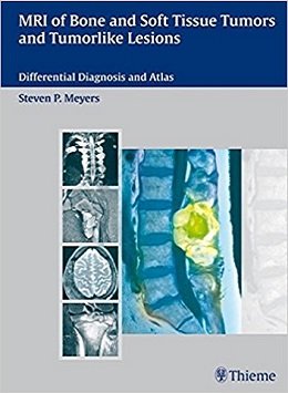
MRI of Bone and Soft Tissue Tumors and Tumorlike Lesions: Differential Diagnosis and Atlas, 1ed
Practical. In-depth. Invaluable.
A guide to the diagnosis of tumors and tumorlike lesions of bone and soft tissue using MRI.
This unique encyclopedic guide takes the same approach you apply in clinical practice. It features fully illustrated differential diagnosis tables organized according to MRI findings and the locations of tumors. An in-depth reference section provides information on each lesion. In addition, almost 3000 high quality images make this practical text an invaluable tool in the diagnosis of common and rare tumors and other disorders of the musculoskeletal system.
Features:
- ۲۰ differential diagnosis tables based on anatomic locations of lesions rather than disease
- Fully illustrated reference chapters containing concise, detailed information for each lesion – from relative frequency and age ranges to MRI findings, treatment, and prognosis
- Over 2900 state-of-the-art illustrations covering the wide range of imaging features for various lesions
- An exceptional level of detail, helping you to differentiate between diseases and conditions that have similar appearances
- Extensive cross-referencing to further up-to-the minute resources
This is the definitive guide to MRI of musculoskeletal tumors. Whether you need a practical guide for day-to-day use or a comprehensive preparation tool for board examinations – keep this text close to the workstation.
Review
Contents
۱ Tumors and tumorlike lesions involving the skull and facial bones
۲ Tumors and tumorlike lesions involving the spine
۳ Paraspinal tumors and tumorlike lesions
۴ Lesions involving the outer surface of bone
۵ Lesions associated with thickening of bone cortex
۶ Intramedullary lesions associated with expansion of intact cortical margins
۷ Intramedullary lesions associated with cortical destruction and extraosseous extension
۸ Solitary intramedullary lesions with well-circumscribed margins
۹ Solitary intramedullary lesions with poorly defined margins of abnormal marrow signal
۱۰ Solitary intramedullary lesions located near the ends of tubular bones
۱۱ Solitary intramedullary metadiaphyseal lesions
۱۲ Solitary intramedullary diaphyseal lesions
۱۳ Osseous tumors and tumorlike lesions at the hands and feet
۱۴ Diffuse,, multiple, poorly defined and/or multifocal zones of abnormal marrow signal
۱۵ Lesions that contain cartilage
۱۶ Tumors and tumorlike lesions within joints
۱۷ Solitary tumors and tumorlike lesions of the soft tissues located mostly deep to the subcutaneous fat
۱۸ Tumors and tumorlike lesions of the superficial soft tissues including subcutaneous fat
۱۹ Lesions involving peripheral nervous tissue
۲۰ Lesions that contain fat
A1 Adamantinoma (Also Referred to as ExtragnathicAdamantinoma, Adamantinoma of Long Bones,Juvenile Intracortical Adamantinoma)
A2 Aneurysmal Bone Cyst
A3 Bone Cyst (Also Referred to as Simple Bone Cyst, Unicameral Bone Cyst, Solitary Bone Cyst)
A4 Angiofibroma (Also Referred to as Juvenile Nasopharyngeal Angiofibroma)
A5 Angiomatoid Fibrous Histiocytoma (Also Referred to as Angiomatoid Malignant Fibrous Histiocytoma)
A6 Angiosarcoma (Also Referred to as Malignant Hemangioendothelioma, High-Grade Hemangioendothelioma, Hemangiosarcoma, Angioendothelioma, Angiofibrosarcoma, and Hemangioendotheliosarcoma)
A7 Chondroblastoma
A8 Chondroma, Intramedullary Type: Enchondroma (Also Referred to as Intra-osseous Chondroma or Central Chondroma)
A9 Chondroma, Periosteal or Juxtacortical Type (Also Referred to as Periosteal or Juxtacortical Chondroma, Surface Chondroma, and Juxtacortical/Pamsteal Chondroma)
A10 Chondromyxoid Fibroma
A11 Chondrosarcoma
A12 Chordoma
A13 Dermatofibrosarcoma and Dermatofibrosarcoma Protuberans
A14 Dermatomyositis
A15 Dermoid and Epidermoid
A16 Desmoid Tumor (Also Referred to as Fibromatosis, Superficial and Deeo Tvoes Involvino Soft Tissues: Desmoplastic Fibroma: Desmoid Tumor within Bone!
A17 Elastofibroma
A18 Eosinophilic Granuloma (Also Referred to as Langerhans Cell Histiocytosis, Formerly Histiocytosis X)
A19 Erdheim-Chester Disease (Also Referred to as Chester-Erdheim Disease, Lipoid Granulomatosis, and Lipogranulomatosis)
A20 Ewing Sarcoma
A21 Nodular Fasciitis (Also Referred to as Pseudosarcomatous Fasciitis, Proliferative Fasciitis, Infiltrative Fasciitis, and Pseudosarcomatous Fibromatosis)
A22 Fibrolipomatous Hamartoma (Also Referred to as Nerve Lipoma, Neural Fibrolipoma, Lipofibromatous Hamartoma, Perineural Lipoma, Intraneural Lipoma, Lipofibroma, and Lipomatous Hamartoma)
A23 Fibroma of the Tendon Sheath
A24 Solitary Fibrous Tumor
A25 Fibrosarcoma
A26 Fibrous Cortical Defect and Nonossifying Fibroma (Also Referred to as Metaphyseal Fibrous Defect), Cortical Desmoid (Also Referred to as Periostea! Desmoid or Distal Femoral Cortical Irregularity), and Fibroxanthoma
A27 Fibrous Dysplasia (Also Referred to as Fibro- osseous Dysplasia, Fibrocartilaginous Dysplasia, and Lichtenstein-Jaffe Disease)
A28 Geode (Also Referred to as Subchondral Cyst and Osteoarthritic Cyst); Soft Tissue and Intra-osseous Ganglion (Also Referred to as Intra-osseous Ganglion, Juxta-articular Bone Cyst, and Periosteal Ganglion)
A29 Giant Cell Tumor of Bone
A30 Giant Cell Tumor of the Tendon Sheath and/or Soft Tissue (Also Referred to as Nodular Tenosynovitis, Fibrous Xanthoma, Tenosynovial Giant Cell Tumor, and Benign Synovioma)
A31 Glomus Tumor (Also Referred to as Glomangioma and Angioglomoid Tumor)
A32 Gout
A33 Hemangioendothelioma (Also Referred to as Low-grade Hemangioendothelioma, Low-grade Hemangioendothelial Sarcoma, Low-grade Angiosarcoma, and Myxoid Angio blastoma)
A34 Hemangiomas (Also Referred to as Vascular Hamartomas)
A35 Hemangiopericytoma
A36 Hematoma, More1-Lavaliee Lesion, and Hemophilic Pseudotumor
A37 Bone and Muscle Infarct (Also Referred to as Avascular Necrosis and Osteonecrosis)
A38 Kaposi Sarcoma (also Referred to as Angiosarcoma Multiplex, Kaposi Disease, Idiopathic Multiple Pigmented Sarcoma of the Skin, and Granuloma Multiplex Hemorrhagicum)
A39 Leiomyoma
A40 Leiomyosarcoma
A41 Leukemia
A42 Lipoblastoma
A43 Lipoma, Atypical Lipoma, and Hibernoma
A44 Liposarcoma
A45 Liposclerosing Myxofibrous Tumor (Also Referred to as Polymorphic Fibro- osseous Lesion of Bone and Polymorphic Fibrocystic Disease of Bone)
A46 Lymphangioma (Also Referred to as Cystic Hygroma)
A47 Lymphoma
A48 Malignant Fibrous Histiocytoma (Also Referred to as Malignant Histiocytoma, Xanthosarcoma. Malignant Fibrous Xanthoma, and Fibroxanthosarcoma]
A49 Meningioma
A50 Metastatic Lesions
A51 Morton Neuroma
A52 Multiple Myeloma (Also Referred to as Myeloma, Kahler Disease, and Plasma Cell Neoplasm; Plasmacytoma Represents a Solitary Neoplastic Variant)
A53 Myositis Ossificans (Also Referred to as Heterotopic Ossification, Reactive Mesenchymal Proliferation, Ossifying Hematoma, and Pseudomalignant Osseous Tumor of Soft Tissues)
A54 Myxoma
A55 Neuroblastoma, Ganglioneuroblastoma, and Ganglioneuroma
A56 Neurofibroma and Malignant Peripheral Nerve Sheath Tumor
A57 Traumatic Neuroma
A58 Osteochondroma (Also Referred to as Osteocartilaginous Exostosis)
A59 Osteofibrous Dysplasia (Also Referred to as Ossifying Fibroma
A60 Osteoid Osteoma
A61 Osteoblastoma (Also Referred to as Ossifying Giant Cell Tumor and Giant Osteoid Osteoma)
A62 Osteoma, Enostosis, Osteopoikilosis, and Melorheostosis
A63 Osteomyelitis
A64 Osteosarcoma (Also Referred to as Osteogenic Sarcoma and Osteoblastic Sarcoma)
A65 Paget Disease (Also Referred to as Osteitis Deformans)
A66 Paraganglioma (Also Referred to as Chemodectoma for Carotid Body Tumor and Glomus Jugulare Tumor for Jugulotympanic Paragangliomas)
A67 Pigmented Villonodular Synovitis
A68 Pleomorphic Hyalinizing Angiectatic Tumor
A69 Rhabdomyosarcoma (Also Referred to as Myosarcoma, Malignant Rhabdomyoma, Rhabdosarcoma, and Embryonal Sarcoma]
A70 Rheumatoid Arthritis
A71 Sarcoid
A72 Schwannoma (Also Referred to as Neurilemoma and Neurinoma)
A73 Synovial Chondromatosis (Also Referred to as Primary and Secondary Chondromatosis, and Synovial Chondrometaplasia), and Synovial Osteochondromatosis
A74 Synovial Cyst
A75 Synovial Sarcoma (Also Referred to as Carcinosarcoma and Spindle Cell Carcinoma of Soft Tissue)
A76 Teratoma
A77 Xanthoma
لینک کوتاه : https://bookbaz.ir/?p=58332
نویسنده : Steven Meyers
ناشر : TPS; 1 edition
سال انتشار : 2009
زبان کتاب : انگلیسی
نوع فایل : PDF
تعداد صفحات : 816
(ISBN) شابک : 3131354216
قیمت کتاب درآمازون : $170.35
حجم فایل : 100 MB
کتاب های مرتبط:
 دانلود کتاب بورد و بخش برای USMLE مراحل ۲ و ۳
Boards & Wards for USMLE Steps 2 & 3, 5ed
 دانلود کتاب روشهای اسکلتی عضلانی هدایت شده با سونوگرافی در پزشکی ورزشی
Ultrasound Guided Musculoskeletal Procedures in Sports Medicine, 1ed
 دانلود کتاب پردازش تصویربرداری پزشکی: تکنیک های پیشرفته تئوری فازی
Medical Image Processing: Advanced Fuzzy Set Theoretic Techniques, 1ed
 دانلود کتاب ملزومات رادیولوژی اسکلتی یاچم و رو (۲ جلدی) + CD
Yochum and Rowe’s Essentials of Skeletal Radiology, 2-Vol, 3ed + CD
 دانلود کتاب MRI کمی مغز: اصول اندازه گیری فیزیکی
Quantitative MRI of the Brain: Principles of Physical Measurement, 2ed
 دانلود کتاب تشخیص افتراقی در تصویربرداری عصبی: ستون فقرات
Differential Diagnosis in Neuroimaging: Spine, 1ed
 دانلود کتاب سرطان پستان: تشخیص زودرس با ماموگرافی
Breast Cancer: Early Detection with Mammography: Casting Type Calcifications Sign of a Subtype with Deceptive Features, 1ed
 دانلود کتاب تشخیص و درمان زود هنگام سرطان (۵ جلدی)
Early Diagnosis and Treatment of Cancer Series, 5-Vol, 1ed
 دانلود کتاب راهنمای روش های رادیولوژی چپمن و ناکیِلنی
Chapman & Nakielny’s Guide to Radiological Procedures, 6ed
 دانلود کتاب آدنوکارسینوما ریوی: رویکردهای درمانی
Pulmonary Adenocarcinoma: Approaches to Treatment, 1ed
 دانلود کتاب مرور بورد پزشکی هسته ای: سوال و جواب های خود ارزیابی
Nuclear Medicine Board Review: Questions and Answers for Self-Assessment, 3ed
 دانلود کتاب FRCR نهایی: یادداشت های بازبینی کامل
The Final FRCR: Complete Revision Notes, 1ed
 دانلود کتاب تشخیص افتراقی در پاتولوژی جراحی پستان
Differential Diagnoses in Surgical Pathology: Breast, 1ed
 دانلود کتاب سونوگرافی در ICU: برنامه های کاربردی عملی
Ultrasonography in the ICU: Practical Applications, 2015th
 دانلود کتاب تصویربرداری قفسه سینه (سری تشخیص مستقیم در رادیولوژی)
Thoracic Imaging (Dx-Direct), 1ed
 دانلود کتاب اطلس تشخیص افتراقی در هماتوپاتولوژی نئوپلاستیک
Atlas of Differential Diagnosis in Neoplastic Hematopathology, 4ed
 دانلود کتاب بررسی معاینه سونوگرافی: فیزیک، شکم، زنان و زایمان
Sonography Exam Review: Physics, Abdomen, Obstetrics and Gynecology, 2ed
 دانلود کتاب تومور و سرطان: سر – گردن – قلب – ریه – گلو
Tumors and Cancers: Head – Neck – Heart – Lung – Gut, 1ed
 دانلود کتاب تومور و سرطان: پوست – بافت نرم – استخوان – اوروژنیتال
Tumors and Cancers: Skin – Soft Tissue – Bone – Urogenitals, 1ed
 دانلود کتاب آناتومی تصویربرداری: ریه ها، مدیاستن و قلب (جلد ۱)
Imaging Anatomy: Text and Atlas Volume 1, Lungs, Mediastinum, and Heart, 1ed
 دانلود کتاب مشاور بالینی فری ۲۰۱۶ (راه حل های پزشکی فری)
Ferri’s Clinical Advisor 2016: 5 Books in 1, 1ed
 دانلود کتاب پرتودرمانی عملی: فیزیک و تجهیزات
Practical Radiotherapy: Physics and Equipment, 3ed
 دانلود کتاب اصول تشخیص مولکولی و شخصی پزشکی سرطان
Principles of Molecular Diagnostics and Personalized Cancer Medicine, 1ed
 دانلود کتاب اصول انکولوژی انجمن سرطان آمریکا
The American Cancer Society’s Principles of Oncology, 1ed
 دانلود کتاب راهنمای انکولوژی پزشکی MD اندرسون
The MD Anderson Manual of Medical Oncology, 4ed
 دانلود کتاب پزشکی هسته ای: روش شناسی و کاربردهای بالینی
Nuclear Medicine Textbook: Methodology and Clinical Applications, 1ed
 دانلود کتاب غربالگری سرطان پستان
Breast Cancer Screening, 1ed
 دانلود کتاب راهنمای اولیه برای رادیوگرافی دندان
 Basic Guide to Dental Radiography, 1ed
 دانلود کتاب سونوگرافی مراقبت اورژانسی
Emergency Point-of-Care Ultrasound, 2ed
 دانلود کتاب تصویربرداری تشخیصی: دهان و فک و صورت
Diagnostic Imaging: Oral and Maxillofacial, 2ed
 دانلود کتاب طبقه بندی سیگنال مغزی EEG برای تشخیص اختلال تشنج صرع
 EEG Brain Signal Classification for Epileptic Seizure Disorder Detection, 1ed
 دانلود کتاب پزشکی دقیق در انکولوژی
 Precision Medicine in Oncology, 1ed
 دانلود کتاب حل مشکلات در انکولوژی حاد
Problem Solving in Acute Oncology
 دانلود کتاب جراحی آندوسکوپی سینوس: آناتومی، بازسازی سه بعدی و روش جراحی + ویدئو
Endoscopic Sinus Surgery: Anatomy, Three-Dimensional Reconstruction, and Surgical Technique, 3ed + Videos
 دانلود کتاب اصول و عمل مراقبت تسکین دهنده و آنکولوژی حمایتی
Principles and Practice of Palliative Care and Supportive Oncology, 4ed
 دانلود کتاب مراقبت از بیمار در رادیوگرافی: همراه با معرفی تصویربرداری پزشکی
Patient Care in Radiography: With an Introduction to Medical Imaging, 9ed
 دانلود کتاب راهنمای جیبی رادیوگرافی مریل
Merrill’s Pocket Guide to Radiography, 14ed
 دانلود کتاب اولتراسوند تشخیصی عروقی
Diagnostic Ultrasound: Vascular, 1ed
 دانلود کتاب راهنمای OCT شبکیه: توموگرافی انسجام نوری
Handbook of Retinal OCT: Optical Coherence Tomography, 2ed
 دانلود کتاب طبقه بندی تومورهای پستان WHO
WHO Classification of Tumours of the Breast, 4ed
 دانلود کتاب تصویربرداری دستگاه گوارش (سری تشخیص مستقیم در رادیولوژی)
Gastrointestinal Imaging (Direct Diagnosis in Radiology: DX-Direct!), 1ed
 دانلود کتاب چشم انداز محاسباتی و پردازش تصویری پزشکی
 Computational Vision and Medical Image Processing V: VipIMAGE 2015
 دانلود کتاب رادیوگرافی PREP (مرور برنامه و آمادگی آزمون)
Radiography PREP (Program Review and Exam Preparation), 8ed
 دانلود کتاب سونوگرافی در کمک به تولید مثل و اوایل بارداری
Ultrasound in Assisted Reproduction and Early Pregnancy, 1ed
 دانلود کتاب تصویربرداری تشخیصی کودکان کافِی (۲ جلدی)
Caffey’s Pediatric Diagnostic Imaging, 2-Vol, 12ed
 دانلود کتاب سونوگرافی تشخیصی: شکم و لگن
 Diagnostic Ultrasound: Abdomen and Pelvis, 1ed
 دانلود کتاب اطلس آناتومی مقطعی، جلد ۳: ستون فقرات، پایانگاه، مفاصل: توموگرافی کامپیوتری و تصویربرداری رزونانس مغناطیسی
Pocket Atlas of Sectional Anatomy, Volume III: Spine, Extremities, Joints: Computed Tomography and Magnetic Resonance Imaging, 2ed
 دانلود کتاب راهنمای مبتدیان برای درمان های سرطانی هدفمند
A Beginner’s Guide to Targeted Cancer Treatments, 1ed
 دانلود کتاب پاتولوژی تشخیصی تومورهای بافت نرم
Diagnostic Pathology: Soft Tissue Tumors, 3ed
 دانلود کتاب اطلس آموزشی ماموگرافی
 Teaching Atlas of Mammography, 4ed
 دانلود کتاب تصویربرداری پستان (سری تشخیص مستقیم در رادیولوژی)
Breast Imaging (Direct Diagnosis in Radiology: DX-Direct!), 1ed
 دانلود کتاب تفسیر تصاویر اسکلتی عضلانی: آناتومی، آسیب شناسی و گزارش اورژانس
Interpreting Musculoskeletal Images: Anatomy, Pathology and Emergency Reporting, 1ed
 دانلود کتاب تصویربرداری قلبی (سری تشخیص مستقیم در رادیولوژی)
Cardiac Imaging (Direct Diagnosis in Radiology: DX-Direct!), 1ed
 دانلود کتاب اطلس رنگی آناتومی سونوگرافی بلاک
Color Atlas of Ultrasound Anatomy, 2ed
 دانلود کتاب مدیریت سرطان پستان، یک موضوع کلینیک های جراحی
 Management of Breast Cancer, An Issue of Surgical Clinics, 1ed
 دانلود کتاب تصویربرداری تشخیصی کودکان
Diagnostic Imaging: Pediatrics, 4ed
 دانلود کتاب سونوگرافی داپلر رنگی در زنان و زایمان
Color Doppler Sonography in Gynecology and Obstetrics, 1ed
 دانلود کتاب پاتولوژی تشخیصی: گره های لنفاوی و لنفوم های خارج گره ای
Diagnostic Pathology: Lymph Nodes and Extranodal Lymphomas, 2ed
 دانلود کتاب تخریب تومور از راه پوست: راهکارها و تکنیک ها
Percutaneous Tumor Ablation: Strategies and Techniques, 1ed
 دانلود کتاب تصویربرداری رزونانس مغناطیسی (۳ جلدی)
Magnetic Resonance Imaging Handbook (MRI), 3-Vol, 1ed
 دانلود کتاب طبقه بندی تومورهای اندام های تولید مثل زن WHO
WHO Classification of Tumours of the Female Reproductive Organs, 4ed
 دانلود کتاب رادیوتراپی والتر و میلر
Walter and Miller’s Textbook of Radiotherapy, 8ed
 دانلود کتاب پاتولوژی و ژنتیک تومورهای غدد اندام درون ریز
Pathology and Genetics of Tumours of Endocrine Organs
 دانلود کتاب آسیب شناسی مولکولی و تشخیص سرطان (رشد سرطان و پیشرفت)
Molecular Pathology and Diagnostics of Cancer
 دانلود کتاب تفسیر رادیوگرافی قفسه سینه در بیماران قلبی کودک
Chest Radiographic Interpretation in Pediatric Cardiac Patients, 1ed
 دانلود کتاب اطلس سونوگرافی اسکلتی عضلانی دست و پاها
Atlas of Musculoskeletal Ultrasound of the Extremities, 1ed
 دانلود کتاب اولتراساوند عمومی و عروقی: مرور موردی
General and Vascular Ultrasound: Case Review, 3ed
 دانلود کتاب CT و MRI از شکم و لگن: فایل آموزشی
CT & MRI of the Abdomen and Pelvis: A Teaching File, 3ed
 دانلود کتاب مرور بورد انکولوژی: راهنمای مطالعه و پرسش و پاسخ
Oncology Board Review: Blueprint Study Guide and Q&A, 2ed
 دانلود کتاب نوروبلاستوما: مداخلات مکانیسم های مولکولی و درمانی
Neuroblastoma: Molecular Mechanisms and Therapeutic Interventions, 1ed
 دانلود کتاب ماموگرافی عملی MR: امآرآی با وضوح بالا پستان
Practical MR Mammography: High-Resolution MRI of the Breast, 2ed
 دانلود کتاب مدیریت سکته مغزی ایسکمیک
Ischemic Stroke Management, 1ed
 دانلود کتاب متاستاز مغز: تصویربرداری عصبی پیشرفته
Brain Metastases: Advanced Neuroimaging, 1ed
 دانلود کتاب تغییرات شریانی در انسان: مرجع کلیدی رادیولوژیست ها و جراحان
Arterial Variations in Humans: Key Reference for Radiologists and Surgeons, 1ed
 دانلود کتاب سرطان پستان: تشخیص زودرس با ماموگرافی
Breast Cancer: Early Detection with Mammography: Crushed Stone-like Calcifications: The Most Frequent Malignant Type, 1ed
 دانلود کتاب رادیوتراپی و رادیوبیولوژی بالینی سرطان سر و گردن
Radiotherapy and Clinical Radiobiology of Head and Neck Cancer, 1ed
 دانلود کتاب رادیولوژی ۱۰۱: مبانی و اصول تصویربرداری
Radiology 101: The Basics & Fundamentals of Imaging, 4ed
 دانلود کتاب تصویربرداری پستان: ضروریات در رادیولوژی
Breast Imaging: The Requisites (Requisites in Radiology), 2ed
 دانلود کتاب سرطان پستان: هنر و علم تشخیص زود هنگام با ماموگرافی
Breast Cancer: The Art and Science of Early Detection with Mammography, 1ed
 دانلود کتاب اصول تصویربرداری کودکان
 Fundamentals of Pediatric Imaging, 2ed
 دانلود کتاب ملزومات عملی شدت مدوله پرتو درمانی
Practical Essentials of Intensity Modulated Radiation Therapy, 3ed
 دانلود کتاب تومورهای استخوانی دورفمن و زرنیاک
Dorfman and Czerniak’s Bone Tumors, 2ed
 دانلود کتاب میکروسکوپی کانفوکال بازتابی تومورهای پوستی
Reflectance Confocal Microscopy of Cutaneous Tumors, 2ed
 دانلود کتاب آنتی بیوتیک و شیمی درمانی فینچ
Antibiotic and Chemotherapy: Expert Consult, 9ed
 دانلود کتاب پوکی استخوان مارکوس و فلدمن (۲ جلدی)
Osteoporosis, 2-Volume Set, Fourth Edition
 دانلود کتاب اطلس آسیب شناسی بافت نرم و استخوان: با بافت شناسی، سیتولوژی و رادیولوژی
Atlas of Soft Tissue and Bone Pathology: With Histologic, Cytologic, and Radiologic Correlations, 1ed
 دانلود کتاب ایمونوهیستوشیمی تشخیصی: ترانوستیک و برنامه های ژنومی
Diagnostic Immunohistochemistry: Theranostic and Genomic Applications, 4ed
 دانلود کتاب تومورهای استخوان و بافت نرم: بررسی چند رشته با ارائه مورد
Bone and Soft Tissue Tumours: A Multidisciplinary Review With Case Presentations, 1ed
 دانلود کتاب سرطان سر و گردن: درمان، توانبخشی و نتایج
Head and Neck Cancer: Treatment, Rehabilitation, and Outcomes, 2ed
 دانلود کتاب تکنیک های پیشرفته در انتقال تصویر جراحی مغز و ستون فقرات
 Advanced Techniques in Image-Guided Brain and Spine Surgery, 1ed
 دانلود کتاب اطلس تعیین سن اسکلت: MRI دست و مچ دست در کودکان
Text-Atlas of Skeletal Age Determination: MRI of the Hand and Wrist in Children, 1ed
 دانلود کتاب تصویربرداری تشخیصی نقایص مادرزادی قلب
Diagnostic Imaging of Congenital Heart Defects, 1ed
 دانلود کتاب جراحی انکولوژی کودکان
Pediatric Surgical Oncology, 1ed
 دانلود کتاب ۳ تفاوت برتر در پزشکی هسته ای
 Top 3 Differentials in Nuclear Medicine, 1ed
 دانلود کتاب تصویربرداری تشخیصی: بیماری اسکلتی عضلانی غیر تروماتیک + ویدئو
Diagnostic Imaging: Musculoskeletal Non-Traumatic Disease, 3ed + Video
 دانلود کتاب تفسیر نمونه برداری از ضایعات کودکان
Biopsy Interpretation of Pediatric Lesions
 دانلود کتاب راهنمای اکوکاردیوگرافی: همگام با کتاب اکوکاردیوگرافی بالینی اوتو
Echocardiography Review Guide: Companion to the Textbook of Clinical Echocardiography, 3ed
 دانلود کتاب هماتولوژی و هماتو-انکولوژی کودکان
Textbook of Pediatric Hematology and Hemato-Oncology, 1ed
 دانلود کتاب MRI اسکلتی عضلانی آسیف سفیودین
Asif Saifuddin’s Musculoskeletal MRI, 2ed
 دانلود کتاب اطلس رنگی سرطان دهان
 Color Atlas of Oral Cancer, 1ed
 دانلود کتاب نورورادیولوژی: تشخیص های افتراقی کلیدی و سوالات بالینی
Neuroradiology: Key Differential Diagnoses and Clinical Questions, 2ed
 دانلود کتاب خط مرزی و عادی اولیه پاتولوژی فین فریشمیت کوهلر/زیمر
Freyschmidt’s ,Koehler/Zimmer, Borderlands of Normal and Early Pathological Fin, 5ed
 دانلود کتاب تصویربرداری در گوش و حلق و بینی
Imaging in Otolaryngology, 1ed
 دانلود کتاب تصویربرداری انکولوژیک: یک رویکرد چند رشته ای
Oncologic Imaging: A Multidisciplinary Approach, 2ed
 دانلود کتاب رادیولوژی حوادث و اورژانس: راهنمای بقا
Accident and Emergency Radiology: A Survival Guide, 3ed
 دانلود کتاب اندام فوقانی کودکان
 The Pediatric Upper Extremity, 2015th
 دانلود کتاب آسیب شناسی مزوتلیوما بدخیم
Pathology of Malignant Mesothelioma
 دانلود کتاب تکنیک های اصلی در جراحی: جراحی قفسه سینه: پیوند، تراشه برداشتن، تومور مدیاستن، برش گسترده قفسه سینه
Master Techniques in Surgery: Thoracic Surgery: Transplantation, Tracheal Resections, Mediastinal Tumors, Extended Thoracic Resections
 دانلود کتاب ژن درمانی سرطان توسط وکتورهای ویروسی و غیر ویروسی
Cancer Gene Therapy by Viral and Non-viral Vectors, 1ed
 دانلود کتاب تصویربرداری سر و گردن مقدماتی
Introductory Head and Neck Imaging, 1ed
 دانلود کتاب سونوگرافی تشخیصی: سر و گردن
Diagnostic Ultrasound: Head and Neck, 1ed
 دانلود کتاب اطلس جیبی اکوکاردیوگرافی
 Pocket Atlas of Echocardiography, 2ed
 دانلود کتاب راهنمای گام به گام اولتراسوند شکمی
 Abdominal Ultrasound: Step by Step, 3ed
 دانلود کتاب نقشه برداری مغز: نشانه ها و تکنیک ها + ویدئو
Brain Mapping: Indications and Techniques, 1ed + Video
 دانلود کتاب روش های کلارک در تصویربرداری تشخیصی
 Clark’s Procedures in Diagnostic Imaging, 1ed
 دانلود کتاب اطلس آندوسکوپی تشخیصی
Atlas of Diagnostic Endoscopy, 3ed
 دانلود کتاب سونوگرافی تشخیصی روماک (۲ جلدی)
Diagnostic Ultrasound, 2-Volume Set 6th Edition
 دانلود کتاب اصول رادیولوژی دهان و فک و صورت
Fundamentals of Oral and Maxillofacial Radiology, 1ed
 دانلود کتاب آنالیز تصویر رادیوگرافی
 Radiographic Image Analysis, 4ed
 دانلود کتاب جراحی عمومی انکولوژی پیچیده
Textbook of Complex General Surgical Oncology, 1ed
 دانلود کتاب اشعه ایکس اسکلتی عضلانی برای دانشجویان و کارآموزان پزشکی
Musculoskeletal X-Rays for Medical Students and Trainees, 1ed
 دانلود کتاب تعویض ترنس کاتتر دریچه آئورت
Transcatheter Aortic Valve Implantation, 1ed
 دانلود کتاب فارماکولوژی سرطان: راهنمای مصور داروهای ضد سرطان
Cancer Pharmacology: An Illustrated Manual of Anticancer Drugs, 1ed
 دانلود کتاب جراحی قفسه سینه بزرگسالان شوگربیکر
Sugarbaker’ Adult Chest Surgery, 2ed
 دانلود کتاب توموگرافی پرتو مخروطی کامپیوتری در ارتودنسی: موارد مصرف، بینش و نوآوری
Cone Beam Computed Tomography in Orthodontics: Indications, Insights, and Innovations, 1ed
 دانلود کتاب پاتولوژی نئوپلاستیک گوارشی
Neoplastic Gastrointestinal Pathology, 1ed
 دانلود کتاب سونوگرافی عروقی عملی: راهنمای مصور
Practical Vascular Ultrasound: An Illustrated Guide, 1ed
 دانلود کتاب تصویربرداری شکم: الزامات اصلی
Abdominal Imaging: The Core Requisites, 1ed
 دانلود کتاب پرتو درمانی والتر و میلر: فیزیک پرتو، درمان و انکولوژی
Walter and Miller’s Textbook of Radiotherapy: Radiation Physics, Therapy and Oncology, 7ed
 دانلود کتاب اطلس رادیولوژی اورژانس: سیستم عروقی، قفسه سینه، شکم و لگن و سیستم تولید مثل
Atlas of Emergency Radiology: Vascular System, Chest, Abdomen and Pelvis, and Reproductive System 2015th
 دانلود کتاب آندوسونوگرافی + ویدئو
Endosonography, 3ed + Video
 دانلود کتاب پیشگیری و غربالگری سرطان
 Cancer Prevention and Screening, 1ed
 دانلود کتاب سونوگرافی کودکان
 Pediatric Ultrasound, 1ed
 دانلود کتاب توانبخشی سرطان ریه
Lung Cancer Rehabilitation, 1ed
 دانلود کتاب هماتولوژی و انکولوژی نوزادی و کودکی ناتان و اوسکی (۲ جلدی)
Nathan and Oski’s Hematology and Oncology of Infancy and Childhood 2Vol, 8ed
 دانلود کتاب سونوگرافی تشخیصی: راهنما و اطلس مقدماتی
Endoscopic Ultrasound: An Introductory Manual and Atlas, 2ed
 دانلود کتاب ساختار سر و گردن ویکر
Structures of the Head and Neck, 1ed
 دانلود کتاب مداخلات در پزشکی ریه
 Interventions in Pulmonary Medicine, 2ed
 دانلود کتاب اطلس بیهوشی منطقه ای هدایت اولتراسوند
Atlas of Ultrasound-Guided Regional Anesthesia, 3ed
 دانلود کتاب تصویربرداری تشخیصی پستان
Diagnostic Imaging: Breast, 3ed
 دانلود کتاب فناوری اولتراسوند برای پزشکان بالینی
Ultrasound Technology for Clinical Practitioners, 1ed
 دانلود کتاب ۱۰۱ راه حل MRI مغز
 ۱۰۱MRI Brain Solutions, 1ed
 دانلود کتاب رادیولوژی پا و مچ پا
Foot and Ankle Radiology, 2ed
 دانلود کتاب فیوژن ژنها و سرطان
 Fusion Genes and Cancer, 1ed
 دانلود کتاب تصویربرداری رزونانس مغناطیسی: اصول فیزیکی و زیست شناختی
Magnetic Resonance Imaging: Physical and Biological Principles, 4ed
 دانلود کتاب راهنمای مراحل سرطان AJCC (ویرایش ۲۰۱۷)
AJCC Cancer Staging Manual, 8ed
 دانلود کتاب اطلس جیبی آناتومی مقطعی: سر و گردن
Pocket Atlas of Sectional Anatomy: Head and Neck, 3ed
دانلود کتاب سونوگرافی مراقبت اورژانسی + ویدئوEmergency Point-of-Care Ultrasound, 2ed + Video
 دانلود کتاب اطلس آموزشی تصویربرداری شکم
Teaching Atlas of Abdominal Imaging, 1ed
 دانلود کتاب موارد رادیولوژی مغز و اعصاب
Neuroradiology Cases