
دانلود کتاب اطلس سر، گردن و مغز روتون: تصاویر دو بعدی و سه بعدی
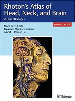
Rhoton’s Atlas of Head, Neck, and Brain: 2D and 3D Images, 1ed
Masterful 2D and 3D head, neck, and brain dissections provide unsurpassed insights into head, neck, and brain anatomy
An internationally renowned and beloved author, educator, brain anatomist, and neurosurgeon, Professor Albert Rhoton has a special place in medical history. He was revered by students and colleagues and is regarded as one of the fathers of modern microscopic neurosurgery. A driving principle in his anatomy lab was the simple phrase, “Every Second.” This was embraced in his philosophy that every second of every day, a patient’s life was improved by a surgeon assisted by the anatomic knowledge his lab helped elucidate and distribute.
Rhoton’s Atlas of Head, Neck, and Brain is the visually exquisite crowning achievement of Dr. Rhoton’s brilliant career and unwavering dedication to the intertwined pursuits of surgical anatomy and neurosurgery. The atlas reflects the unparalleled contributions Dr. Rhoton made to the contemporary understanding of neurosurgical anatomy. Dr. Peris-Celda, with the collaboration of an impressive cadre of international multidisciplinary experts, worked closely under Dr. Rhoton’s tutelage on this project. This book is the culmination of 5 years of work and experience gleaned from more than 40 years of surgical anatomy research and exquisite dissection techniques performed in Dr. Rhoton’s laboratory.
Special Features
- Each anatomic dissection meticulously labeled with English and Latin descriptors for easy cross referencing with other resources.
- Multiple views of the most complex regions of the head, neck, and brain provide a deeper understanding of anatomy.
- More than 600 anatomical images systematically organized in four major sections: Osteology of the Head and Neck; Face and Neck; Ear, Nose, Pharynx, Larynx, and Orbit; and Neuroanatomy and Cranial Base.
- Superb 2D images presented in a large printed format to optimize the viewing experience.
- ۳D digital images fully realize the beauty of the dissections and enhance the learning process.
- Specimens injected with colored silicone provide better visualization of arteries and veins.
Breathtakingly stunning, this atlas is certain to be a treasured reference for medical students, residents, and clinicians specializing in neurosurgery, facial plastic surgery, otolaryngology, maxillofacial surgery, and craniofacial surgery for many years to come.
Contents
۱ Adult and Fetal Skull
۲ Bones of the Skull and Skull Bone Articulations
۳ Cervical Vertebrae
۴ Face: Superficial Dissection
۵ Face: Deep Dissection
۶ Anterior Aspect of the Neck: Superfcial Dissection
۷ Anterior Aspect of the Neck: Deep Dissection
۸ Posterior Aspect of the Neck
۹ Parapharyngeal Dissection
۱۰ External and Middle Ear
۱۱ Internal Ear
۱۲ Nose: Sagittal Dissection
۱۳ Nose: Coronal Dissection
۱۴ Endonasal Endoscopy
۱۵ Pharynx
۱۶ Larynx
۱۷ Orbit
۱۸ Eye and Orbital Contents
۱۹ Internal Structure of the Eyeball
۲۰ Cerebrum
۲۱ Cerebellum and Brainstem
۲۲ Brain, Meninges, and Sutures
۲۳ Cerebrovascular and Intraventricular Dissection
۲۴ Cranial Base and CraniocervicaI Junction
۲۵ Sagittal and Endoscopic Dissection of the Cranial Base
۲۶ Cranial Nerves
۲۷ Brain Sections
۲۸ Fiber Dissection of the Brain
جهت مشاهده نسخه همراه با مجموعه تصاویر کامل کتاب کلیک کنید
لینک کوتاه : https://bookbaz.ir/?p=70923
نویسنده : Maria Peris-Celda , Francisco Martinez-Soriano
ناشر : Thieme; 1 edition
سال انتشار : 2018
زبان کتاب : انگلیسی
نوع فایل : PDF (نسخه اصلی)
تعداد صفحات : 648
(ISBN) شابک : 1604069007
قیمت کتاب درآمازون : $299.99
حجم فایل : 155 MB
کتاب های مرتبط:
 دانلود کتاب پزشکی گوش و حلق و بینی و جراحی سر و گردن اسکات-براون (۳ جلدی)
Scott-Brown’s Otorhinolaryngology and Head and Neck Surgery, 3-Vol, 8ed
 دانلود کتاب تصویربرداری در گوش و حلق و بینی
Imaging in Otolaryngology, 1ed
 دانلود کتاب اختلالات خواب در نورولوژی: یک رویکرد عملی
Sleep Disorders in Neurology: A Practical Approach, 2ed
 دانلود کتاب جراحی رباتیک سر و گردن: اطلس تشریحی و جراحی
Robotic Head and Neck Surgery: An Anatomical and Surgical Atlas, 1ed
 دانلود کتاب تکنیک های اصلی در جراحی سر و گردن: تیروئید، پاراتیروئید، غدد بزاقی، پارانازال
Master Techniques in Otolaryngology – Head and Neck Surgery: Thyroid, Parathyroid, Salivary Glands, Paranasal, 1ed
 دانلود کتاب راهنمای پایندگی چشم پزشکی عصبی + ویدئو
The Neuro-Ophthalmology Survival Guide, 2ed + Video
 دانلود کتاب راهنمای پزشکی هریسون
Harrisons Manual of Medicine, 19ed
 دانلود کتاب به دام افتادن اعصاب محیطی: تشخیص و مدیریت بالینی
Peripheral Nerve Entrapments: Clinical Diagnosis and Management, 1ed
 دانلود کتاب جوانسازی سنتروفشیال + ویدئو
Centrofacial Rejuvenation, 1ed + Video
 دانلودکتاب آناتومی بالینی ستون فقرات، نخاع و ANS
Clinical Anatomy of the Spine, Spinal Cord, and ANS, 3ed
 دانلود کتاب تکنیک های اصلی در گوش و حلق و بینی – جراحی سر و گردن: حنجره، هایپوفارنکس، اوروفارنکس، دهان و گردن
Master Techniques in Otolaryngology – Head and Neck Surgery: Larynx, Hypopharynx, Oropharynx, Oral Cavity and Neck, 1ed
 دانلود کتاب آناتومی جراحی سر و گردن پرویز جانفزا
Janfaza Surgical Anatomy of the Head and Neck, 1ed
 دانلود کتاب جراحی مغز و اعصاب گرینبرگ
Handbook of Neurosurgery, 9ed
 دانلود کتاب تصمیم گیری در نورولوژی بزرگسالان + ویدئو
Decision-Making in Adult Neurology, 1ed + Video
 دانلود کتاب تکنیک های اصلی در جراحی زیبایی بینی عزیز زاده
Master Techniques in Rhinoplasty, 1ed
 دانلود کتاب دوره های جراحی مغز و اعصاب: پرسش و پاسخ
Neurosurgery Rounds: Questions and Answers
 دانلود کتاب تشخیص افتراقی در تصویربرداری عصبی: ستون فقرات
Differential Diagnosis in Neuroimaging: Spine, 1ed
 دانلود کتاب مرور موردی جراحی مغز و اعصاب: سؤالات و پاسخها
Neurosurgery Case Review: Questions and Answers, 2ed
 دانلود کتاب تسلط بر عکاسی پزشکی سر و گردن
Mastering Medical Photography of the Head and Neck, 1ed
 دانلود کتاب نوروپاتی محیطی: رویکرد عملی
Peripheral Neuropathies: A Practical Approach, 1ed
 دانلود کتاب مرور جراحی مغز و اعصاب: برای استفاده روزانه بالینی و آماده سازی بورد شفاهی
Neurosurgical Review: For Daily Clinical Use and Oral Board Preparation, 1ed
 دانلود کتاب جامع گوش و حلق و بینی ساتالوف: حنجره شناسی (جلد ۴)
Sataloff’s Comprehensive Textbook of Otolaryngology: Laryngology, Vol-4, 1ed
دانلود کتاب تسلط بر عکاسی پزشکی سر و گردن + ویدئوMastering Medical Photography of the Head and Neck, 1ed + Video
 دانلود کتاب نوروتوکسین بوتولینوم برای اختلالات سر و گردن
Botulinum Neurotoxin for Head and Neck Disorders, 2ed
 دانلود کتاب فلپ های موضعی در بازسازی سر و گردن جکسون
Jackson’s Local Flaps in Head and Neck Reconstruction, 3ed
 دانلود کتاب رادیولوژی بیماری های عفونی و التهابی: مغز و نخاع (جلد ۱)
Radiology of Infectious and Inflammatory Diseases – Volume 1: Brain and Spinal Cord, 1ed
 دانلود کتاب پزشکی گوش و حلق و بینی عملی: جراحی سر و گردن (۲ جلدی)
Operative Otolaryngology: Head and Neck Surgery, 2-Vol, 3ed
 دانلود کتاب پزشکی گوش و حلق و بینی و جراحی سر و گردن اسکات-براون: جراحی سر و گردن، جراحی پلاستیک (جلد ۳)
Scott-Brown’s Otorhinolaryngology and Head and Neck Surgery: Head and Neck Surgery, Plastic Surgery, Vol-3, 8ed
 دانلود کتاب پزشکی گوش و حلق و بینی کامینگز: جراحی سر و گردن (۳ جلدی)
Cummings Otolaryngology: Head and Neck Surgery, 3-Vol, 7ed
 دانلود کتاب جراحی محل اتصال مهره جمجمه + ویدئو
Surgery of the Craniovertebral Junction, 2ed + Videos
 دانلود کتاب درمان سلولی برای آسیب مغز و اعصاب
Cellular Therapy for Neurological Injury, 1ed
 دانلود کتاب آناتومی عصبی عروقی در نورورادیولوژی مداخله ای
Neurovascular Anatomy in Interventional Neuroradiology: A Case-Based Approach, 1ed
 دانلود کتاب مغز انسان در عکس ها و نمودارها
The Human Brain in Photographs and Diagrams, 4ed
 دانلود کتاب توانبخشی سکته مغزی: رویکرد مبتنی بر عملکرد
Stroke Rehabilitation: A Function-Based Approach, 4ed
 دانلود کتاب بینش هایی در مورد نورولوژی بالینی
Insights into Clinical Neurology, 1ed
 دانلود کتاب اطلس ویدیویی پایش نوروفیزیولوژیک در جراحی تومورهای نفوذی مغز
Video Atlas of Neurophysiological Monitoring in Surgery of Infiltrating Brain Tumors, 1ed
 دانلود کتاب حفاظت مغز در اسکیزوفرنی ، اختلالات خلقی و شناختی
Brain Protection in Schizophrenia, Mood and Cognitive Disorders
 دانلود کتاب پزشکی گوش و حلق و بینی بالنگر: جراحی سر و گردن
Ballenger’s Otorhinolaryngology: Head and Neck Surgery, 18ed
 دانلود کتاب جراحی ستون فقرات حداقل تهاجمی + ویدئو
Minimally Invasive Spine Surgery, 1ed + Video
 دانلود کتاب نورولوژی کودک و فراتر از آن
Child Neurology and Beyond 2023
 دانلود کتاب اندوسکوپی ترانس نازال قاعده جمجمه و جراحی مغز
Transnasal Endoscopic Skull Base and Brain Surgery, 1ed
 دانلود کتاب مغز و اعصاب بالینی هانکی
Clinical Neurology Hankey, 1ed
 دانلود کتاب سرطان سر و گردن: درمان، توانبخشی و نتایج
Head and Neck Cancer: Treatment, Rehabilitation, and Outcomes, 2ed
 دانلود کتاب ابزار دقیق ستون فقرات: تکنیک های جراحی
Spinal Instrumentation: Surgical Techniques, 1ed
 دانلود کتاب تکنیک های پیشرفته در انتقال تصویر جراحی مغز و ستون فقرات
 Advanced Techniques in Image-Guided Brain and Spine Surgery, 1ed
 دانلود کتاب الکترومیوگرافی و اختلالات عصبی عضلانی
Electromyography and Neuromuscular Disorders, 4ed
 دانلود کتاب تفسیر نوار مغز
 Handbook of EEG Interpretation, 3ed
 دانلود کتاب چشم پزشکی عصبی لیو، والپ و گالتا: تشخیص و مدیریت + ویدئو
Liu, Volpe, and Galetta’s Neuro-Ophthalmology: Diagnosis and Management, 3ed + Video
 دانلود کتاب جراحی آندوسکوپی کرانیوسینوستوز
Endoscopic Craniosynostosis Surgery, 1ed
 دانلود کتاب اطلس عمل جراحی مغز و اعصاب (۸ جلدی)
AANS’ Neurosurgical Operative Atlas, 8-Vol
 دانلود کتاب بازسازی عروق شریانی سر و گردن
Arterial Revascularization of the Head and Neck, 1ed
 دانلود کتاب سری کارشناسی ستون فقرات AO: ناهنجاری های ستون فقرات کودکان (جلد ۹)
AOSpine Masters Series, Volume 9: Pediatric Spinal Deformities, 1ed
 دانلود کتاب نورورادیولوژی: تشخیص های افتراقی کلیدی و سوالات بالینی
Neuroradiology: Key Differential Diagnoses and Clinical Questions, 2ed
 دانلود کتاب تصویربرداری سر و گردن مقدماتی
Introductory Head and Neck Imaging, 1ed
 دانلود کتاب مغز و اعصاب شناختی و زوال عقل آکسفورد
Oxford Textbook of Cognitive Neurology and Dementia, 1ed
 دانلود کتاب اپیدوروسکوپی: اطلس روشها
Epiduroscopy: Atlas of Procedures, 1ed
 دانلود کتاب پزشکی گوش و حلق و بینی کودکان کامینگز
Cummings Pediatric Otolaryngology, 1ed
 دانلود کتاب ضروریات بیهوشی اعصاب و مراقبت ویژه اعصاب گوپتا و گلب
Gupta and Gelb’s Essentials of Neuroanesthesia and Neurointensive Care, 2ed
 دانلود کتاب اختلالات مرتبط با ورزش: تشخیص و مدیریت
Sports-Related Concussion: Diagnosis and Management, 2ed
 دانلود کتاب نقشه برداری مغز: نشانه ها و تکنیک ها + ویدئو
Brain Mapping: Indications and Techniques, 1ed + Video
دانلود کتاب الکترومیوگرافی و اختلالات عصبی عضلانی + ویدئوElectromyography and Neuromuscular Disorders, 4ed + Video
 دانلود کتاب سکته مغزی: پاتوفیزیولوژی، تشخیص و مدیریت
Stroke: Pathophysiology, Diagnosis, and Management, 7ed
 دانلود کتاب نورولوژی نتر
Netter’s Neurology, 3ed
 دانلود کتاب اسکولیوز ایدیوپاتیک
 Idiopathic Scoliosis, 2ed
 دانلود کتاب جراحی پلاستیک – اصول و عمل + ویدئو
Plastic Surgery – Principles and Practice, 1ed + Video
 دانلود کتاب راهنمای نوروپاتولوژی عمومی اسکرول و پوریِر
Escourolle & Poirier’s Manual of Basic Neuropathology, 5ed
 دانلود کتاب استادی جراحی زیبایی بینی: اطلس جامع تکنیک های جراحی
Mastering Rhinoplasty: A Comprehensive Atlas of Surgical Techniques, 2ed
 دانلود کتاب علوم اعصاب شناختی سیستم های بشری: کار و زندگی روزمره
Cognitive Neuroscience of Human Systems: Work and Everyday Life
 دانلود کتاب کالبدشناسی اعصاب بالینی گری: اساس آناتومی علوم اعصاب بالینی
Gray’s Clinical Neuroanatomy: The Anatomic Basis for Clinical Neuroscience, 1ed
 دانلود کتاب غددشناسی عصبی رفتاری
Behavioral Neuroendocrinology, 1ed
 دانلود کتاب تکنیک های تسلط در جوان سازی صورت عزیز زاده + ویدئو
Master Techniques in Facial Rejuvenation, 2ed + Video
 دانلود کتاب بیومکانیک ستون فقرات: مفاهیم اساسی، اختلالات نخاعی و درمان
Biomechanics of the Spine: Basic Concepts, Spinal Disorders and Treatments, 1ed
 دانلود کتاب ۱۰۱ راه حل MRI مغز
 ۱۰۱MRI Brain Solutions, 1ed
 دانلود کتاب اطلس ضایعات سلار، سوپراسلار و پاراسلار
Atlas of Sellar, Suprasellar, and Parasellar Lesions, 1ed
 دانلود کتاب مغز و اعصاب بالینی لانگه (امینف، نسخه اصلی)
Aminoff Clinical Neurology, 8ed
 دانلود کتاب اطلس عصب صورت و ساختارهای مرتبط + آلبوم تصویری
Atlas of the Facial Nerve and Related Structures, 1ed + Image Album
 دانلود کتاب مراقبت زوال عقل: رویکرد عملی
Dementia Care: A Practical Approach, 1ed
 دانلود کتاب جراحی زاویه سربلوپونتین
 Surgery of the Cerebellopontine Angle, 1ed
 دانلود کتاب رویکردهای بیولوژیکی برای ترمیم و بازسازی دیسک ستون فقرات برای پزشکان
Biological Approaches to Spinal Disc Repair and Regeneration for Clinicians, 1ed
 دانلود کتاب اطلس رنگی جراحی ساقه مغز
 Color Atlas of Brainstem Surgery, 1ed
 دانلود کتاب علوم اعصاب رفتاری
Behavioral Neuroscience, 8ed
 دانلود کتاب از دست دادن حافظه، آلزایمر و دمانس: راهنمای عملی برای پزشکان + ویدئو
Memory Loss, Alzheimer’s Disease, and Dementia: A Practical Guide for Clinicians, 2ed + Video
 دانلود کتاب ناهنجاری های شریانی وریدی داخل جمجمه: موارد ضروری برای بیماران و پزشکان
Intracranial Arteriovenous Malformations: Essentials for Patients and Practitioners, 1ed
 دانلود کتاب اطلس جراحی آندوسکوپیک حنجره
Atlas of Endoscopic Laryngeal Surgery, 1ed
 دانلود کتاب سری کارشناسی ستون فقرات AO: تغییر شکل ستون فقرات بزرگسالان (جلد ۴)
AOSpine Masters Series, Volume 4: Adult Spinal Deformities, 1ed
 دانلود کتاب راهنمای درمان فیزیکی ستون فقرات اولسون
Manual Physical Therapy of the Spine, 2ed
 دانلود کتاب اطلس میکروجراحی قاعده جمجمه جانبی
Atlas of Microsurgery of the Lateral Skull Base, 2ed
 دانلود کتاب اطلس آناتومی زوبوتا: سر، گردن و نوروآناتومی (جلد ۳)
Sobotta Atlas of Anatomy: Head, Neck and Neuroanatomy, Vol-3, 16ed
 دانلود کتاب یک رویکرد تشریحی برای جراحی ستون فقرات کم تهاجمی
An Anatomic Approach to Minimally Invasive Spine Surgery, 2ed
 دانلود کتاب جراحی مغز و اعصاب کودکان کوهن
Pediatric Neurosurgery: Tricks of the Trade, 1ed
 دانلود کتاب اصول علوم اعصاب برای علوم و اختلالات ارتباطاتی
Neuroscience Fundamentals for Communication Sciences and Disorders, 1ed
 دانلود کتاب معاینه عصبی دژانگ
DeJong’s The Neurologic Examination, 8ed
 دانلود کتاب جراحی پلاستیک چشم قسمت فوقانی صورت + ویدئو
Ophthalmic Plastic Surgery of the Upper Face, 1ed + Video
 دانلود کتاب انکولوژی سر و گردن: راهنمای مختصر
Head and Neck Oncology: A Concise Guide, 1ed
 دانلود کتاب نورولوژی بالینی کودکان فنیچل: رویکرد علائم و نشانه ها
Fenichel’s Clinical Pediatric Neurology: A Signs and Symptoms Approach, 8ed
 دانلود کتاب تب مغزی: چگونه واکسن ها از مننژیت و سایر بیماری های کشنده جلوگیری می کنند
Brain Fever: How Vaccines Prevent Meningitis And Other Killer Diseases, 1ed
 دانلود کتاب ضایعات زبان: چالش های تشخیصی و راهبردهای درمانی
Tongue Lesions: Diagnostic Challenges and Therapeutic Strategies, 1ed
 دانلود کتاب مانیتورینگ مراقبت حاد مغز و اعصاب
Neurocritical Care Monitoring, 1ed
 دانلود کتاب پزشکی گوش و حلق و بینی علائم محور: جراحی سر و گردن و حنجره شناسی (۳ جلدی)
Symptom Oriented Otolaryngology – Head and Neck Surgery and Laryngology, 3-Vol, 1ed
 دانلود کتاب راهنمای بالینی عصب شناسی
 Neurology: A Clinical Handbook, 1ed
 دانلود کتاب پایه آناتومیک عصب شناسی تشخیصی
Anatomic Basis of Neurologic Diagnosis, 1ed
 دانلود کتاب مغز و اعصاب بین المللی
International Neurology, 2ed
 دانلود کتاب راهنمای ابزار جراحی مغز و اعصاب
 The Neurosurgical Instrument Guide, 1ed
 دانلود کتاب آنژیوگرافی مغزی کاربردی: آناتومی و پاتولوژی عروقی نرمال
Applied Cerebral Angiography: Normal Anatomy and Vascular Pathology, 1ed
 دانلود کتاب اطلس مدیریت چند رشته ای بیماری های حدقه ای
Interdisciplinary Management of Orbital Diseases: Textbook and Atlas, 1ed
 دانلود کتاب مغز انسان در عکس ها و نمودارها نولته
Nolte’s The Human Brain in Photographs and Diagrams, 5ed
 دانلود کتاب جراحی قاعده جمجمه جانبی: اطلس کلینیک هاوس
Lateral Skull Base Surgery: The House Clinic Atlas, 1ed
 دانلود کتاب مغز و اعصاب بالینی امینوف
Lange Clinical Neurology, 10ed
 دانلود کتاب فلپ موضعی در بازسازی صورت بِیکر + ویدئو
Local Flaps in Facial Reconstruction, 3ed + Video
 دانلود کتاب بیوپسی عضلانی: یک رویکرد عملی
Muscle Biopsy: A Practical Approach, 5ed
 دانلود کتاب موارد رادیولوژی مغز و اعصاب
Neuroradiology Cases
 دانلود کتاب اطلس ویدئویی روشهای نورواندوواسکولار
 Video Atlas of Neuroendovascular Procedures, 1ed
 دانلود کتاب اطلس ویدئویی جراحی ستون فقرات
 Video Atlas of Spine Surgery, 1ed
 دانلود کتاب اطلس جراحی تیروئید: اصول، تمرین و موارد بالینی
Atlas of Thyroid Surgery: Principles, Practice and Clinical Cases, 1ed
 دانلود کتاب اطلس ویدئویی جراحی آنوریسم داخل جمجمه
Video Atlas of Intracranial Aneurysm Surgery, 1ed
 دانلود کتاب بیماری صرع کودکان پیلاک: تشخیص و درمان
Pellock’s Pediatric Epilepsy: Diagnosis and Therapy, 4ed
 دانلود کتاب تصویربرداری استخوان گیجگاهی
 Imaging of the Temporal Bone, 4ed
 دانلود کتاب تکنیک های جراحی مغز و اعصاب اشمیدک و سویت (۲ جلدی) + ویدئو
Schmidek and Sweet Operative Neurosurgical Techniques, 2-Vol, 7ed + Video
 دانلود کتاب عصب شناسی، روانپزشکی و اختلالات سیستمیک مرتبط اجمالی
Synopsis of Neurology, Psychiatry and Related Systemic Disorders, 1ed
 دانلود کتاب مرواریدهای پزشکی خواب + ویدئو
Sleep Medicine Pearls, 3ed + Video
 دانلود کتاب هنر جراحی زیبایی نهایی: اصول و فنون (۳ جلدی) + ویدئو
The Art of Aesthetic Surgery: Principles and Techniques, 3-Vol, 2ed + Video
 دانلود کتاب نورومدولاسیون خلاقانه
Innovative Neuromodulation, 1ed
 دانلود کتاب مجموعه تصاویر پزشکی نتر: سیستم عصبی (جلد هفتم، ۲ جلدی)
The Netter Collection of Medical Illustrations: Nervous System Package, 2ed
 دانلود کتاب اطلس جیبی آناتومی مقطعی: سر و گردن، توموگرافی کامپیوتری و تصویربرداری رزونانس مغناطیسی
Pocket Atlas of Sectional Anatomy: Head and Neck, Computed Tomography and Magnetic Resonance Imaging, 4ed
 دانلود کتاب بورد و بخش برای USMLE مراحل ۲ و ۳
Boards & Wards for USMLE Steps 2 & 3, 5ed
 دانلود کتاب تمرین مراقبت نورولوژیک
 The Practice of Neurocritical Care, 1ed
 دانلود کتاب علوم اعصاب رفتاری: ضروریات و فراتر از آن
Behavioral Neuroscience: Essentials and Beyond, 1ed
 دانلود کتاب مجموعه تصاویر پزشکی نتر: سیستم اسکلتی عضلانی (جلد ششم، ۳ جلدی)
The Netter Collection of Medical Illustrations: Musculoskeletal System Package, 2ed
 دانلود کتاب الکترودیاگنوز در مغز و اعصاب بالینی امینف
Aminoff’s Electrodiagnosis in Clinical Neurology, 6ed
 دانلود کتاب مراقبت سکته مغزی حاد
Acute Stroke Care, 3ed
 دانلود کتاب جراحی آندوسکوپی سینوس: آناتومی، بازسازی سه بعدی و تکنیک جراحی
Endoscopic Sinus Surgery: Anatomy, Three-Dimensional Reconstruction, and Surgical Technique, 4ed
 دانلود کتاب سرگیجه و عدم تعادل: راهنمای عملی برای تشخیص و مدیریت + ویدئو
Vertigo and Disequilibrium: A Practical Guide to Diagnosis and Management, 2ed + Video
 دانلود کتاب تعادل ساژیتال ستون فقرات: از عادی به پاتولوژی
Sagittal Balance of the Spine: From Normal to Pathology, 1ed
 دانلود کتاب تصویربرداری از مغز با MRI و CT: رویکرد الگوی تصویری
Brain Imaging with MRI and CT: An Image Pattern Approach, 1ed
 دانلود کتاب رویکرد موردی برای رینوپلاستی ساختار باز
A Case Approach to Open Structure Rhinoplasty, 1ed
 دانلود کتاب اطلس توپوگرافی و پاتوتوپوگرافی آناتومی سر و گردن
Atlas of Topographical and Pathotopographical Anatomy of the Head and Neck, 1ed
 دانلود کتاب اطلس عمل جراحی مغز و اعصاب: جراحی مغز و اعصاب کاربردی
Neurosurgical Operative Atlas: Functional Neurosurgery, 3ed
 دانلود کتاب راهنمای سم شناسی عصبی رشد
Handbook of Developmental Neurotoxicology, 2ed
 دانلود کتاب اطلس سر، گردن و مغز روتون: تصاویر دو بعدی و سه بعدی + تصاویر
Rhoton’s Atlas of Head, Neck, and Brain: 2D and 3D Images, 1ed + Images
 دانلود کتاب مغز و اعصاب کودکان
 Handbook of Pediatric Neurology, 1ed
 دانلود کتاب سری کارشناس ستون فقرات AO: تومورهای نخاعی اولیه (جلد ۲)
AOSpine Masters Series Volume 2: Primary Spinal Tumors, 1ed
 دانلود کتاب تغییر شکل ستون فقرات: ملزومات
Spinal Deformities: The Essentials, 2ed
 دانلود کتاب مغز و اعصاب کودکان (ویرایش ۲۰۱۷)
Pediatric Neurology, 2ed
دانلود کتاب نقشه برداری مغز: نشانه ها و تکنیک هاBrain Mapping: Indications and Techniques, 1ed
 دانلود کتاب جراحی ترانس نازال آندوسکوپیک قاعده جمجمه و مغز
Transnasal Endoscopic Skull Base and Brain Surgery, 2ed
 دانلود کتاب اطلس علوم اعصاب نتر + ویدئو
Netter’s Atlas of Neuroscience, 3ed + Video
 دانلود کتاب سیستم عصبی یکپارچه: روش تشخیص سیستماتیک مبتنی بر مورد
The Integrated Nervous System: A Systematic Diagnostic Case-Based Approach, 2ed
 دانلود کتاب آسیب های مغزی تروماتیک: روش های ارزیابی بالینی و قانونی اعصاب روان
Traumatic Brain Injury: Methods for Clinical and Forensic Neuropsychiatric Assessment, 3ed
 دانلود کتاب بازسازی صورت پس از جراحی ماز + ویدئو
Facial Reconstruction After Mohs Surgery, 1ed + Video
 دانلود کتاب معاینه مغز و اعصاب دی مِیِر
DeMyer’s The Neurologic Examination, 7ed