
دانلود کتاب تشریح پاتولوژی جراحی: راهنمای تصویری
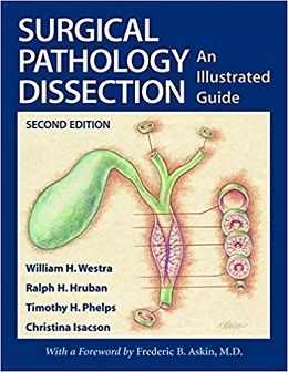
Surgical Pathology Dissection: An Illustrated Guide, 2ed
FROM REVIEWS OF 1E: “Hruban, Westra and Isacson, working with a superb medical illustrator did an admirable job in taking the Johns Hopkins’ gross room manual and translating it into a practical, concise, and easily accessible guide to contemporary practice in the surgical pathology laboratory.” -Modern Path
Our goal in writing the first edition of Surgical Pathology Dissection: An Illustrated Guide was to create a user-friendly, hands-on guide for the dissection of surgical pathology specimens. To do this, we brought a team of surgical pathologists with a broad range of expertise together with Timothy H. Phelps, one of the leading medical illustrators in the United States. In so doing, we believe we created a manual that provides a logical, concise approach to the most commonly encountered specimens. In the years since the first edition was published, Surgical Pathology Dissection: An Illustrated Guide has emerged as the standard in the field, and in 1996, Timothy H. Phelps was awarded the Illustrated Book Award from the Association of Medical Illustrators for his artwork in the book.
We have made a number of significant improvements in the second edition of Surgical Pathology Dissection: An Illustrated Guide. First, new coauthors were asked to join the existing team to add a fresh perspective to key chapters. For example, Elizabeth Montgomery, Robb E. Wilentz, Michael Torbenson, Susan Abraham, E. Rene Rodriguez, and Pedram Argani have helped update key chapters on the digestive system, heart, and breast. Second, new chapters, including chapters on transplantation and sentinel lymph nodes, have been added, reflecting emerging trends in surgical pathology practice. Importantly, these new chapters retain the user-friendly style characteristic of the first edition. Third, new illustrations, including those for dissection of an explanted heart, craniofacial bones, and sentinel lymph nodes, have been added. In addition, a number of the original illustrations, such as for the dissection of breast specimens, have been significantly revised and improved. Fourth, updates to existing chapters, particularly where they were needed to conform to the more recently published College of American Pathologists (CAP) and Association of Directors of Anatomic and Surgical Pathology (ADASP) guidelines, have been made. These changes were made with the goal of keeping Surgical Pathology Dissection: An Illustrated Guide user-friendly and up to date. Each chapter therefore continues to include descriptions and illustrations of the mechanics involved when handling each specimen as well as a conceptual framework for questions to keep in mind during the dissection. At the end of each chapter, the section entitled “Important Issues to Address in Your Pathology Report” helps guide the user to the key information needed to stage most tumors accurately.
Review
Contents
I. General Approach and Techniques
II. Lymph Nodes for Metastatic Tumors
III. The Head and Neck
IV. The Digestive System
V. The Cardiovascular/Respiratory System
VI. Bonef Soft Tissue, and Skin
VII. The Breast
VIII. The Female Genital System
IX. The Urinary Tract and Male Cental System
X. The Ocular System
XI. The Endocrine System
XII. Pediatric Tumors
XIII. The Central Nervous System
XIV. The Hematopoietic and Lymphatic System
XV. Odds and Ends
لینک کوتاه : https://bookbaz.ir/?p=67911
نویسنده : William H. Westra , Ralph H. Hruban
ناشر : Springer; 2nd edition
سال انتشار : 2004
زبان کتاب : انگلیسی
نوع فایل : PDF
تعداد صفحات : 261
(ISBN) شابک : 0387955593
قیمت کتاب درآمازون : $70.74
حجم فایل : 5 MB
کتاب های مرتبط:
 دانلود کتاب تشخیص افتراقی در پاتولوژی جراحی: سیستم گوارشی
 Differential Diagnoses in Surgical Pathology: Gastrointestinal System, 1ed
 دانلود کتاب سیتولوژی: اصول تشخیصی و همبستگی بالینی + ویدئو
Cytology: Diagnostic Principles and Clinical Correlates, 5ed + Video
 دانلود کتاب تشخیص افتراقی در پاتولوژی جراحی پستان
Differential Diagnoses in Surgical Pathology: Breast, 1ed
 دانلود کتاب راهنمای جامع آزمایشگاهی و آزمایشات تشخیصی با مفاهیم پرستاری دیویس
Davis’s Comprehensive Manual of Laboratory and Diagnostic Tests With Nursing Implications, 9ed
 دانلود کتاب انکولوژی عملی سر و گردن
Practical Head and Neck Oncology, 1ed
 دانلود کتاب پیشرفت در پاتولوژی جراحی: سرطان آندومتر
 Advances in Surgical Pathology: Endometrial Carcinoma, 1ed
 دانلود کتاب پاتولوژی و ژنتیک تومورهای سر و گردن
Pathology and Genetics of Head and Neck Tumours
 دانلود کتاب آناتومی بالینی: یک رویکرد مطالعه موردی
Clinical Anatomy: A Case Study Approach, 1ed
 دانلود کتاب اصول پاتولوژی کلیه
Fundamentals of Renal Pathology, 2ed
 دانلود کتاب تشخیص افتراقی در پاتولوژی جراحی گتوزو
Gattuso’s Differential Diagnosis in Surgical Pathology, 4ed
 دانلود کتاب پاتولوژی پستان دابس
Breast Pathology, 3ed
 دانلود کتاب پاتولوژی آندروود: یک رویکرد بالینی
Underwood’s Pathology: A Clinical Approach, 7ed
 دانلود کتاب پاتولوژی پایه رابینز و کومار
Robbins & Kumar Basic Pathology, 11ed
 دانلود کتاب متاژنومیک برای میکروب شناسی
Metagenomics for Microbiology
 دانلود کتاب مباحث کلی آسیب شناسی دهان بالینی
Clinical Outline of Oral Pathology, 4ed
 دانلود کتاب پاتولوژی تشخیصی: انکولوژی مولکولی
Diagnostic Pathology: Molecular Oncology, 1ed
 دانلود کتاب پاتولوژی تشخیصی: قفسه سینه
Diagnostic Pathology: Thoracic, 2ed
 دانلود کتاب پاتولوژی تشخیصی: استخوان
Diagnostic Pathology: Bone, 2ed
 دانلود کتاب پاتولوژی تشخیصی: درماتوپاتولوژی غیرنئوپلاستیک
Diagnostic Pathology: Nonneoplastic Dermatopathology, 2ed
 دانلود کتاب پاتولوژی تشخیصی: پستان
Diagnostic Pathology: Breast, 2ed
 دانلود کتاب هماتوپاتولوژی
 Hematopathology, 2ed
 دانلود کتاب روش های اگزوسیتوز
Exocytosis Methods
 دانلود کتاب پاتولوژی تشخیصی: نوروپاتولوژی
 Diagnostic Pathology: Neuropathology, 2ed
 دانلود کتاب پاتولوژی تشخیصی: کالبد گشایی بیمارستانی
Diagnostic Pathology: Hospital Autopsy, 1ed
 دانلود کتاب درماپاتولوژی الستون
Dermatopathology, 3ed
 دانلود کتاب پاتولوژی استخوان و بافت نرم
Bone and Soft Tissue Pathology, 2ed
 دانلود کتاب اصول پاتوفیزیولوژی بولاک
Principles of Pathophysiology, 2ed
 دانلود کتاب پاتولوژی و ژنتیک تومورهای غدد اندام درون ریز
Pathology and Genetics of Tumours of Endocrine Organs
 دانلود کتاب پاتولوژی تشخیصی: سر و گردن
Diagnostic Pathology: Head and Neck, 2ed
 دانلود کتاب تشخیص های دشوار در پاتولوژی پستان
 Difficult Diagnoses in Breast Pathology, 1ed
 دانلود کتاب عفونت و التهاب قرنیه
 Corneal Infection and Inflammation, 1ed
 دانلود کتاب سیتوپاتولوژی جامع (ویرایش ۲۰۱۵) + ویدئو
Comprehensive Cytopathology, 4ed + Video
 دانلود کتاب آسیب شناسی مولکولی و تشخیص سرطان (رشد سرطان و پیشرفت)
Molecular Pathology and Diagnostics of Cancer
 دانلود کتاب هماتوپاتولوژی: مبانی در آسیب شناسی تشخیصی
Hematopathology: Foundations in Diagnostic Pathology, 2ed
 دانلود کتاب آسیب شناسی پوست ویدون
Weedon’s Skin Pathology: Expert Consult, 3ed
 دانلود کتاب نوروپاتولوژی جراحی عملی: یک رویکرد تشخیصی
Practical Surgical Neuropathology: A Diagnostic Approach, 2ed
 دانلود کتاب پاتولوژی تشخیصی استخوان
Diagnostic Pathology: Bone, 3ed
 دانلود کتاب پاتولوژی بیماری های کبدی کانل
Pathology of Liver Diseases, 1ed
 دانلود کتاب فلبوتومی: یک رویکرد مبتنی بر شایستگی
Phlebotomy: A Competency Based Approach, 5ed
 دانلود کتاب بورد بررسی تخصصی پاتولوژی تشریحی مک هیل
McGraw-Hill Specialty Board Review Anatomic Pathology, 1ed
 دانلود کتاب میکروب شناسی پزشکی جاوتز ملنک و ایدلبرگز (ویرایش ۲۰۱۶)
Jawetz Melnick & Adelbergs Medical Microbiology, 27ed
 دانلود کتاب پاتولوژی سر و گردن
Head and Neck Pathology, 1ed
 دانلود کتاب اطلس تشخیصی نئوپلازی مزانشیمی پوستی
Diagnostic Atlas of Cutaneous Mesenchymal Neoplasia, 1ed
 دانلود کتاب پاتولوژی ملانوسیتیک سطحی
Superficial Melanocytic Pathology, 1ed
 دانلود کتاب مرور پاتولوژی پزشکی قانونی: پرسش و پاسخ
Forensic Pathology Review: Questions and Answers, 1ed
 دانلود کتاب پاتولوژی کالبد شکافی: راهنما و اطلس + ویدئو
Autopsy Pathology: A Manual and Atlas, 3ed + Video
 دانلود کتاب پاتوفیزیولوژی پورت: مفاهیم وضعیت های سلامت تغییر یافته
Porth’s Pathophysiology: Concepts of Altered Health States, 9ed
 دانلود کتاب ملزومات در خون شناسی و آسیب شناسی بالینی
Essentials in Hematology and Clinical Pathology, 1ed
 دانلود کتاب پاتولوژی سم شناسی هاشک و روسو (۳ جلدی)
Haschek and Rousseaux’s Handbook of Toxicologic Pathology, 3-Vol, 4ed
 دانلود کتاب آسیب شناسی دهان و فک و صورت: یک دلیل منطقی برای تشخیص و درمان
Oral and Maxillofacial Pathology: A Rationale for Diagnosis and Treatment, 2ed
 دانلود کتاب درماتوپاتولوژی: مبانی آسیب شناسی تشخیصی
Dermatopathology: Foundations in Diagnostic Pathology, 2ed
 دانلود کتاب موارد در پزشکی بالینی کومار و کلارک
Kumar & Clark’s Cases in Clinical Medicine, 4ed
 دانلود کتاب سیتولوژی آسپیراسیون با سوزن ظریف اورل و استرت
Orell and Sterrett’s Fine Needle Aspiration Cytology, 5ed
 دانلود کتاب پاتولوژی تشخیصی تومورهای بافت نرم
Diagnostic Pathology: Soft Tissue Tumors, 3ed
 دانلود کتاب بخش های منجمد حین عمل جراحی: بروز اشتباه در تشخیص
Intraoperative Frozen Sections: Diagnostic Pitfalls
 دانلود کتاب مرجع تست تشخیصی و آزمایشگاهی موزبی
Mosby’s Diagnostic and Laboratory Test Reference, 15ed
 دانلود کتاب اولین کمک برای USMLE مرحله ۳
First Aid for the USMLE Step 3, 5ed
 دانلود کتاب ضروریات سیتوپاتولوژی تشخیصی
Diagnostic Cytopathology Essentials, 1ed
 دانلود کتاب راهنمای میکروب شناسی بالینی (۲ جلدی، ویرایش ۲۰۱۵)
Manual of Clinical Microbiology, 2-Vol, 11ed
 دانلود کتاب شیمی بالینی، ایمونولوژی و کنترل کیفیت آزمایشگاه
Clinical Chemistry, Immunology and Laboratory Quality Control: A Comprehensive Review for Board Preparation, Certification and Clinical Practice
 دانلود کتاب مدیریت بخش اورژانس اشتراوس و مایر
Strauss and Mayer’s Emergency Department Management, 1ed
 دانلود کتاب پاتو فلش: فلش کارت های پاتوفیزیولوژی
 Patho Phlash!: Pathophysiology Flash Cards, 1ed
 دانلود کتاب ۱۰۰ مورد در پاتولوژی بالینی و پزشکی آزمایشگاهی
۱۰۰Cases in Clinical Pathology and Laboratory Medicine, 2ed
 دانلود کتاب پاتولوژی دهان وو
Oral Pathology, 3ed
 دانلود کتاب تشخیص آزمایشگاهی بیماری ها در آزمایشگاه بالینی
Laboratory Medicine Diagnosis of Disease in Clinical Laboratory, 2ed
 دانلود کتاب طبقه بندی تومورهای پستان WHO
WHO Classification of Tumours of the Breast, 4ed
 دانلود کتاب اختلالات معمول بافت نرم ABC
ABC of Common Soft Tissue Disorders, 1ed
 دانلود کتاب ایمونوهیستوشیمی تشخیصی: کاربردهای ترانوستیک و ژنومیک
Diagnostic Immunohistochemistry: Theranostic and Genomic Applications, 6ed
 دانلود کتاب راهنمای پاتولوژی بالینی آکسفورد
Oxford Handbook of Clinical Pathology, 2ed
 دانلود کتاب هماتوپاتولوژی نئوپلاستیک نولز
Knowles Neoplastic Hematopathology, 3ed
 دانلود کتاب پیشرفت در پاتولوژی جراحی: مزوتلیوما
Advances in Surgical Pathology: Mesothelioma, 1ed
 دانلود کتاب پاتولوژی تشخیصی: پاتولوژی پیوند اعضا
Diagnostic Pathology: Transplant Pathology, 2ed
 دانلود کتاب پاتولوژی جراحی روسای و اکرمن (۲ جلدی، ویرایش ۲۰۱۸)
Rosai and Ackerman’s Surgical Pathology, 2-Vol, 11ed
 دانلود کتاب آسیب شناسی قلبی عروقی عملی
Practical Cardiovascular Pathology, 2ed
 دانلود کتاب پاتولوژی سوء مصرف مواد مخدر کارچ
Karch’s Pathology of Drug Abuse, 5ed
 دانلود کتاب پاتولوژی قفسه سینه عملی: بیماری های ریه، قلب و تیموس
Practical Thoracic Pathology: Diseases of the Lung, Heart, and Thymus, 1ed
 دانلود کتاب اطلس تشخیصی پاتولوژی کلیه
 Diagnostic Atlas of Renal Pathology, 2ed
The Nature of Disease: Pathology for the Health Professions, 2ed
 دانلود کتاب پاتولوژی عملی پستان: روش تشخیصی
Practical Breast Pathology: A Diagnostic Approach, 1ed
 دانلود کتاب پاتولوژی کبدی عملی: یک رویکرد تشخیصی
Practical Hepatic Pathology: A Diagnostic Approach, 2ed
 دانلود کتاب مرور سریع پاتولوژی (ویرایش ۲۰۱۹)
Rapid Review Pathology, 5ed
 دانلود کتاب اطلس پاتولوژی آناتومیک با تصویربرداری
Atlas of Anatomic Pathology with Imaging
 دانلود کتاب اصول پاتولوژی سم شناسی
 Fundamentals of Toxicologic Pathology, 3ed
 دانلود کتاب سارکوما بافت نرم: رویکرد مبتنی بر الگو برای تشخیص
Soft Tissue Sarcomas: A Pattern-Based Approach to Diagnosis, 1ed
 دانلود کتاب اسرار پزشکی
 Medical Secrets, 6ed
 دانلود کتاب نرم ژنومی در آسیب شناسی
Genomic Applications in Pathology
 دانلود کتاب اطلس رنگی طب داخلی یوسَتین
Color Atlas of Internal Medicine, 1ed
 دانلود کتاب پاتولوژی ریوی عملی: یک رویکرد تشخیصی
Practical Pulmonary Pathology: A Diagnostic Approach, 3ed
 دانلود کتاب پاتولوژی تومورهای ملانوسیتیک
 Pathology of Melanocytic Tumors, 1ed
 دانلود کتاب راهنمای پاتولوژی سر و گردن (۲ جلدی)
Textbook of Head and Neck Pathology, 2-Vol, 1ed
 دانلود کتاب آسیب شناسی دستگاه گوارش لوین، وینستن و ریدل (۲ جلدی)
Lewin, Weinstein and Riddell’s Gastrointestinal Pathology and its Clinical Implications 2-Vol, 2ed
 دانلود کتاب پاتولوژی زنان و زایمان
 Gynecologic Pathology, 2ed
 دانلود کتاب تشخیص های افتراقی در پاتولوژی جراحی: سیتوپاتولوژی
Differential Diagnoses in Surgical Pathology: Cytopathology, 1ed
 دانلود کتاب آسیب شناسی کالبد شکافی کودکان
Handbook of Pediatric Autopsy Pathology
 دانلود کتاب بیوتکنولوژی مولکولی: اصول و کاربرد DNA نوترکیب
Molecular Biotechnology: Principles and Applications of Recombinant DNA, 4ed
 دانلود کتاب کاهش درد، بیهوشی و بارداری: راهنمای عملی
Analgesia, Anaesthesia and Pregnancy: A Practical Guide, 3ed
 دانلود کتاب بیماری پستان (۲ جلدی)
Breast Disease, 2-Vol, 2ed
 دانلود کتاب اطلس پاتولوژی رابینز و کوتران
Robbins and Cotran Atlas of Pathology, 3ed
 دانلود کتاب پاتولوژی تشخیصی: بافت شناسی طبیعی
Diagnostic Pathology: Normal Histology, 2ed
 دانلود کتاب آسیب شناسی وابسته به سم شناسی هاشک و روسو (۳ جلدی)
Haschek and Rousseaux’s Handbook of Toxicologic Pathology, 3ed
 دانلود کتاب زیست شناسی مولکولی سلول
Molecular Biology of the Cell, 6ed
 دانلود کتاب یادداشت های پزشکی USMLE مرحله ۱ ۲۰۱۸: مجموعه ۷ جلدی
USMLE Step 1 Lecture Notes 2018: 7-Book Set, 1ed
 دانلود کتاب راهنمای ایمنی آزمایشگاهی
Handbook for Laboratory Safety, 1ed
 دانلود کتاب بررسی موارد آسیب شناسی چشمی
 Ocular Pathology Case Reviews: Expert Consult, 1ed
 دانلود کتاب هماتوپاتولوژی: مبانی پاتولوژی تشخیصی
Hematopathology: Foundations in Diagnostic Pathology, 3ed
 دانلود کتاب آسیب شناسی: مفاهیم برای فیزیوتراپیست
Pathology: Implications for the Physical Therapist, 4ed
 دانلود کتاب آسیب شناسی قلبی عروقی
Cardiovascular Pathology, 4ed
 دانلود کتاب راهنمای آزمایشگاهی سیتوژنتیک AGT
The AGT Cytogenetics Laboratory Manual, 4ed
 دانلود کتاب یادداشت های فلبوتومی: راهنمای جیبی برای جمع آوری خون
Phlebotomy Notes: Pocket Guide to Blood Collection, 2ed
 دانلود کتاب پاتولوژی ارتوپدیک
 Orthopaedic Pathology, 5ed
 دانلود کتاب مفاهیم کلیدی در انکولوژی: موارد با تفسیر متخصص
Challenging Concepts in Oncology: Cases with Expert Commentary, 1ed
 دانلود کتاب طبقه بندی تومورهای هماتوپویسیس و بافت لنفوئید WHO
WHO Classification of Tumours of Haematopoietic and Lymphoid Tissues, 4ed
 دانلود کتاب سیتوپاتولوژی عملی: روش تشخیصی با سوزن ظریف آسپیراسیون بیوپسی
Practical Cytopathology: A Diagnostic Approach to Fine Needle Aspiration Biopsy, 1ed
 دانلود کتاب نوروپاتولوژی جراحی کانونی صرع
Surgical Neuropathology of Focal Epilepsies, 1ed
 دانلود کتاب مبانی در میکروبیولوژی تالارو
Foundations in Microbiology, 10ed
 دانلود کتاب برنامه درمانی در انکولوژی تابشی خان
Khan’s Treatment Planning in Radiation Oncology, 4ed
 دانلود کتاب میکروبیولوژی و ایمونولوژی پاریجا
Textbook of Microbiology and Immunology, 2ed
 دانلود کتاب پاتولوژی تشخیصی گرینسون: دستگاه گوارش
Diagnostic Pathology: Gastrointestinal, 2ed
 دانلود کتاب راهنمای مولکولی و بالینی ایمونولوژی آزمایشگاهی
Manual of Molecular and Clinical Laboratory Immunology, 8ed
 دانلود کتاب پاتولوژی مثانه
 Bladder Pathology, 1ed
 دانلود کتاب مبانی پاتولوژی پاتوما ۲۰۲۱
Pathoma: Fundamentals of Pathology 2021
 دانلود کتاب راهنمای تشخیصی غدد درون ریز
 Handbook of Diagnostic Endocrinology, 3ed
 دانلود کتاب نقد و بررسی میکروبیولوژی و ایمونولوژی
 Review of Microbiology and Immunology, 4ed
 دانلود کتاب پاتوفیزیولوژی بیماری قلبی: یک پروژه مشترک دانشجویان پزشکی و دانشکده
Pathophysiology of Heart Disease: A Collaborative Project of Medical Students and Faculty, 6ed
 دانلود کتاب هماتولوژی آزمایشگاهی بالینی مک کنزی
Clinical Laboratory Hematology: Pearson New International Edition, 2ed
 دانلود کتاب DNA در شیمی ابرمولکولی و فناوری نانو
DNA in Supramolecular Chemistry and Nanotechnology, 1ed
 دانلود کتاب مدیریت آزمایشگاه بالینی
 Clinical Laboratory Management, 2ed
 دانلود کتاب پپتیدهای درمانی و پروتئین ها: فرمولاسیون، فرایند و سیستم های انتقال
Therapeutic Peptides and Proteins: Formulation, Processing, and Delivery Systems, 3ed
 دانلود کتاب پیشرفت در پاتولوژی جراحی: کارسینوم کولورکتال و تومورهای کرمی ضمیمه
Advances in Surgical Pathology: Colorectal Carcinoma and Tumors of the Vermiform Appendix, 1ed
 دانلود کتاب تست های آزمایشگاهی و تشخیصی
Textbook of Laboratory and Diagnostic Testing, 1ed
 دانلود کتاب درماتوپاتولوژی عملی راپینی
Practical Dermatopathology, 3ed
 دانلود کتاب دانشنامه ویروس شناسی (۵ جلدی)
Encyclopedia of Virology, 5-Volume Set, 3ed
 دانلود کتاب اطلس هماتوپاتولوژی: مورفولوژی، ایمونوفنوتیپ، سیتوژنتیک و روشهای مولکولی
Atlas of Hematopathology: Morphology, Immunophenotype, Cytogenetics, and Molecular Approaches
 دانلود کتاب آسیب شناسی عملی ریوی: روش تشخیصی
Practical Pulmonary Pathology: A Diagnostic Approach, 2ed
 دانلود کتاب آسیب شناسی پستان: مبانی آسیب شناسی تشخیصی
Breast Pathology: Foundations in Diagnostic Pathology, 2ed
 دانلود کتاب آسیب شناسی دهان و فک و صورت نویل
Oral and Maxillofacial Pathology, 4ed
 دانلود کتاب ملزومات پاتولوژی تشریحی
Essentials of Anatomic Pathology, 4ed
 دانلود کتاب تفسیر و سیتولوژی تنفسی و مشکلات تشخیصی با سوزن ظریف
Fine Needle Aspiration Cytology Interpretation and Diagnostic Difficulties, 2ed
 دانلود کتاب پاتولوژی تشخیصی: واسکولار
Diagnostic Pathology: Vascular, 1ed
 دانلود کتاب بیماری های قلبی برانوالد: کتاب پزشکی قلب و عروق (۲ جلدی)
Braunwald’s Heart Disease: A Textbook of Cardiovascular Medicine, 2-Vol, 11ed
 دانلود کتاب یادداشت های پزشکی USMLE مرحله ۱ ۲۰۲۰: مجموعه ۷ کتاب
USMLE Step 1 Lecture Notes 2020: 7-Book Set , 1ed
 دانلود کتاب پاتولوژی مو با همبستگی های تریکوسکوپی
Hair Pathology with Trichoscopic Correlations, 1ed
 دانلود کتاب هماتولوژی آزمایشگاهی بالینی
Clinical Laboratory Hematology, 4ed
 دانلود کتاب پاتولوژی دهان: پاتولوژی بالینی مرتبط
Oral Pathology: Clinical Pathologic Correlations, 6ed
 دانلود کتاب تقلید نئوپلاستیک در آسیب شناسی گوارش و کبد
Neoplastic Mimics in Gastrointestinal and Liver Pathology, 1ed
 دانلود کتاب پاتولوژی نئوپلاستیک گوارشی
Neoplastic Gastrointestinal Pathology, 1ed
 دانلود کتاب تفسیر بیوپسی: مقاطع منجمد
Biopsy Interpretation: The Frozen Section, 2ed
 دانلود کتاب هماتوپاتولوژی پوست
 Hematopathology of the Skin, 1ed
 دانلود کتاب ملزومات پاتوفیزیولوژی پورث
Porth’s Essentials of Pathophysiology, 5ed
 دانلود کتاب بررسی تشخیصی آسیب شناسی جراحی استرنبرگ
Sternberg’s Diagnostic Surgical Pathology Review