- صفحه اصلی سایت
- فهرست کتاب ها
- پزشکی قلب و عروق، قفسه سینه
- نورولوژی، جراحی مغز و اعصاب و علوم اعصاب
- چشم پزشکی، بینایی سنجی
- دندان پزشکی، دهان و فک و صورت
- پزشکی دستگاه گوارشی، کبد و صفراوی
- پزشکی گوش، حلق و بینی
- پزشکی ریه و مجاری تنفسی
- ارولوژی و نفرولوژی
- ارتوپدی و روماتولوژی
- پوست و مو و ناخن (درماتولوژی)
- جراحی (عمومی،زیبایی و ..)
- پزشکی زنان و زایمان
- پزشکی بازسازی و احیا
- بیماریهای عفونی و قارچی
- غدد و متابولیسم
- انکولوژی، سرطان و تومور شناسی
- معاینات و علائم تشخیصی
- بیهوشی (عمومی و موضعی)
- رادیولوژی، پرتو نگاری، سونوگرافی، اسکن، MRI، تصویربرداری، ماموگرافی
- تروما (ارتوپدی، فک و صورت، مغزی)
- آموزش پزشکی، آمادگی آزمون و منابع پزشکی
- آناتومی و فیزیولوژی
- ایمونولوژی و آلرژی
- پاتولوژی، سیتوپاتولوژی و سیتولوژی
- پروتز و اندام مصنوعی
- پزشکی کودکان، نوزادان و جنین شناسی
- سلول های بنیادی
- هماتولوژی (خون شناسی) و پزشکی انتقال خون
- چاقی، تغذیه و رژیم غذایی
- اخلاق پزشکی
- پزشک قانونی
- پیرپزشکی و پزشکی سالمندان
- مراقبت های ویژه، حاد و بحرانی
- پیوند اعضا
- داروسازی، داروشناسی و دارودرمانی
- روانپزشکی
- دامپزشکی
- پرستاری و دستیاری پزشک
- علوم آزمایشگاهی
- سم شناسی
- انگل شناسی
- ژن شناسی و ژن درمانی (ژنتیک)
- بافت شناسی و مهندسی بافت
- تکنولوژی و بیوتکنولوژی پزشکی
- توان بخشی، کاردرمانی، ورزش درمانی
- پزشکی سنتی و گیاهی و پزشکی جایگزین
- بهداشت و درمان
- مهندسی شیمی
- مهندسی پزشکی
- مهندسی صنایع غذایی
- فیزیک و بیوفیزیک
- زیست شناسی (بیولوژی سلولی و مولکولی)
- شیمی
- بیوشیمی (پزشکی، دارویی و …) و شیمی بالینی
- بیو انفورماتیک، انفورماتیک پزشکی
- نرم افزارهای مورد نیاز
- پشتیبانی بوک باز
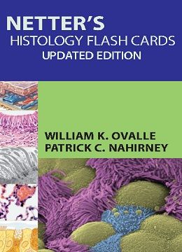
دانلود کتاب فلش کارت بافت شناسی نتر
Netter’s Histology Flash Cards, Updated Edition, 1ed
Take your understanding of histology one step further with Netter’s Histology Flash Cards. Specially designed to help you master what you need to know, these 200 flash cards are designed to reinforce your understanding of how the human body works in health as well as illness and injury. Classic anatomy illustrations from medical illustrator Frank H. Netter, MD provide strong visual aids and memorable diagrams to facilitate learning. Extensive additional images, including light and electron micrographs, provide an even more comprehensive study experience.
- Review salient microscopic features of cells, tissues, and organs of the body thanks to beautiful Netter illustrations and histology slides.
- Study basic biological and histological principles as well as the clinical relevance of histological structure and function through concise textual summaries.
- Recognize and interpret microscopic sections quickly thanks to an at-a-glance organization.
- Master the histology knowledge you need to know by using these flash cards in conjunction with Netter’s Essential Histology, 2nd Edition, also by Drs. Ovalle and Nahirney. They’re completely cross referenced to the Netter’s Essential Histology text so it’s easy to find more in-depth information on any topic.
Review
“This is a valuable study aid in histology, particularly in preparation for board review. The explanatory text is succinct, and the visual identification and clinical relevance of topics are emphasized. The quality of illustrations is exceptional and the combination of gross anatomic views with microscopic images presents histology in the context of gross anatomy, which helps students understand the correlation of structure and function. In the modern learning environment that employs problem-based and integrated curricula for medical and dental students, clinical information is highly valuable. To enhance the value of the flashcards even more, the names of the cells, tissues, structures or organs could be moved from the front to the back of the card. Currently, users do not have to identify what is presented on a card, unless they make a special effort to cover the top of a card. In addition, the number of microscopic images could be increased to replace some of the Netter’s illustrations.”-Joanna Goral, PhD(Midwestern Universty) Doody Review: 77/100
Contents
۱. The Cell
۲. Epithelium and Exocrine Glands
۳. Connective Tissue
۴. Muscle Tissue
۵. Nervous Tissue
۶. Cartilage and Bone
۷. Blood and Bone Marrow
۸. Cardiovascular System
۹. Lymphoid System
۱۰. Endocrine System
۱۱. Integumentary System
۱۲. Upper Digestive System
۱۳. Lower Digestive System
۱۴. Liver, Gallbladder, and Exocrine Pancreas
۱۵. Respiratory System
۱۶. Urinary System
۱۷. Male Reproductive System
۱۸. Female Reproductive System
۱۹. Eye and Adnexa
۲۰. Special Senses
لینک کوتاه : https://bookbaz.ir/?p=60467
نویسنده : William K. Ovalle PhD , Patrick C. Nahirney
ناشر : Saunders; 1st edition
سال انتشار : 2013
زبان کتاب : انگلیسی
نوع فایل : PDF
تعداد صفحات : 467
(ISBN) شابک : 1455776564
قیمت کتاب درآمازون : $36.61
حجم فایل : 40 MB
کتاب های مرتبط:
 دانلود کتاب بافت شناسی: یک کتاب ضروری + ویدئو
Histology – An Essential Textbook 1st Edition + Video
 دانلود کتاب آمادگی آزمون Thieme برای USMLE: پرسش و پاسخ بافت شناسی و جنین شناسی پزشکی
Thieme Test Prep for the USMLE: Medical Histology and Embryology Q&A, 1ed
 دانلود کتاب بافت شناسی: یک کتاب ضروری
Histology: An Essential Textbook, 1ed
 دانلود کتاب بافت شناسی ضروری نتر
Netter’s Essential Histology, 3ed
 دانلود کتاب پزشکی مراقبتهای ویژه: اصول تشخیص و مدیریت در بزرگسالان
Critical Care Medicine: Principles of Diagnosis and Management in the Adult, 4ed
 دانلود کتاب فلش کارت بافت شناسی عمومی لانگه
Lange Basic Histology Flash Cards, 1ed
 دانلود کتاب آناتومی جراحی و رویکردهای آن نتر
Netter’s Surgical Anatomy and Approaches, 1ed
 دانلود کتاب آناتومی، بافت شناسی، زیست شناسی سلولی: ارزیابی پیش آزمون و بررسی
Anatomy, Histology, & Cell Biology: PreTest Self-Assessment & Review, 4ed
 دانلود کتاب تشخیص کنونی و جراحی درمانی
Current Diagnosis and Treatment Surgery, 14ed
 دانلود کتاب بورد بررسی جامع جراحی مغز و اعصاب
 Comprehensive Neurosurgery Board Review, 2ed
 دانلود کتاب تکنیک های اصلی در جراحی: جراحی فتق
 Master Techniques in Surgery: Hernia, 1ed
 دانلود کتاب دانشنامه پزشکی گیل (۹ جلدی)
Gale Encyclopedia of Medicine, 9-Vol, 2015th
 دانلود کتاب پزشکی تورنتو نوت (نسخه ۲۰۱۵)
Toronto Notes 2015 (Essential Med Notes), 31ed
 دانلود کتاب اطلس عمل جراحی اس آر بی
SRB’s Surgical Operations: Text and Atlas
 دانلود کتاب پزشکی بلوپرینتز
Blueprints Medicine, 5ed
 دانلود کتاب ۲۵۰ مورد در پزشکی بالینی
 ۲۵۰Cases in Clinical Medicine, 4ed
 دانلود کتاب راهنمای صحیح پزشکی صحرایی
 Field Guide to Wilderness Medicine, 4ed
 دانلود کتاب ارزیابی پیشرفته: تفسیر یافته ها و تدوین تشخیص افتراقی
 Advanced Assessment: Interpreting Findings and Formulating Differential Diagnoses, 3ed
 دانلود کتاب راهنمای آزمایشگاهی آناتومی و فیزیولوژی: ایجاد ارتباطات
Human Anatomy & Physiology Laboratory Manual: Making Connections
 دانلود کتاب آسیب شناسی استخوان و بافت نرم: پاتولوژی عملکرد بالا
Bone and Soft Tissue Pathology: A Volume in the High Yield Pathology, 1ed
 دانلود کتاب آسیب شناسی آندروود: یک رویکرد بالینی
Underwood’s Pathology: a Clinical Approach, 6ed
 دانلود کتاب تجزیه و تحلیل پیشگویانه سیستم های تصمیم گیری پزشکی
Practical Predictive Analytics and Decisioning Systems for Medicine
 دانلود کتاب تکنیک های اعمال جراحی ستون فقرات
 Operative Techniques in Spine Surgery, 1ed
 دانلود کتاب آسیب شناسی میور
Muir’s Textbook of Pathology, 15ed
 دانلود کتاب زیست شناسی مولکولی سلول
Molecular Biology of the Cell, 6ed
 دانلود کتاب آلرژی میدلتون: اصول و تمرین (۲ جلدی)
Middleton’s Allergy 2-Vol: Principles and Practice, 8ed
 دانلود کتاب درسی بین المللی دیابت شیرین (۲ جلدی)
International Textbook of Diabetes Mellitus, 2Vol, 4ed
 دانلود کتاب عمل جراحی عمومی فارکهارسون
Farquharson’s Textbook of Operative General Surgery, 10ed
 دانلود کتاب استراتژی عمل در جراحی روده بزرگ و رکتوم شاسین
Chassin’s Operative Strategy in Colon and Rectal Surgery
 دانلود کتاب ساختار و عملکرد بدن
 Body Structures and Functions, 12ed
 دانلود کتاب مقدمه ای بر جراحی فک و صورت میچل
An Introduction to Oral and Maxillofacial Surgery, 2ed
 دانلود کتاب FRCA مقدماتی: OSCEs در بیهوشی
Primary FRCA: OSCEs in Anaesthesia
 دانلود کتاب تمرین کوتاه جراحی بیلی و لاو
Bailey & Love’s Short Practice of Surgery, 26ed
 دانلود کتاب راهنمای بالینی اورولوژی پن
Penn Clinical Manual of Urology, 2ed
 دانلود کتاب درسنامه ارتوپدی با روش معاینه بالینی در ارتوپدی
Textbook of Orthopedics with Clinical Examination Methods in Orthopedics, 4ed
 دانلود کتاب موفقیت در مصاحبه دانشکده پزشکی
Succeed in Your Medical School Interview: Stand Out from the Crowd and Get into Your Chosen Medical School, 2ed
 دانلود کتاب راهنمای نفرولوژی شرییر
Manual of Nephrology, 8ed
 چگونه در BMAT خبره شویم: آماده سازی بی نظیر برای موفقیت در آزمون پذیرش پزشکی
How to Master the BMAT: Unbeatable Preparation for Success in the BioMedical Admissions Test, 3ed
 دانلود کتاب تصویربرداری عصبی: یک پرونده آموزشی
Neuroimaging: A Teaching File
 دانلود کتاب راهنمای مصور اصطلاحات پزشکی
Illustrated Guide to Medical Terminology, 2ed
 دانلود کتاب درسی رادیولوژی و تصویربرداری ساتون (۲ جلدی)
Sutton Textbook of Radiology and Imaging: 2-Vol, 7ed
 دانلود کتاب ICD-9-CM 2015 برای بیمارستان (۳ جلدی)
ICD-9-CM 2015 for Hospitals, Volumes 1, 2 and 3, Professional Edition
 دانلود کتاب مروارید جراحی بالینی
 Clinical Surgery Pearls, 2ed
 دانلود کتاب پزشکی موبایل هریت لین
The Harriet Lane Handbook: Mobile Medicine Series, 20ed
 دانلود کتاب روش های مراقبت های اولیه فنینگر و فاولر
Pfenninger and Fowler’s Procedures for Primary Care, 3ed
 دانلود کتاب بافت شناسی برای پاتولوژیست ها
Histology for Pathologists, 4ed
 دانلود کتاب پزشکی سیسیل گلدمن (۲ جلدی)
Goldman-Cecil Medicine, 2-Vol, 25ed
 دانلود کتاب اطلس جراحی ترمیمی و جوی پیشرفته
Atlas of Advanced Operative Surgery, 1ed
 دانلود کتاب دوره های جراحی مغز و اعصاب: پرسش و پاسخ
Neurosurgery Rounds: Questions and Answers
 دانلود کتاب بورد بررسی شفاهی جراحی مغز و اعصاب
 Neurosurgery Oral Board Review, 2ed
 دانلود کتاب علائم و نشانه های بالینی
 Lww, Handbook of Signs & Symptoms 2015, 5ed
 دانلود کتاب راهنمای تشخیص و درمان مرک
The Merck Manual, 19ed
 دانلود کتاب مشاور بالینی فری ۲۰۱۵ (۵ جلدی)
Ferri’s Clinical Advisor 2015
 دانلود کتاب نقد و بررسی پاتولوژی رابینز و کوتران
Robbins and Cotran Review of Pathology, 4ed
 دانلود کتاب راهنمای ارتوپدی تاراسکون
Tarascon Pocket Orthopaedica, 3ed
 دانلود کتاب راهنمای مراقبت های ویژه پزشکی ایروین و ریپه
Irwin & Rippe’s Manual of Intensive Care Medicine, 6ed
 دانلود کتاب کمک های اولیه برای آزمون USMLE مرحله ۱
First Aid for the USMLE Step 1, 25ed
 دانلود کتاب کراش استپ ۳ بروشرت
Brochert’s Crush Step 3: The Ultimate USMLE Step 3 Review, 4ed
 دانلود کتاب راهنمای بالینی و آزمایشگاهی ایمپلنت اوردنچر
Clinical and Laboratory Manual of Implant Overdentures
 دانلود کتاب آناتومی و تکنیک جراحی
 Surgical Anatomy and Technique: A Pocket Manual, 4ed
 دانلود کتاب تشخیص پزشکی و درمان کارنت
CURRENT Medical Diagnosis and Treatment 2015, 54ed
 دانلود کتاب درسی فیزیولوژی
Textbook of Physiology, 3ed
 دانلود کتاب خود ارزیابی و بورد بررسی کودکان رودولف
Rudolphs Pediatrics Self-Assessment and Board Review
 دانلود کتاب درسی عملی بیوشیمی برای دانشجویان پزشکی
Practical Textbook of Biochemistry for Medical Students, 2ed
 دانلود کتاب نقد و بررسی داروسازی برای ناپلکس موزبی
Mosby’s Pharmacy Review for the NAPLEX®, ۱ed
 دانلود کتاب فلش کارت اصطلاحات پزشکی موزبی
Mosby’s Medical Terminology Flash Cards, 3ed
 دانلود کتاب آموزش دوره درسی تروما
Instructional Course Lectures Trauma 2
 دانلود کتاب بورد بررسی و ارزیابی اصول طب داخلی هریسون
Harrisons Principles of Internal Medicine Self-Assessment and Board Review, 18ed
 دانلود کتاب اسرار مراقبتهای ویژه
 Critical Care Secrets, 5ed
 دانلود کتاب راهنمای بالینی و آزمایشگاهی ایمپلنت اباتمنت دندانی
Clinical and Laboratory Manual of Dental Implant Abutments
 دانلود کتاب جراحی قلب ، کتابچه تخصص در جراحی آکسفورد
Cardiothoracic Surgery, 2ed (Oxford Specialist Handbooks in Surgery)
 دانلود کتاب پروتکل های پژوهشی استخوان
 Bone Research Protocols, 2ed
 دانلود کتاب پیشرفت هایی در درمان سرطان
Advances in Cancer Therapy
 دانلود کتاب اصول پزشکی داخلی هریسون ، چاپ نوزدهم (۲ جلدی)
Harrison’s Principles of Internal Medicine 2Vol, 19ed
 دانلود کتاب پزشکی کودکان نلسون (۲ جلدی، ویرایش ۲۰)
Nelson Textbook of Pediatrics, 2-Volume Set, 20ed
 دانلود کتاب اسرار مرحله ۱ آزمونUSMLE
 USMLE Step 1 Secrets, 3ed
 دانلود کتاب اسرار مرحله ۲ آزمون USMLE
 USMLE Step 2 Secrets, 4ed
 دانلود کتاب اسرار مرحله ۳ آزمونUSMLE
 USMLE Step 3 Secrets, 1ed
 دانلود کتاب آزمون بورد استادی USMLE گام ۳
Master the Boards USMLE Step 3, 3ed
 دانلود کتاب راهنمای درمان ضد میکروبی سانفورد
The Sanford Guide to Antimicrobial Therapy, 40 Spi
 دانلود کتاب بهداشت عمومی نوین
The New Public Health, 3ed
 دانلود کتاب درسی جنین شناسی بالینی
Textbook of Clinical Embryology
 دانلود کتاب معرفی فارماکولوژی: برای پرستاری و بهداشت و درمان
Introducing Pharmacology: For Nursing and Healthcare
 دانلود کتاب مراقبت اورژانسی و بهداشت عمومی
Emergency Care and the Public’s Health
 دانلود کتاب میدانهای الکترومغناطیسی در زیست شناسی و پزشکی
Electromagnetic Fields in Biology and Medicine
 دانلود کتاب انکولوژی بالینی و کاهش خطا: راهنمای پزشکان
Clinical Oncology and Error Reduction: A Manual for Clinicians
 دانلود کتاب اعتبار سنجی نشانگر زیستی: جنبه های بالینی، فنی و تجاری
Biomarker Validation: Technological, Clinical and Commercial Aspects
 دانلود کتاب اندازه گیری فیزیولوژیک (فیزیک پزشکی و مهندسی پزشکی)
The Physiological Measurement Handbook
 دانلود کتاب روش های بالینی در طب اورژانسی رابرتز و هجز
Roberts and Hedges’ Clinical Procedures in Emergency Medicine, 6ed
 دانلود کتاب اصول مهندسی بافت (نسخه اصلی)
Principles of Tissue Engineering, 4ed
 دانلود کتاب اطلس رنگی آناتومی پا و مچ پا مک مین
McMinn’s Color Atlas of Foot and Ankle Anatomy, 4ed
 دانلود کتاب برنامه ریزی شغلی در پزشکی و علوم زندگی: انتخاب آگاهانه
Planning a Career in Biomedical and Life Sciences: Making Informed Choices
 دانلود کتاب دیکشنری پزشکی و زیست شناسی الزویر
Elsevier’s Dictionary of Medicine and Biology
 دانلود کتاب درسی پزشکی API (دو جلدی)
API Textbook of Medicine 2-Vol, 9ed
 دانلود کتاب موارد طولانی در پزشکی بالینی، پرسش و پاسخ
Long Cases in Clinical Medicine: Concerned With Long Cases, Their Presentation, Related Questions, Answers and Discussions
 دانلود کتاب موارد پزشکی بالینی کومار و کلارک
Kumar & Clark’s Cases in Clinical Medicine, 3ed
 دانلود کتاب سلول های بنیادی خون بند ناف و پزشکی احیا
Cord Blood Stem Cells and Regenerative Medicine
 دانلود کتاب ۱۰۰ مورد در پزشکی بالینی
۱۰۰ Cases in Clinical Medicine, 3ed
 دانلود کتاب بازسازی و ترمیم زخم
Wound Regeneration and Repair: Methods and Protocols
 دانلود کتاب صدا درمانی: مطالعات بالینی
Voice Therapy: Clinical Case Studies, 4ed
 دانلود کتاب راهنمای مطالعه برای پرستاری جراحی داخلی: ارزیابی و مدیریت مشکلات بالینی
Study Guide for Medical-Surgical Nursing: Assessment and Management of Clinical Problems, 9ed
 دانلود کتاب اصطلاحات پزشکی برای خنگ ها
Medical Terminology For Dummies, 2ed
 دانلود کتاب هسته موارد بالینی در جراحی و تخصص جراحی
Core Clinical Cases in Surgery and Surgical Specialties, 2ed
 دانلود کتاب راهنمای معاینه فیزیکی و شرح حال بیتس
Bates’ Pocket Guide to Physical Examination and History Taking, 7ed
 دانلود کتاب زیست شناسی سلول های بنیادی و مهندسی بافت در علوم دندانپزشکی
Stem Cell Biology and Tissue Engineering in Dental Sciences
 دانلود کتاب مبانی پزشکی احیا: برنامه های کاربردی بالینی و درمانی
Foundations of Regenerative Medicine: Clinical and Therapeutic Applications
 دانلود کتاب گذشتن از امتحان بورد تخصصی جراحی عمومی
Passing the General Surgery Oral Board Exam, 2ed
 دانلود کتاب مدیریت عملی درد
 Practical Management of Pain, 5ed
 دانلود کتاب تومور بافت نرم انزینگر و ویس
Enzinger and Weiss’s Soft Tissue Tumors, 6ed
 دانلود کتاب آسیب شناسی عملی بافت نرم: روش تشخیصی
Practical Soft Tissue Pathology: A Diagnostic Approach, 1ed
 دانلود کتاب اطلس عمل جراحی زولینگر
Zollinger’s Atlas of Surgical Operations, 9th Edition
 دانلود کتاب اطلس رنگی حشره شناسی پزشکی چپمن و هال
A Colour Atlas of Medical Entomology: Chapman & Hall Medical Atlas
 دانلود کتاب راهنمای معاینه فیزیکی موزبی
Mosby’s Physical Examination Handbook, 7ed
 دانلود کتاب پزشکی عمومی
Crash Course General Medicine, 4ed
 دانلود کتاب بنیاد راهنمای دکتر به پزشکی و جراحی
Crash Course: Foundation Doctor’s Guide to Medicine and Surgery, 2ed
 دانلود کتاب ملزومات بیوشیمی برای دانشجویان پزشکی
Essentials of Biochemistry for Medical Students, 2ed
 دانلود کتاب ۵۰ درسی که هر جراح پلاستیک باید بداند
۵۰ Studies Every Plastic Surgeon Should Know
 دانلود کتاب درسی جراحی سابیستون: بنیان بیولوژیکی تمرین جراحی مدرن
Sabiston Textbook of Surgery: The Biological Basis of Modern Surgical Practice, 19ed
 دانلود کتاب پزشکی سیسیل گلدمن
Goldman’s Cecil Medicine: Premium Edition, 24ed
 دانلود کتاب اصول جراحی شوارتز
 Schwartz’s Principles of Surgery, 10ed
 دانلود کتاب آزمون خود ارزیابی و نقد و بررسی پزشک خانواده
Family Medicine PreTest Self-Assessment And Review, 3ed
 دانلود کتاب بافت شناسی: مرتبط با بیولوژی سلولی و مولکولی
Histology: With Correlated Cell and Molecular Biology, 7ed
 دانلود کتاب درمان جایگزین در عمومی، سوختگی، و جراحی پلاستیک: مهندسی بافت در عمل بالینی
Dermal Replacements in General, Burn, and Plastic Surgery: Tissue Engineering in Clinical Practice
 دانلود کتاب واکسن ها: کارشناس مشورت
Vaccines: Expert Consult 6ed
 دانلود کتاب درمان متداول کان
Conn’s Current Therapy 2015, 1ed
 دانلود کتاب راهنمای پزشکی هریسون
 Harrisons Manual of Medicine, 18th Edition
 دانلود کتاب بورد بررسی و آزمون فارماکولوژی کاتزونگ
Katzung & Trevor’s Pharmacology Examination and Board Review, 10ed
 دانلود کتاب اصول و تمرین پزشکی دیویدسون
Davidson’s Principles and Practice of Medicine, 22ed
 دانلود بسته کامل آموزش آیلتس ۱ تا ۹ همراه با جواب
Cambridge IELTS 1 ~ 9 With Answer
 دانلود کتاب اصول و تمرین بیماری های عفونی مندل، داگلاس، و بنت
Mandell, Douglas, and Bennett’s Principles and Practice of Infectious Diseases: 2-Volume Set, 8e


