دانلود کتاب تشریح بالینی عکاسی بدن انسان گری + ویدئو
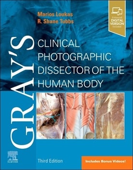
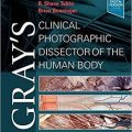
Gray’s Clinical Photographic Dissector of the Human Body 3rd Edition + Video
Gray’s Clinical Photographic Dissector of the Human Body The perfect hands-on reference, Gray’s Clinical Photographic Dissector of the Human Body, 3rd Edition, is a practical resource in the anatomy lab, on surgical rotations, during clerkship and residency and beyond! This fully revised third edition uses a unique, step-by-step presentation of full-color cadaveric photographs to orient you more quickly in the anatomy lab, and points out the clinical relevance of each structure and every dissection. Each photograph depicts clearly labeled anatomical structures, including muscles, bones, nerves, blood vessels, and organs―making this one-of-a-kind resource ideal for preparing for laboratory sessions and as a useful reference during dissections.
- Contains nearly 1,100 full-color photographs for comparison to the cadavers you study, helping you become more proficient and confident in your understanding of the intricacies of the human body.
- Guides you through each dissection step-by-step, using a unique, real-world photographic presentation.
- Includes complementary high-quality schematic drawings throughout to help orientate you and aid understanding.
- Contains superb corresponding Gray’s illustrations to add clarity to key anatomical structures.
- Helps you easily relate anatomical structures to clinical conditions and procedures.
- Features new explanatory videos of human cadaveric dissection for each chapter.
- Depicts the pertinent anatomy for more than 30 common clinical procedures such as prosthetic hip replacements, intravenous catheters, lumbar puncture, and knee joint aspiration, including where to make the relevant incisions.
- Reflects the same level of accuracy and thoroughness that has made the Gray’s ‘family’ of products the most trusted learning resources in anatomy.
- Prepared by an expert author team―highly experienced educators and leading authorities in clinical anatomy.
Review
Contents
۱. Dissection laboratory materials, tools, and techniques
۲. Muscles of the back and scapula
۳. Suboccipital triangle and spinal cord
۴. Pectoral region and female breast
۵. Lungs, removal of heart, and posterior mediastinum
۶. Heart
۷. Axilla and arm
۸. Forearm (antebrachium)
۹. Hand
۱۰. Anterolateral abdominal wall and inguinal region
۱۱. Peritoneal cavity
۱۲. Gastrointestinal tract
۱۳. Posterior abdominal wall
۱۴. Pelvis
۱۵. Perineum
۱۶. Gluteal region
۱۷. Thigh and leg
۱۸. Leg and ankle
۱۹. Foot
۲۰. Neck
۲۱. Face
۲۲. Infratemporal fossa
۲۳. Calvaria, dural venous sinuses, brain, and cranial nerves
۲۴. Orbit
۲۵. Ear
۲۶. Nasal cavity
۲۷. Pharynx and oral cavity
۲۸. Larynx
۲۹. Retropharyngeal region and pharynx
Video Contents
۱. ۲.۱ Back and Posterior Scapula Muscles
۲. ۲.۲ Posterior Scapula and Arm
۳. ۳.۱ Spinal Cord In Situ (Laminectomy)
۴. ۳.۲ Spinal Cord (Removed)
۵. ۳.۳ Suboccipital Triangle
۶. ۴.۱ Pectoral Region and Anterior Thoracic Wall
۷. ۵.۱ Lung
۸. ۵.۲ Posterior Mediastinum
۹. ۶.۱ Heart In Situ
۱۰. ۶.۲ Heart Anatomy (Exterior): Overview I
۱۱. ۶.۳ Heart Anatomy (Exterior): Overview II – Coronary Arteries
۱۲. ۶.۴ Heart Anatomy III: Atria, Ventricles, and Valves
۱۳. ۶.۵ Heart Anatomy IIII: Right Atrium and Right Ventricle
۱۴. ۶.۶ Heart Anatomy IIIII: Left Ventricle and Left Atrium
۱۵. ۷.۱ Axilla and Pectoral Region (In Situ): Axillary Artery and Brachial Plexus
۱۶. ۷.۲ Axilla, Pectoral Region, and Anterior Arm
۱۷. ۷.۳ Axilla and Arm: Brachial Plexus
۱۸. ۸.۱ Anterior Forearm
۱۹. ۸.۲ Posterior Forearm
۲۰. ۸.۳ Elbow Joint I
۲۱. ۸.۴ Elbow Joint II
۲۲. ۸.۵ Dorsum of the Hand
۲۳. ۹.۱ Palmar Hand
۲۴. ۱۰.۱ Anterolateral Abdominal Wall (External View)
۲۵. ۱۰.۲ Anterior Thoracic Wall (Internal View)
۲۶. ۱۰.۳ Male Inguinal Region (In Situ): Inguinal Ligament, Spermatic Cord, and Testes
۲۷. ۱۱.۱ Female Pelvic Contents & Peritoneal Cavity
۲۸. ۱۲.۱ Abdominal Cavity Overview I: Stomach, Spleen, Liver, and Celiac Trunk
۲۹. ۱۲.۲ Abdominal Cavity Overview II: Superior Mesenteric Artery, Small and Large Intestines
۳۰. ۱۲.۳ Abdominal Cavity Overview III: Inferior Mesenteric Artery, Gonadal Veins, and Sigmoid Colon
۳۱. ۱۳.۱ Posterior Abdominal Wall
۳۲. ۱۳.۲ Kidney
۳۳. ۱۴.۱ Male Pelvis (Hemisection)
۳۴. ۱۴.۲ Female Pelvis (Hemisection)
۳۵. ۱۴.۳ Pelvis (Hemisection): Floor, Vasculature and Sacral Plexus
۳۶. ۱۶.۱ Gluteal Regional I
۳۷. ۱۶.۲ Gluteal Regional II
۳۸. ۱۶.۳ Posterior Thigh
۳۹. ۱۷.۱ Anterior Thigh
۴۰. ۱۸.۱ Dorsum of the Foot
۴۱. ۱۸.۲ Anterolateral Foot
۴۲. ۱۸.۳ Sole of the Foot
۴۳. ۱۸.۴ Knee Joint
۴۴. ۱۸.۵ Anterolateral Leg
۴۵. ۱۸.۶ Posterior Leg
۴۶. ۲۰.۱ Neck
۴۷. ۲۱.۱ Face
۴۸. ۲۲.۱ Infratemporal Fossa
۴۹. ۲۳.۱ Brain I: Inferior Surface
۵۰. ۲۳.۲ Brain II: Midsagittal
۵۱. ۲۳.۳ Midsagittal Head I
۵۲. ۲۳.۴ Midsagittal Head II
۵۳. ۲۳.۵ Midsagittal Head III
۵۴. ۲۴.۱ Orbit I
۵۵. ۲۴.۲ Orbit II
۵۶. ۲۶.۱ Nasal Cavity Lateral Wall
۵۷. ۲۶.۲ Pterygopalatine Fossa
۵۸. ۲۷.۱ Oral Cavity
۵۹. ۲۹.۱ Posterior Pharynx
۶۰. ۲۹.۲ Skull: Surface Anatomy
۶۱. ۲۹.۳ Skull: Cranial Floor
۶۲. ۲۹.۴ Head
تمامی ویدئوها با کیفیت اچ دی و فول اچ دی از سورس اصلی الزویر پلاس میباشد
لینک کوتاه : https://bookbaz.ir/?p=467121
نویسنده : Marios Loukas MD PhD
ناشر : Elsevier; 3rd edition
سال انتشار : 2025
زبان کتاب : انگلیسی
نوع فایل : MP4 + PDF (کیفیت اصلی)
تعداد صفحات : 525 به همراه کانتنت و ایندکس
(ISBN) شابک : 0443107092
قیمت کتاب درآمازون : $62.99
حجم فایل : -
