دانلود کتاب چشم پزشکی عصبی مصور + ویدئو
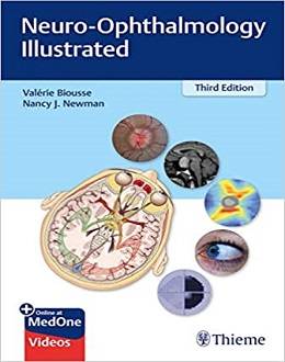
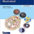
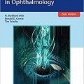
Neuro-Ophthalmology Illustrated, 3ed + Video
highly praised, well written, and richly illustrated modern classic in neuro-ophthalmology now in an exciting new edition with video clips!
Neuro-ophthalmology is an “overlap” specialty, encompassing all disorders that affect the parts of the central nervous system related to vision. Neuro-Ophthalmology Illustrated, Third Edition by world-renowned neuro-ophthalmologists and professors Valérie Biousse and Nancy J. Newman expands on the widely acclaimed prior editions, lauded with awards by the Association of American Publishers and the British Medical Association. The updated text reflects diagnostic advances such as optical coherence tomography and features new high-quality images and videos.
The text starts with neuro-ophthalmic and funduscopic examinations, visual fields, commonly used ancillary testing methods, and an overview of visual loss. Subsequent chapters detail a wide array of conditions including retinal vascular diseases, optic neuropathies, disc edema, disorders of higher cortical function, abnormal visual perceptions, diplopia, orbital syndromes, cavernous sinus and orbital vascular disorders, nystagmus/other ocular oscillations, and disorders of the eyelid. Final chapters are dedicated to nonorganic neuro-ophthalmic symptoms and signs, diagnosis of headache and facial pain, neurologic and systemic disorders that commonly have neuro-ophthalmologic manifestations, and an approach to management of the visually impaired patient.
Key Highlights
- Concise text coupled with more than 600 high-quality images enhances understanding of complex concepts
- Easily accessible within pertinent sections, ۶۹ video clips cover a wide spectrum of topics such as examination techniques, normal eye movements, eye movement disorders, and pupil findings
- Clinical pearls, flow charts, boxes, tables, and a highly detailed index at the end of the text improve retention and assimilation of knowledge
Neuro-Ophthalmology Illustrated book is essential reading for students, ophthalmology, neurology, and neurosurgery residents, as well as seasoned clinicians in these specialties. At once authoritative and easy to read, this resource provides readers with all the tools they need to diagnose and manage neuro-ophthalmologic disorders.
Contents
۱ The Neuro-Ophthalmic Examination
۲ Funduscopic Examination
۳ Visual Fields
۴ Ancillary Testing Commonly Used in Neuro-Ophthalmology
۵ Visual Loss: An Overview
۶ Transient Visual Loss
۷ Retinal Vascular Diseases
۸ Optic Neuropathies
۹ Disc Edema
۱۰ Disorders of Higher Cortical Function
۱۱ Abnormal Visual Perceptions: Hallucinations and Illusions
۱۲ The Pupil
۱۳ Diplopia
۱۴ Orbital Syndrome
۱۵ Cavernous Sinus and Orbital Vascular Disorders
۱۶ Nystagmus and Other Ocular Oscillations
۱۷ Disorders of the Eyelid
۱۸ Nonorganic Neuro-Ophthalmic Symptoms and Signs
۱۹ Diagnosis of Headache and Facial Pain
۲۰ Disorders Commonly Encountered in Neuro-Ophthalmology
۲۱ The Visually Impaired Patient
Video Contents
Video 1.1 Left relative afferent pupillary defect (RAPD) A dense left RAPD is demonstrated by the swinging flashing test in a patient with a left optic neuropathy
Video 1.2 Quantification of the relative afferent pupillary defect with neutral density filters
Video 1.3 Normal optokinetic nystagmus response in both eyes
Video 9.1 Cranial nerve sixth palsy in idiopathic intracranial hypertension
Video 12.1 Examination of the normal pupils
Video 12.2 Normal pupillary constriction to near stimulus
Video 12.3 Bilateral light-near dissociation in dorsal midbrain syndrome
Video 12.4 Right Horner syndrome
Video 12.5 Demonstration of dilation lag in left Horner syndrome
Video 12.6 Left Horner syndrome associated with a left cranial nerve six palsy, revealing a left cavernous sinus mass
Video 12.7 Right tonic pupil
Video 12.8 Left mydriasis in the setting of a left third nerve palsy
Video 13.1 Normal eye movement examination with demonstration of each step of the examination
Video 13.2 Eye movements showing a left cranial nerve six palsy
Video 13.3 Peripheral facial palsy showing an intact Bell phenomenon
Video 13.4 Normal uncover–cover test and normal cross-cover test in an orthophoric patient
Video 13.5 Large exotropia on cover-uncover and cross-cover test
Video 13.6 Three-step test in a left superior oblique (fourth cranial nerve) palsy, with demonstration of cross-cover test
Video 13.7 Neutralization of vertical ocular deviation with prisms
Video 13.8 Hirshberg and Krimsky tests
Video 13.9 Abnormal eye movements in the setting of thyroid eye disease
Video 13.10 Left Brown syndrome (Courtesy of Jason Peragallo, MD )
Video 13.11 Pulsatile proptosis in the right eye secondary to right sphenoid wing dysplasia
Video 13.12 Chronic progressive external ophthalmoplegia
Video 13.13 Bilateral fluctuating ptosis secondary to myasthenia gravis
Video 13.14 Right fluctuating ptosis and abnormal eye movements in a baby with myasthenia gravis (Courtesy of Jason Peragallo, MD )
Video 13.15 Bilateral ophthalmoplegia triggered by fatigue (sustained upgaze) in myasthenia gravis
Video 13.16 Right gaze palsy and right facial palsy related to a right sixth cranial nerve nucleus lesion
Video 13.17 Left sixth cranial nerve palsy with left Horner syndrome localizing to the left cavernous sinus
Video 13.18 Bilateral type I Duane syndrome
Video 13.19 Thalamic esotropia and upgaze deficit secondary to a thalamic hemorrhage
Video 13.20 Demonstration of normal ocular intorsion (intact fourth cranial nerve) in a complete third nerve palsy
Video 13.21 Partial third nerve palsy
Video 13.22 Complete third nerve palsy with pupil involvement
Video 13.23 Bilateral nuclear third nerve palsies secondary to a midbrain stroke
Video 13.24 Complete third nerve palsy with pupil sparing
Video 13.25 Orbital apex syndrome with ophthalmoplegia and optic neuropathy
Video 13.26 Cavernous sinus syndrome with complete ophthalmoplegia and mild chemosis
Video 13.27 Bilateral ophthalmoplegia from Miller-Fisher syndrome
Video 13.28 Upbeat nystagmus from Wernicke encephalopathy
Video 13.29 Unilateral left internuclear ophthalmoplegia with skew deviation
Video 13.30 Bilateral internuclear ophthalmoplegia
Video 13.31 Left one-and-a-half syndrome
Video 13.32 Convergence insufficiency
Video 13.33 Dorsal midbrain syndrome
Video 13.34 Doll’s head maneuver in dorsal midbrain syndrome
Video 13.35 Neuromyotonia
Video 14.1 Bilateral asymmetric proptosis with normal eye movements from thyroid eye disease
Video 15.1 Carotid–cavernous fistula
Video 16.1 Oscillopsia as seen by a patient with severe pendular nystagmus
Video 16.2 Congenital nystagmus (Courtesy of Janet Rucker, MD)
Video 16.3 Latent nystagmus (Courtesy of Caroline Tilikete, MD)
Video 16.4 Spasmus nutans (Courtesy of Jason Peragallo, MD)
Video 16.5 Right eye vertical oscillations from severe monocular visual loss (Courtesy of Jason Peragallo, MD)
Video 16.6 Gaze-evoked nystagmus
Video 16.7 Downbeat nystagmus
Video 16.8 Periodic alternating nystagmus
Video 16.9 Acquired pendular nystagmus (Courtesy of Caroline Tilikete, MD)
Video 16.10 Seesaw nystagmus
Video 16.11 Oculopalatal myoclonus
Video 16.12 Superior oblique myokymia
Video 16.13 Ocular flutter
Video 16.14 Opsoclonus
Video 16.15 Square-wave jerks and ocular dysmetria
Video 17.1 Marcus Gunn jaw-winking syndrome (Courtesy of Jason Peragallo, MD)
Video 17.2 Blepharospasm
Video 18.1 Optokinetic nystagmus present in one eye with nonorganic visual loss
Video 18.2 Spasm of convergence
Video 18.3 Voluntary nystagmus
لینک کوتاه : https://bookbaz.ir/?p=154917
نویسنده : Valerie Biousse , Nancy Newman
ناشر : Thieme; 3 edition
سال انتشار : 2020
زبان کتاب : انگلیسی
نوع فایل : MP4 + PDF (کیفیت اصلی)
تعداد صفحات : 696
(ISBN) شابک : 1684200741
قیمت کتاب درآمازون : $103.79
حجم فایل : 1450 MB
