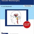
دانلود کتاب اطلس عمل جراحی مغز و اعصاب: جراحی مغز و اعصاب عروقی + ویدئو
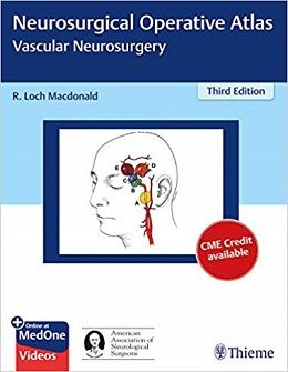
Neurosurgical Operative Atlas: Vascular Neurosurgery, 3ed + Video
A state-of-the-art neurovascular surgery atlas from internationally renowned neurosurgeon R. Loch Macdonald
Neurosurgical Operative Atlas: Vascular Neurosurgery, Third Edition, by R. Loch Macdonald and expert contributors, reflects the latest advances in endoscopic, endovascular, microsurgical, and bypass techniques used in the treatment of cerebrovascular disease. The entire atlas has been streamlined and updated with new content, including 38 videos that complement the concise step-by-step guidance in the text.
The book begins with five chapters on vascular and microsurgical instrumentation and equipment, clipping versus coiling, aneurysm surgery techniques, the pterional approach, and minimally invasive approaches. Disease and procedure-specific chapters are organized by three sections: aneurysms and subarachnoid hemorrhage, vascular malformations, and ischemic and other cerebrovascular disease. Every chapter includes salient tips on patient selection and procedural indications, preoperative information and tests, patient positioning, operative nuances, and postoperative complications.
Key Highlights
- Nearly 300 high-quality color illustrations detail impacted anatomy and procedures
- The latest techniques for treating a full spectrum of aneurysms, such as ophthalmic segment, supraclinoid internal carotid artery, middle and anterior cerebral artery, basilar and posterior cerebral artery, and others
- Treatment of vascular abnormalities including arteriovenous malformations, superficial and brainstem cavernous malformations, arteriovenous fistulae, Moyamoya disease, and more
Neurosurgical residents will benefit from the firsthand knowledge shared by international masters, while veteran neurosurgeons will glean invaluable insights on cutting-edge endovascular techniques to enhance clinical practice.
Contents
۱. Vascular and Microsurgical Instrumentation and Equipment
۲. How to Repair the Intracranial Aneurysm: Clipping or Coiling Decision Making
۳. Aneurysm Surgery Techniques
۴. Pterional Approach
۵. Minimally Invasive Approaches to Aneurysms
۶. Ophthalmic Segment Aneurysms
۷. Supraclinoid Internal Carotid Artery Aneurysms
۸. Anterior Communicating Artery Aneurysms
۹. Middle Cerebral Artery Aneurysms 56
۱۰. Distal Anterior Cerebral Artery Aneurysms: Anterior Interhemispheric Approach
۱۱. Pterional Transsylvian and Extended Approaches for Upper Basilar Aneurysms
۱۲. Orbitocranial Zygomatic Approach for Upper Basilar Artery Aneurysms
۱۳. Subtemporal and Pretemporal Approaches for Basilar and Posterior Cerebral Artery Aneurysms
۱۴. Transsylvian Transclinoidal and Transcavernous Approach for Basilar Bifurcation Aneurysms
۱۵. Vertebral Artery and Posterior Inferior Cerebellar Artery Aneurysms
۱۶. Retrolabyrinthine Transsigmoid and Extreme Lateral Infrajugular Transcondylar-Transtubercular Exposures for Aneurysms
۱۷. Vertebral Confluence and Midbasilar Aneurysms Including Transpetrosal Approach
۱۸. Fusiform, Dolichoectatic, and Dissecting Aneurysms
۱۹. Endoscopic Approaches to Intracranial Aneurysms.
۲۰. Microsurgical Treatment of Previously Endovascularly Treated Aneurysms
۲۱. Pterional Craniotomy for Exposure of Contralateral Aneurysms
۲۲. Infectious Intracranial Aneurysms
۲۳. Arteriovenous Malformations of the Cerebral Convexities
۲۴. Arteriovenous Malformations of the Basal Ganglia and Thalamus
۲۵. Intraventricular and Deep Arteriovenous Malformations
۲۶. Vein of Galen Malformations
۲۷. Posterior Fossa Arteriovenous Malformations
۲۸. Superficial Cavernous Malformations
۲۹. Brainstem Cavernous Malformations
۳۰. Spinal Vascular Malformations
۳۱. Carotid Cavernous Fistulas
۳۲. Transverse and Sigmoid Dural Arteriovenous Fistula
۳۳. Tentorial and Posterior Fossa Dural Arteriovenous Fistulas
۳۴. Anterior Fossa, Superior Sagittal Sinus, and Convexity Dural Arteriovenous Malformations
۳۵. Carotid Endarterectomy
۳۶. Superficial Temporal Artery to Middle Cerebral Artery Bypass
۳۷. Indirect Bypasses for Moyamoya Disease
۳۸. Positional Compression of the Vertebral Arteries
۳۹. Minimally Invasive Approaches for Spontaneous Intracerebral Hemorrhage
Video Contents
۱.۱ Positioning.
۱.۲ Arachnoid dissection.
۱.۳ Vascular anastomosis.
۲.۱ ۳D rotational angiography of right middle cerebral artery aneurysm.
۲.۲ ۳D rotational angiography of right posterior communicating artery aneurysm.
۲.۳ ۳D rotational angiography of bilobulated anterior communicating artery aneurysm.
۲.۴ ۳D rotational angiography of pericallosal artery aneurysm.
۲.۵ PICA-PICA side-to-side bypass for proximal left PICA aneurysm.
۲.۶ ۳D rotational angiography of left blister-like carotid artery aneurysm.
۴.۱ Right pterional craniotomy and clipping of an unruptured anterior communicating artery aneurysm in a 66-year-old woman.
۷.۱ Clipping of a left posterior communicating artery aneurysm (PcomA aneurysm).
۷.۲ Clipping of multiple aneurysm (left PcomA and anterior choroidal artery aneurysm).
۷.۳ Clipping of a right unruptured internal carotid artery bifurcation aneurysm.
۸.۱ Clipping of an unruptured anterior communicating artery aneurysm.
۸.۲ Clipping of a ruptured anteriorly-projecting anterior communicating artery aneurysm.
۸.۳ Clipping of a ruptured inferiorly-projecting anterior communicating artery aneurysm.
۸.۴ Clipping of a ruptured ectatic anterior communicating artery segment aneurysm.
۹.۱ Craniotomy and clipping of an unruptured middle cerebral artery aneurysm.
۱۶.۱ Left retrolabyrinthine transsigmoid approach for clipping of a ruptured basilar trunk aneurysm.
۲۳.۱ Intraoperative demonstrating microneurosurgical techniques for resection of a right frontotemporal AVM.
۲۴.۱ of the case shown in Fig. 24.3.
۲۴.۲ of the case shown in Fig. 24.6.
۲۵.۱ A medial occipital AVM is demonstrated to highlight the transfalcine approach.
۲۵.۲ Demonstration of dissecting the artery from proximal to distal and ensuring that the distal normal artery is demonstrated before ligating feeding arteries to the AVM.
۲۵.۳ Bipolar techniques, clean diathermy, microclips, and dirty diathermy techniques.
۲۵.۴ This shows an example of going to the cone first.
۲۵.۵ Resection of the AVM shown in Fig. 25.3.
۲۵.۶ Sylvian fissure dissection for insular AVM.
۲۷.۱ Resection of cerebellar arteriovenous malformation with associated acute intracerebellar hemorrhage (Fig. 27.4).
۲۹.۱ Left lateral supracerebellar infratentorial approach to brainstem CM
۳۰.۱ of a case of a patient with two spinal dural arteriovenous fistulas.
۳۲.۱ Intra-operative of a suboccipital craniotomy for disconnection of Borden III DAVF arising from tentorium to superior vermis.
۳۳.۱ This demonstrates a torcular craniotomy for a previously ruptured galenic dural arteriovenous fistula.
۳۴.۱ Endoscopic endonasal approach to an ophthalmic artery branch supplied arteriovenous fistula, as described in the preceding text.
۳۶.۱ STA-MCA bypass.
۳۶.۲ Double barrel STA-MCA bypass.
۳۷.۱ Pial synangiosis for pediatric moyamoya disease.
۳۸.۱ Transcranial Doppler ultrasound of a patient with positional VBI showing reduced cerebral blood flow velocity with head turning.
لینک کوتاه : https://bookbaz.ir/?p=111809
نویسنده : R. Loch Macdonald
ناشر : Thieme/AANS; 3 edition
سال انتشار : 2019
زبان کتاب : انگلیسی
نوع فایل : MP4 + PDF (کیفیت اصلی)
تعداد صفحات : 322
(ISBN) شابک : 1626231109
قیمت کتاب درآمازون : $200.00
حجم فایل : 2200 MB
