
دانلود کتاب اطلس رنگی میکرو جراحی مغز و اعصاب: میکرو آناتومی، روش ها و تکنیک ها (۳ جلدی)
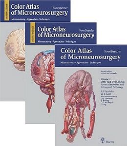
Color Atlas of Microneurosurgery: Microanatomy, Approaches and Techniques, 3-Vol, 2ed
Refinements in the neurosurgical armamentarium continue to push the borders of neurosurgery forward. Lesions considered inoperable a few years ago can now be resected, especially in the region of the skull base. These new developments, plus rapid technological innovations in microneurosurgery, have dramatically altered the scope of modern neurosurgery.
Now, with Volume 2 of the acclaimed Color Atlas of Microneurosurgery, the distinguished authors provide detailed descriptions of surgical anatomy and the major neurosurgical approaches to cerebrovascular lesions. You will find coverage of aneurysms, arteriovenuous malformations, cerebrovascular malformations, and vascular compression- all derived from a wide range of etiologies. Divided into three sections on anatomy, surgical approaches, and underlying pathology, the book demonstrates the most innovative new techniques, procedures and approaches as performed in hundreds of clinical cases. The result is the most detailed and comprehensive microneurosurgical atlas ever compiled, an ideal reference for practicing neurosurgeons and residents-in-training.
From the Back Cover
Refinements in the neurosurgical armamentarium continue to push the borders of neurosurgery forward. Lesions considered inoperable a few years ago can now be resected, especially in the region of the skull base. These new developments, plus rapid technological innovations in microneurosurgery, have dramatically altered the scope of modern neurosurgery.
Now, with Volume 2 of the acclaimed Color Atlas of Microneurosurgery, the distinguished authors provide detailed descriptions of surgical anatomy and the major neurosurgical approaches to cerebrovascular lesions. You will find coverage of aneurysms, arteriovenuous malformations, cerebrovascular malformations, and vascular compression- all derived from a wide range of etiologies. Divided into three sections on anatomy, surgical approaches, and underlying pathology, the book demonstrates the most innovative new techniques, procedures and approaches as performed in hundreds of clinical cases. The result is the most detailed and comprehensive microneurosurgical atlas ever compiled, an ideal reference for practicing neurosurgeons and residents-in-training.
Refinements in the neurosurgical armamentarium continue to push the borders of neurosurgery forward. Lesions considered inoperable a few years ago can now be resected, especially in the region of the skull base. These new developments, plus rapid technological innovations in microneurosurgery, have dramatically altered the scope of modern neurosurgery.
Now, with Volume 2 of the acclaimed COLOR ATLAS OF MICRONEUROSURGERY, the distinguished authors provide detailed descriptions of surgical anatomy and the major neurosurgical approaches to cerebrovascular lesions. You will find coverage of aneurysms, arteriovenuous malformations, cerebrovascular malformations, and vascular compression- all derived from a wide range of etiologies. Divided into three sections on anatomy, surgical approaches, and underlying pathology, the book demonstrates the most innovative new techniques, procedures and approaches as performed in hundreds of clinical cases. The result is the most detailed and comprehensive microneurosurgical atlas ever compiled, an ideal reference for practicing neurosurgeons and residents-in-training.
From reviews of previous volumes:
Ranks with the very best previous attempts at codifying neurosurgical operations. The attention to detail is excellent… -The New England Journal of Medicine
A valuable addition to any library…I would recommend it to all neurosurgeons with an interest in cerebrovascular disease…The operative photographs are of extremely high quality.-Chicago Medicine
The final volume in the acclaimed series provides coverage of the anatomy, surgical approaches, and techniques involved in performing cerebral revascularization. Filled with over 2,000 vibrant images, it provides the visual instruction neurosurgeons need.
Highlights include:
- A complete section detailing intracranial vasculature and anatomy of the spinal cord
- A case material section featuring a rich diversity of clinical situations to illustrate a variety of microsurgical techniques
- Thorough coverage of bypasses, reconstructions, and the use of endarterectomy to achieve revascularization
- Presentation of both surgical and endovascular techniques for re-establishing blood flow through the carotid and cerebral arteries
- Information on tumors of the spinal cord and spinal vascular malformations, particularly cavernous and arteriovenous malformations
Volume 1: Intracranial Tumors: Microanatomy, Approaches and Techniques
Volume 2: Cerebrovascular Lesions: Microanatomy, Approaches and Techniques
Volume 3: Intra- und Extracranial Revascularization and Intraspinal Pathology: Microanatomy, Approaches and Techniques
لینک کوتاه : https://bookbaz.ir/?p=10501
نویسنده : Wolfgang T. Koos , Robert F. Spetzler
ناشر : Thieme; 2nd , Revised And Expanded Edition
سال انتشار : 2013
زبان کتاب : انگلیسی
نوع فایل : PDF
تعداد صفحات : 1763
(ISBN) شابک : 0865779007
قیمت کتاب درآمازون : $995
حجم فایل : 325 MB
کتاب های مرتبط:
 دانلود کتاب توانبخشی سکته مغزی: رویکرد مبتنی بر عملکرد
Stroke Rehabilitation: A Function-Based Approach, 4ed
 دانلود کتاب مقدمه ای بر جراحی فک و صورت میچل
An Introduction to Oral and Maxillofacial Surgery, 2ed
 دانلود کتاب درمان فعلی در جراحی دهان و فک و صورت
 Current Therapy In Oral and Maxillofacial Surgery, 1ed
 دانلود کتاب مغز و اعصاب بالینی لانگه (امینف، نسخه اصلی)
Aminoff Clinical Neurology, 8ed
 دانلود کتاب اطلس رنگی جراحی ساقه مغز
 Color Atlas of Brainstem Surgery, 1ed
 دانلود کتاب نقشه برداری مغز: نشانه ها و تکنیک ها + ویدئو
Brain Mapping: Indications and Techniques, 1ed + Video
 دانلود کتاب کالبدشناسی اعصاب بالینی گری: اساس آناتومی علوم اعصاب بالینی
Gray’s Clinical Neuroanatomy: The Anatomic Basis for Clinical Neuroscience, 1ed
 دانلود کتاب اطلس تکنیک های جراحی عمومی
 Atlas of General Surgical Techniques Expert Consult, 1ed
 دانلود کتاب اطلس تکنیک های جراحی غدد درون ریز
 Atlas of Endocrine Surgical Techniques: A Volume in the Surgical Techniques Atlas Series, 1ed
 دانلود کتاب روش های جراحی الکساندر
Alexander’s Surgical Procedures, 1ed
 دانلود کتاب جراحی
 Textbook of Surgery, 4ed
 دانلود کتاب جیبی ارتوپدی تروما و روماتولوژی چرچیل
Churchill’s Pocketbook of Orthopaedics, Trauma and Rheumatology, 2ed
 دانلود کتاب موارد رادیولوژی مغز و اعصاب
Neuroradiology Cases
 دانلود کتاب تکنیک های اصلی در جراحی: جراحی قفسه سینه: پیوند، تراشه برداشتن، تومور مدیاستن، برش گسترده قفسه سینه
Master Techniques in Surgery: Thoracic Surgery: Transplantation, Tracheal Resections, Mediastinal Tumors, Extended Thoracic Resections
 دانلود کتاب جراحی کولورکتال: همگام با تمرین متخصص جراحی
Colorectal Surgery: A Companion to Specialist Surgical Practice, 5ed
 دانلود کتاب راهنمای نوروپاتولوژی عمومی اسکرول و پوریِر
Escourolle & Poirier’s Manual of Basic Neuropathology, 5ed
 دانلود کتاب علوم اعصاب شناختی سیستم های بشری: کار و زندگی روزمره
Cognitive Neuroscience of Human Systems: Work and Everyday Life
 دانلود کتاب مراقبت زوال عقل: رویکرد عملی
Dementia Care: A Practical Approach, 1ed
 دانلود کتاب جراحی زاویه سربلوپونتین
 Surgery of the Cerebellopontine Angle, 1ed
 دانلود کتاب مغز و اعصاب بالینی امینوف
Lange Clinical Neurology, 10ed
 دانلود کتاب سری کارشناسی ستون فقرات AO: ناهنجاری های ستون فقرات کودکان (جلد ۹)
AOSpine Masters Series, Volume 9: Pediatric Spinal Deformities, 1ed
 دانلود کتاب جراحی اعصاب عروق مغزی
Cerebrovascular Neurosurgery, 1ed
 دانلود کتاب ۱۰۱ راه حل MRI مغز
 ۱۰۱MRI Brain Solutions, 1ed
 دانلود کتاب اصول عصب شناسی آدامز و ویکتور
Adams and Victor’s Principles of Neurology, 11ed
 دانلود کتاب جراحی قفسه سینه بزرگسالان شوگربیکر
Sugarbaker’ Adult Chest Surgery, 2ed
 دانلود کتاب رویکردهای بیولوژیکی برای ترمیم و بازسازی دیسک ستون فقرات برای پزشکان
Biological Approaches to Spinal Disc Repair and Regeneration for Clinicians, 1ed
 دانلود کتاب پایه آناتومیک عصب شناسی تشخیصی
Anatomic Basis of Neurologic Diagnosis, 1ed
 دانلود کتاب اجازه برای دیالیز: روشهای جراحی و رادیولوژیک
Access for Dialysis: Surgical and Radiologic Procedures, 2ed
 دانلود کتاب جراحی عروق آکسفورد
Vascular Surgery (Oxford Specialist Handbooks in Surgery), 2ed
 دانلود کتاب غددشناسی عصبی رفتاری
Behavioral Neuroendocrinology, 1ed
 دانلود کتاب جراحی زیبایی و بازسازی بدشکلی های گوش
Cosmetic and Reconstructive Surgery of Congenital Ear Deformities, 1ed
 دانلود کتاب طرح های جراحی مغز و اعصاب
 Neurosurgery Outlines, 1ed
 دانلود کتاب آناتومی و رویکردهای جراحی نتر
Netter’s Surgical Anatomy and Approaches, 2ed
 دانلود کتاب پاتولوژی جراحی لوله گوارش، کبد، مجرای صفراوی و پانکراس اودزه و گلدبلوم
Odze and Goldblum Surgical Pathology of the GI Tract, Liver, Biliary Tract and Pancreas, 3ed
 دانلود کتاب تکنیک های جراحی پلاستیک زیبایی: جوان سازی صورت با فیلر ها
Techniques in Aesthetic Plastic Surgery Series: Facial Rejuvenation with Fillers, 1ed
 دانلود کتاب جراحی آندوسکوپی سینوس: آناتومی، بازسازی سه بعدی و تکنیک جراحی
Endoscopic Sinus Surgery: Anatomy, Three-Dimensional Reconstruction, and Surgical Technique, 4ed
 دانلود کتاب جراحی ستون فقرات حداقل تهاجمی + ویدئو
Minimally Invasive Spine Surgery, 1ed + Video
 دانلود کتاب عمل جراحی اضطراری دست (ویرایش ۲۰۱۷)
Emergency Surgery of the Hand, 1ed
 دانلود کتاب جراحی مغز و اعصاب معاصر (۲ جلدی)
Textbook of Contemporary Neurosurgery, 2-Vol, 1ed
 دانلود کتاب اختلالات حرکتی در دوران کودکی + ویدئو
Movement Disorders in Childhood, 2ed + Video
 دانلود کتاب جراحی و انکولوژی سر و گردن استل و ماران
Stell & Maran’s Textbook of Head and Neck Surgery and Oncology, 5ed
 دانلود کتاب نورولوژی نتر
Netter’s Neurology, 3ed
 دانلود کتاب ساده سازی جراحی بالینی
 Clinical Surgery Made Easy, 1ed
 دانلود کتاب تکنیک های اصلی در جراحی ارتوپدی: ارتوپدی انکولوژی و بازسازی های پیچیده
Master Techniques in Orthopaedic Surgery: Orthopaedic Oncology and Complex Reconstruction, 1ed
 دانلود کتاب مبانی تکنیک های جراحی میکروسکوپی و بای پس + ویدئو
Microsurgical Basics and Bypass Techniques, 1ed + Video
 دانلود کتاب دایره المعارف علوم اعصاب محاسباتی
Encyclopedia of Computational Neuroscience, 2ed
 دانلود کتاب اختلالات خواب در نورولوژی: یک رویکرد عملی
Sleep Disorders in Neurology: A Practical Approach, 2ed
 دانلود کتاب سکته مغزی: پاتوفیزیولوژی، تشخیص و مدیریت
Stroke: Pathophysiology, Diagnosis, and Management, 6ed
 دانلود کتاب جراحی دهان و فک و صورت عملی راب و اسمیت
Rob & Smith’s Operative Oral and Maxillofacial Surgery, 2ed
 دانلود کتاب تکنیک های اصلی در جراحی ارتوپدی: پا و مچ پا
Master Techniques in Orthopaedic Surgery: The Foot and Ankle, 3ed
 دانلود کتاب بورد بررسی مغز و اعصاب: راهنمای مطالعه مصور
Neurology Board Review: An Illustrated Study Guide
 دانلود کتاب راهنمای درمان سردرد کلینیک کلیولند
The Cleveland Clinic Manual of Headache Therapy, 2ed
 دانلود کتاب تکنیک های اصلی در جراحی ارتوپدی: جراحی ترمیمی زانو
Master Techniques in Orthopaedic Surgery: Reconstructive Knee Surgery, 3ed
 دانلود کتاب تکنیک های اصلی در جراحی ارتوپدی: شانه
Master Techniques in Orthopaedic Surgery: Shoulder, 3ed
دانلود کتاب مبانی تکنیک های جراحی میکروسکوپی و بای پسMicrosurgical Basics and Bypass Techniques, 1ed
 دانلود کتاب اختلالات عصبی عضلانی: درمان و مدیریت
Neuromuscular Disorders: Treatment and Management, 2ed
 دانلود کتاب جراحی زیبایی بینی غیوران
Rhinoplasty: Expert Consult Premium Edition, 1ed
 دانلود کتاب جراحی بالینی اطفال جونز
Jones’ Clinical Paediatric Surgery, 7ed
 دانلود کتاب اطلس آناتومی زوبوتا: سر، گردن و نوروآناتومی (جلد ۳)
Sobotta Atlas of Anatomy: Head, Neck and Neuroanatomy, Vol-3, 16ed
 دانلود کتاب معاینه عصبی دژانگ
DeJong’s The Neurologic Examination, 8ed
 دانلود کتاب تکنیک های عملی جراحی ستون فقرات
 Operative Techniques: Spine Surgery, 3ed
 دانلود کتاب جراحی زیبایی و ترمیمی پستان: حل عوارض و اجتناب از نتایج نامطلوب
Aesthetic and Reconstructive Breast Surgery: Solving Complications and Avoiding Unfavorable Results, 1ed
 دانلود کتاب تکنیک های پیشرفته در انتقال تصویر جراحی مغز و ستون فقرات
 Advanced Techniques in Image-Guided Brain and Spine Surgery, 1ed
 دانلود کتاب بین المللی جراحی زیبایی (۲ جلدی)
International Textbook of Aesthetic Surgery, 1ed
 دانلود کتاب جراحی مغز و اعصاب کودکان کوهن
Pediatric Neurosurgery: Tricks of the Trade, 1ed
 دانلود کتاب بازسازی پیشرفته: زانو
 Advanced Reconstruction: Knee, 1ed
 دانلود کتاب راهنمای درمان فیزیکی ستون فقرات اولسون
Manual Physical Therapy of the Spine, 2ed
 دانلود کتاب خلاصه ای از جراحی ستون فقرات
 Synopsis of Spine Surgery, 3ed
 دانلود کتاب رسیدن به تشخیص جراحی
 Arriving at a Surgical Diagnosis
 دانلود کتاب نورورادیولوژی: تشخیص های افتراقی کلیدی و سوالات بالینی
Neuroradiology: Key Differential Diagnoses and Clinical Questions, 2ed
 دانلود کتاب اطلس ویدئویی جراحی آنوریسم داخل جمجمه
Video Atlas of Intracranial Aneurysm Surgery, 1ed
 دانلود کتاب کرانیوفارنژیوما: تشخیص، درمان و پیامد جامع
Craniopharyngiomas: Comprehensive Diagnosis, Treatment and Outcome, 1ed
 دانلود کتاب تکنیک های اجرایی ضروری و آناتومی اسکات-کانر و داوسون
Scott-Conner & Dawson: Essential Operative Techniques and Anatomy, 4ed
 دانلود کتاب پیشرفت در پاتولوژی جراحی: کارسینوم کولورکتال و تومورهای کرمی ضمیمه
Advances in Surgical Pathology: Colorectal Carcinoma and Tumors of the Vermiform Appendix, 1ed
 دانلود کتاب اطلس روشهای هدایت تصویری ستون فقرات
 Atlas of Image-Guided Spinal Procedures, 2ed
 دانلود کتاب اتصال کرانیو ورتبرال: تشخیص، پاتولوژی، تکنیک های جراحی
The Craniovertebral Junction: Diagnosis – Pathology – Surgical Techniques, 1ed
 دانلود کتاب مشکلات بالینی در جراحی هانت و مارشال (ویرایش ۲۰۱۶)
Hunt & Marshall’s Clinical Problems in Surgery, 3ed
 دانلود کتاب سکته مغزی: پاتوفیزیولوژی، تشخیص و مدیریت + ویدئو
Stroke: Pathophysiology, Diagnosis, and Management, 7ed + Video
 دانلود کتاب علائم بالینی در عصب شناسی
 Clinical Signs in Neurology, 1ed
 دانلود کتاب استادی جراحی زیبایی بینی: اطلس جامع تکنیک های جراحی
Mastering Rhinoplasty: A Comprehensive Atlas of Surgical Techniques, 2ed
 دانلود کتاب جراحی عمومی انکولوژی پیچیده
Textbook of Complex General Surgical Oncology, 1ed
 دانلود کتاب جراحی پلاستیک و بازسازی پستان بوستویک + ویدئو
Bostwick’s Plastic and Reconstructive Breast Surgery, 4ed + Video
 دانلود کتاب رادیولوژی اعصاب: مرور کلی
Neuroradiology: A Core Review, 1ed
 دانلود کتاب ملزومات در جراحی مغز و اعصاب سمیعی
Samii’s Essentials in Neurosurgery, 2ed
 دانلود کتاب آرنج و مچ دست: تکنیک های جراحی آرتروسکوپی پیشرفته AANA
The Elbow and Wrist: AANA Advanced Arthroscopic Surgical Techniques, 1ed
 دانلود کتاب از دست دادن حافظه، آلزایمر و دمانس: راهنمای عملی برای پزشکان + ویدئو
Memory Loss, Alzheimer’s Disease, and Dementia: A Practical Guide for Clinicians, 2ed + Video
 دانلود کتاب اطلس جراحی عمومی رباتیک
Atlas of Robotic General Surgery, 1ed
 دانلود کتاب مفصل ران: تکنیک های جراحی آرتروسکوپی پیشرفته AANA
The Hip: AANA Advanced Arthroscopic Surgical Techniques, 1ed
 دانلود کتاب زانو: تکنیک های جراحی آرتروسکوپی پیشرفته AANA
The Knee: AANA Advanced Arthroscopic Surgical Techniques, 1ed
 دانلود کتاب بررسی تشخیصی آسیب شناسی جراحی استرنبرگ
Sternberg’s Diagnostic Surgical Pathology Review
 دانلود کتاب اطلس جراحی آندوسکوپیک حنجره
Atlas of Endoscopic Laryngeal Surgery, 1ed
 دانلود کتاب سری کارشناسی ستون فقرات AO: عفونت های نخاعی (جلد ۱۰)
AOSpine Masters Series, Volume 10: Spinal Infections, 1ed
 دانلود کتاب مغز انسان در عکس ها و نمودارها نولته
Nolte’s The Human Brain in Photographs and Diagrams, 5ed
 دانلود کتاب تکنیک های جراحی مغز و اعصاب اشمیدک و سویت (۲ جلدی) + ویدئو
Schmidek and Sweet Operative Neurosurgical Techniques, 2-Vol, 7ed + Video
 دانلود کتاب جراحی پلاستیک پریودنتال و اطراف ایمپلنت مبتنی بر شواهد
Evidence-Based Periodontal and Peri-Implant Plastic Surgery: A Clinical Roadmap from Function to Aesthetics, 2015th
 دانلود کتاب درمان کنونی تروما و مراقبت های حیاتی جراحی
Current Therapy of Trauma and Surgical Critical Care, 3ed
 دانلود کتاب اطلس ویدئویی جراحی ستون فقرات
 Video Atlas of Spine Surgery, 1ed
 دانلود کتاب جراحی قاعده جمجمه جانبی: اطلس کلینیک هاوس
Lateral Skull Base Surgery: The House Clinic Atlas, 1ed
 دانلود کتاب تکنیک های اصلی در جراحی ارتوپدی: آرتروپلاستی زانو
Master Techniques in Orthopaedic Surgery: Knee Arthroplasty, 3ed
 دانلود کتاب اطلس ویدئویی جراحی کبد، صفراوی و پانکراس
 Video Atlas: Liver, Biliary & Pancreatic Surgery, 1ed
 دانلود کتاب اطلس ویدئویی جراحی کم تهاجمی پیشرفته
Video Atlas of Advanced Minimally Invasive Surgery, 1ed
 دانلود کتاب اطلس جیبی آناتومی مقطعی: سر و گردن، توموگرافی کامپیوتری و تصویربرداری رزونانس مغناطیسی
Pocket Atlas of Sectional Anatomy: Head and Neck, Computed Tomography and Magnetic Resonance Imaging, 4ed
 دانلود کتاب میکرو جراحی بازسازی سر و گردن
Microsurgical Reconstruction of the Head and Neck, 1ed
 دانلود کتاب تکنیک های اصلی در جراحی: جراحی مری
 Master Techniques in Surgery: Esophageal Surgery, 1ed
 دانلود کتاب تکنیک های اصلی در جراحی سر و گردن: تیروئید، پاراتیروئید، غدد بزاقی، پارانازال
Master Techniques in Otolaryngology – Head and Neck Surgery: Thyroid, Parathyroid, Salivary Glands, Paranasal, 1ed
 دانلود کتاب بورد بررسی جامع جراحی مغز و اعصاب
 Comprehensive Neurosurgery Board Review, 2ed
 دانلود کتاب اصول جراحی مغز و اعصاب
Principles of Neurological Surgery, 4ed
 دانلود کتاب جراحی قلب کیرکلین /بارات-بویس (۲ جلدی)
Kirklin/Barratt-Boyes Cardiac Surgery, 2Vol, 4ed
 دانلود کتاب اطلس ویدیویی پایش نوروفیزیولوژیک در جراحی تومورهای نفوذی مغز
Video Atlas of Neurophysiological Monitoring in Surgery of Infiltrating Brain Tumors, 1ed
 دانلود کتاب بورد بررسی شفاهی جراحی مغز و اعصاب
 Neurosurgery Oral Board Review, 2ed
 دانلود کتاب جراحی آندوسکوپی سینوسهای پارانازال و قاعده جمجمه قدامی
Endoscopic Surgery of the Paranasal Sinuses and Anterior Skull Base, 2ed
 دانلود کتاب بیماری های قلبی برانوالد: کتاب پزشکی قلب و عروق (۲ جلدی)
Braunwald’s Heart Disease: A Textbook of Cardiovascular Medicine, 2-Vol, 10ed
 دانلود کتاب همراه ضروری جراحی مغز و اعصاب
 The Essential Neurosurgery Companion, 1ed
 دانلود کتاب عوارض در جراحی مغز و اعصاب
 Complications in Neurosurgery, 1ed
 دانلود کتاب تحریک الکتریکی مغز برای درمان اختلالات عصبی
Electrical Brain Stimulation for the Treatment of Neurological Disorders, 1ed
 دانلود کتاب رادیوسرجری قاعده جمجمه: رویکردی مبتنی بر مورد
Radiosurgery of the Skull Base: A Case-Based Approach, 1ed
دانلود کتاب اطلس روشهای هدایت تصویری ستون فقرات + ویدئوAtlas of Image-Guided Spinal Procedures, 2ed + Video
 دانلود کتاب مباحثه در انکولوژی مغز و اعصاب: بهترین شواهد پزشکی برای جراحی تومور مغزی
 Controversies in Neuro-Oncology: Best Evidence Medicine for Brain Tumor Surgery, 1ed
دانلود کتاب عوارض در جراحی مغز و اعصاب + ویدئوComplications in Neurosurgery, 1ed + Video
 دانلود کتاب راهنمای جراحی مغز و اعصاب
The Neurosurgeon’s Handbook, 1ed
 دانلود کتاب اطلس عمل جراحی مغز و اعصاب (۸ جلدی)
AANS’ Neurosurgical Operative Atlas, 8-Vol
 دانلود کتاب اطلس تروما ستون فقرات: بزرگسالان و کودکان
Atlas of Spine Trauma: Adult & Pediatric, 1ed
 دانلود کتاب تکنیک های اعمال جراحی ستون فقرات
 Operative Techniques in Spine Surgery, 1ed
 دانلود کتاب دانشنامه پزشکی گیل (۹ جلدی)
Gale Encyclopedia of Medicine, 9-Vol, 2015th
 دانلود کتاب کنترل هوموستاتیک عملکرد مغز
Homeostatic Control of Brain Function, 1ed
 دانلود کتاب آناتومی مغزی-جمجمه ای کاربردی
Applied Cranial-Cerebral Anatomy, 1ed
 دانلود کتاب راهنمای تکنیک جراحی: راهنمای واقعی جراح برای هدایت اتاق عمل + ویدئو
Handbook of Surgical Technique: A True Surgeon’s Guide to Navigating the Operating Room, 1ed + Video
 دانلود کتاب نورورادیولوژی جنین، نوزادان و کودکان
Fetal, Neonatal and Pediatric Neuroradiology, 1ed
 دانلود کتاب تکنیک های عمل در جراحی صرع + ویدئو
Operative Techniques in Epilepsy Surgery, 2ed + Video
 دانلود کتاب نشانگرهای زیستی برای آسیب مغزی تروماتیک
Biomarkers for Traumatic Brain Injury, 1ed
 دانلود کتاب سینوس فرونتال: رویکردها و بحث های جراحی
The Frontal Sinus: Surgical Approaches and Controversies, 1ed
 دانلود کتاب نورومدولاسیون ضروری
Essential Neuromodulation, 1ed
 دانلود کتاب اطلس مغز و اعصاب بالینی
Atlas of Clinical Neurology, 3ed
 دانلود کتاب جراحی مغز و اعصاب گرینبرگ
Handbook of Neurosurgery, 7ed
 دانلود کتاب نوروپاتولوژی جراحی کانونی صرع
Surgical Neuropathology of Focal Epilepsies, 1ed
 دانلود کتاب جراحی آندوسکوپی گوش: اصول، موارد مصرف و تکنیک ها + ویدئو
Endoscopic Ear Surgery: Principles, Indications, and Techniques, 1ed + Videos
دانلود کتاب نقشه برداری مغز: نشانه ها و تکنیک هاBrain Mapping: Indications and Techniques, 1ed
 دانلود کتاب کوردوما و کندروسارکوما قاعده جمجمه و ستون فقرات
Chordomas and Chondrosarcomas of the Skull Base and Spine, 1ed
 دانلود کتاب ضایعات عروقی سر و گردن: تشخیص و مدیریت
Vascular Lesions of the Head and Neck: Diagnosis and Management, 1ed
 دانلود کتاب گوش میانی و میکروجراحی ماستوئید
 Middle Ear and Mastoid Microsurgery, 2ed
 دانلود کتاب دیسک بین مهره ای کمر
The Lumbar Intervertebral Disc, 1ed
 دانلود کتاب روشهای بالینی هاچیسون
Hutchison’s Clinical Methods: An Integrated Approach to Clinical Practice, 23ed
 دانلود کتاب دوره های جراحی مغز و اعصاب: پرسش و پاسخ
Neurosurgery Rounds: Questions and Answers
 دانلود کتاب پرسش و پاسخ تمرینی جراحی مغز و اعصاب
 Neurosurgery Practice Questions and Answers, 2ed
 دانلود کتاب سرطان سر و گردن: یک رویکرد چند رشته ای
Head and Neck Cancer: A Multidisciplinary Approach, 4ed
 دانلود کتاب جراحی گوش و حلق و بینی کودکان
Surgical Pediatric Otolaryngology, 2ed
 دانلود کتاب خود ارزیابی جراحی مغز و اعصاب: پرسش و پاسخ
Neurosurgery Self-Assessment: Questions and Answers, 1ed
 دانلود کتاب جراحی اندوسکوپیک مغز و اعصاب
Neuroendoscopic Surgery, 1ed
 دانلود کتاب رادیو جراحی استریوتاکتیک هیپوفرکشن هدایت تصویری: درمان تومور مغزی و ستون فقرات
Image-Guided Hypofractionated Stereotactic Radiosurgery: A Practical Approach to Guide Treatment of Brain and Spine Tumors, 1ed
 دانلود کتاب ملزومات بیهوشی اعصاب کودکان
Essentials of Pediatric Neuroanesthesia, 1ed