دانلود کتاب بیماری های دریچه قلب: همگام با بیماری قلبی براون والد + ویدئو
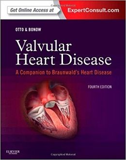
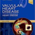
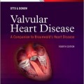
Valvular Heart Disease: A Companion to Braunwald’s Heart Disease, 4ed + Video
۲۰۱۴ BMA Medical Book Awards 1st Prize Award Winner in Cardiology category
Internationally renowned cardiologists Drs. Catherine M. Otto and Robert O. Bonow help you to apply all of the latest scientific knowledge and clinical strategies with Valvular Heart Disease, a companion to Braunwald’s Heart Disease. This indispensable resource for your everyday practice provides all of the knowledge you need to give your patients the most accurate diagnoses, the best possible heart disease treatment options, and the expert care they deserve.
“…This textbook is a key to all of us for rational decision-making and the optimal management of patients with valvular heart disease.” By Perfusion, Apr 2015
- Apply today’s best medical and surgical approaches with comprehensive, expert guidance on today’s full range of clinical heart disease treatments.
- Provide appropriate care to pediatric and pregnant patients with focused chapters on the unique needs of these special populations.
- Update your knowledge of these increasingly common heart diseases with brand-new chapters covering 3-dimensional anatomy of the aortic and mitral valves; clinical and genetic risk factors for calcific valve disease; risk assessment for aortic and mitral valve surgery; image-guided transcatheter valve procedures; optimal timing of valve interventions; state-of-the-art valve surgery; and much more.
- Apply the most current standards of practice with summaries of the ACC/AHA and new ESC valvular heart disease guidelines in each chapter.
- Vividly visualize heart valve anatomy and dysfunction, as well as important techniques and procedures, thanks to a multitude of detailed illustrations, full-color images, and online video content.
- Search the full text and access all the images online at Expert Consult.
Your purchase entitles you to access the web site until the next edition is published, or until the current edition is no longer offered for sale by Elsevier, whichever occurs first. Elsevier reserves the right to offer a suitable replacement product (such as a downloadable or CD-ROM-based electronic version) should access to the web site be discontinued.
Review
“This textbook – the fourth edition of “Valvular Heart Disease” – is the leading textbook in this field, providing the knowledge which conforms to the current guideline recommendations of the American and European Society of Cardiology and Cardio-Thoracic Surgery.
The editors of “Valvular Heart Disease” are among the world’s leaders in this field and they have selected outstanding authors who discuss, in depth, different topics of VHD. They systematically cover the pathogenesis, pathophysiology, clinical findings, imaging, natural history and treatment options.” By Perfusion, Apr 2015
Contents
۱ Epidemiology of Valvular Heart Disease
۲ Three-Dimensional Anatomy of the Aortic and Mitral Valves
۳ Cellular and Molecular Basis of Calcific Aortic Valve Disease
۴ Clinical and Genetic Risk Factors for Calcific Valve Disease
۵ Left Ventricular Adaptation to Pressure and/or Volume Overload
۶ Echocardiographic Evaluation of Valvular Heart Disease
۷ Evaluation of Valvular Heart Disease by Cardiac Catheterization and Angiocardiography
۸ Evaluation of Valvular Heart Disease by Cardiac Magnetic Resonance and Computed Tomography
۹ Basic Principles of Medical Therapy in the Patient with Valvular Heart Disease
۱۰ Risk Assessment for Valvular Heart Disease
۱۱ Aortic Stenosis
۱۲ Aortic Regurgitation
۱۳ The Bicuspid Aortic Valve and Associated Aortic Disease
۱۴ Surgical Approach to Diseases of the Aortic Valve and the Aortic Root
۱۵ Transcatheter Aortic Valve Implantation
۱۶ Imaging Guidance of Transcatheter Valve Procedures
۱۷ Rheumatic Mitral Valve Disease
۱۸ Myxomatous Mitral Valve Disease
۱۹ Secondary Mitral Regurgitation
۲۰ Mitral Regurgitation:
۲۱ Mitral Valve Repair and Replacement
۲۲ Transcatheter Mitral Valve Repair and Replacement
۲۳ Intraoperative Echocardiography for Mitral Valve Surgery
۲۴ Diseases of the Tricuspid and Pulmonic Valves
۲۵ Infective Endocarditis
۲۶ Prosthetic Heart Valves
۲۷ Valvular Heart Disease in Pregnancy
Video Contents
۱. Video 1-1A. Carcinoid
۲. Video 1-1B. Carcinoid
۳. Video 1-1C. Carcinoid
۴. Video 1-2. Aortic valve prolapse associated with mitral prolapse
۵. Video 1-3A. Cabergoline
۶. Video 1-3B. Cabergoline
۷. Video 1-4A. Antiphospholipid syndrome
۸. Video 1-4B. Antiphospholipid syndrome
۹. Video 1-5. Systemic lupus erythematosus
۱۰. Video 1-6. Failing biological valve
۱۱. Video 1-7. Endomyocardial fibrosis
۱۲. Video 1-8A. Traumatic tricuspid prolapse
۱۳. Video 1-8B. Traumatic tricuspid prolapse
۱۴. Video 1-9A. Pacemaker-induced tricuspid stenosis
۱۵. Video 1-9B. Pacemaker-induced tricuspid stenosis
۱۶. Video 1-9C. Pacemaker-induced tricuspid stenosis
۱۷. Video 1-10A. Parachute mitral valve
۱۸. Video 1-10B. Parachute mitral valve
۱۹. Video 1-11. Pacemaker endocarditis
۲۰. Video 1-12. Prosthetic valve endocarditis
۲۱. Video 1-13. Radiation-induced valve disease
۲۲. Video 6-1. Mitral stenosis jet on color-flow Doppler imaging
۲۳. Video 6-2A. Level of outflow obstruction
۲۴. Video 6-2B. Level of outflow obstruction
۲۵. Video 6-3A. Transesophageal echocardiography of a stenotic bicuspid aortic valve
۲۶. Video 6-3B. Transesophageal echocardiography of a stenotic bicuspid aortic valve
۲۷. Video 6-3C. Transesophageal echocardiography of a stenotic bicuspid aortic valve
۲۸. Video 6-4A. Bicuspid aortic valve
۲۹. Video 6-4B. Bicuspid aortic valve
۳۰. Video 6-5A.”Mild rheumatic mitral stenosis
۳۱. Video 6-5B.”Mild rheumatic mitral stenosis
۳۲. Video 6-6A. Planimetry of mitral valve area
۳۳. Video 6-6B. Planimetry of mitral valve area
۳۴. Video 6-7A. Three-dimensional transesophageal echocardiography of mitral stenosis
۳۵. Video 6-7B. Three-dimensional transesophageal echocardiography of mitral stenosis
۳۶. Video 6-8. Spontaneous left atrial contrast
۳۷. Video 6-9A. Mitral valve morphology
۳۸. Video 6-9B. Mitral valve morphology
۳۹. Video 6-10A.”Transesophageal echocardiography-guided balloon mitral commissurotomy
۴۰. Video 6-10B.”Transesophageal echocardiography-guided balloon mitral commissurotomy
More…
۱۵۰. Video 2-3: 3D transesophageal echocardiographic long-axis view video of a mitral valve with ischemic mitral regurgitation.
۱۵۱. Video 2-4 (Left). 3D transesophageal echocardiographic video of rheumatic mitral valve stenosis.
۱۵۲. Video 2-4 (Right). 3D transesophageal echocardiographic video of rheumatic mitral valve stenosis.
۱۵۳. Video 2-5 (Left Cropped). On the left is 3D transesophageal echocardiographic video of a normal aortic valve as seen from the aorta.
۱۵۴. Video 2-5 (Right Cropped). On the left is 3D transesophageal echocardiographic video of a normal aortic valve as seen from the aorta.
۱۵۵. Video 2-5 (Left Uncropped). On the left is 3D transesophageal echocardiographic video of a normal aortic valve as seen from the aorta.
۱۵۶. Video 2-5 (Right Uncropped). On the left is 3D transesophageal echocardiographic video of a normal aortic valve as seen from the aorta.
۱۵۷. Video 18-1. Transthoracic Echocardiogram (Black and White)
۱۵۸. Video 18-2. Transthoracic Echocardiogram (Color Doppler)
۱۵۹. Video 18-3. Transesophageal Echocardiogram
۱۶۰. Video 18-4. Transesophageal Echocardiogram 130-degrees.
۱۶۱. Video 18-5. Transesophageal Echocardiogram 130-degrees with Color Doppler
۱۶۲. Video 18-6. 3D Transesophageal echocardiography 3D
۱۶۳. Video 1. Functional MR Due to Mitral Valve Tenting
۱۶۴. Video 1. Functional MR Due to Ischemic Heart Disease
۱۶۵. Video 1. Pre-Cardiac Resynchronization Therapy (CRT)
۱۶۶. Video 2. Pre-Cardiac Resynchronization Therapy (CRT)
۱۶۷. Video 3. Pre-Cardiac Resynchronization Therapy (CRT)
۱۶۸. Video 4. Post-Cardiac Resynchronization Therapy (CRT)
۱۶۹. Video 5. Post Cardiac Resynchronization Therapy
۱۷۰. Video 1. Pre Atrial Functional MR
۱۷۱. Video 2. Pre-Atrial Functional MR
۱۷۲. Video 3: Post-Atrial Functional MR
۱۷۳. Video 4. Post-Cardiac Resynchronization Therapy (CRT)
۱۷۴. Video 21-1A. Most typical clinical and surigcal differences between fibroelastic deficiency and Barlow disease
۱۷۵. Video 21-1B. Most typical clinical and surigcal differences between fibroelastic deficiency and Barlow disease
۱۷۶. Video 21-2A. Most Typical Clinical And Surgical Differences Between Fibroelastic Deficiency and Barlow Disease
۱۷۷. Video 21-2B. Most Typical Clinical And Surgical Differences Between Fibroelastic Deficiency and Barlow Disease
۱۷۸. Video 21-3A. Most Typical Clinical And Surgical Differences Between Fibroelastic Deficiency and Barlow Disease
۱۷۹. Video 21-3B. Most Typical Clinical And Surgical Differences Between Fibroelastic Deficiency and Barlow Disease
۱۸۰. Video 21-4A. Currently most commonly applied surgical approaches to posterior leaflet prolapse
۱۸۱. Video 21-4B. Currently most commonly applied surgical approaches to posterior leaflet prolapse
۱۸۲. Video 21-4C. Currently most commonly applied surgical approaches to posterior leaflet prolapse
جهت مشاهده نسخه همراه با ویدئوهای کامل ویرایش ۲۰۲۱ این کتاب کلیک کنید
لینک کوتاه : https://bookbaz.ir/?p=85220
نویسنده : Catherine M. Otto MD , Robert O. Bonow MD MS
ناشر : Saunders; 4 edition
سال انتشار : 2014
زبان کتاب : انگلیسی
نوع فایل : MP4 + PDF (کیفیت اصلی)
تعداد صفحات : 494
(ISBN) شابک : 1455748609
قیمت کتاب درآمازون : $144.96
حجم فایل : 600 MB
