- فهرست کتاب ها
- قلب و عروق، قفسه سینه
- نورولوژی، جراحی مغز و اعصاب و علوم اعصاب
- چشم پزشکی، بینایی سنجی
- دندان پزشکی، دهان و فک و صورت
- دستگاه گوارشی و کبد
- گوش، حلق و بینی
- ریه و مجاری تنفسی
- ارولوژی و نفرولوژی
- ارتوپدی و روماتولوژی
- پوست و مو و ناخن (درماتولوژی)
- جراحی (عمومی،زیبایی و ..)
- زنان و زایمان
- پزشکی بازسازی و احیا
- بیماریهای عفونی و قارچی
- غدد و متابولیسم
- سرطان و تومور شناسی
- معاینات و علائم تشخیصی
- بیهوشی (عمومی و موضعی)
- رادیولوژی، پرتو نگاری، سونوگرافی، اسکن، MRI، تصویربرداری، ماموگرافی
- تروما (ارتوپدی، فک و صورت، مغزی)
- آموزش و تحقیقات پزشکی، آمادگی آزمون و منابع پزشکی
- آناتومی و فیزیولوژی
- ایمونولوژی و آلرژی
- پاتولوژی، سیتوپاتولوژی و سیتولوژی
- پروتز و اندام مصنوعی
- پزشکی کودکان، نوزادان و جنین شناسی
- سلول های بنیادی
- هماتولوژی (خون شناسی) و پزشکی انتقال خون
- چاقی، تغذیه و رژیم غذایی
- اخلاق پزشکی
- پزشک قانونی
- پیرپزشکی و پزشکی سالمندان
- مراقبت های ویژه، حاد و بحرانی
- پیوند اعضا
- داروسازی، داروشناسی و دارودرمانی
- روانپزشکی
- دامپزشکی
- پرستاری و دستیاری پزشک
- علوم آزمایشگاهی
- سم شناسی
- انگل شناسی
- ژن شناسی و ژن درمانی (ژنتیک)
- بافت شناسی و مهندسی بافت
- تکنولوژی و بیوتکنولوژی پزشکی
- توان بخشی، کاردرمانی، ورزش درمانی
- پزشکی سنتی و گیاهی و پزشکی جایگزین
- بهداشت و درمان
- مهندسی شیمی
- مهندسی پزشکی
- مهندسی صنایع غذایی
- فیزیک و بیوفیزیک
- زیست شناسی (بیولوژی سلولی و مولکولی)
- شیمی
- بیوشیمی (پزشکی، دارویی و …) و شیمی بالینی
- بیو انفورماتیک، انفورماتیک پزشکی
- ثبت سفارش کتاب
- نرم افزارهای مورد نیاز
- پشتیبانی بوک باز
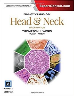
دانلود کتاب پاتولوژی تشخیصی: سر و گردن
Diagnostic Pathology: Head and Neck, 2ed
Part of the highly regarded Diagnostic Pathology series, this updated volume is a visually stunning, easy-to-use reference covering all aspects of head and neck pathology. Outstanding images―including gross and microscopic pathology, a wide range of stains, and detailed medical illustrations―make this an invaluable diagnostic aid for every practicing pathologist, resident, or fellow. This second edition incorporates the most recent clinical, pathological, histological, and molecular knowledge in the field to provide a comprehensive overview of all key issues relevant to today’s practice.
- Thoroughly updated content throughout including new coverage of oropharyngeal carcinoma; HPV-associated, mammary analogue secretory carcinoma; EWSR1 driven tumors; molecular pathways as targets for salivary duct carcinoma; and much more
- High-quality, carefully annotated color images (۵۰% new!) provide clinically and diagnostically important information on more than 315 new and evolving entities of the head and neck and endocrine organs
- State-of-the-art coverage of tumors, tumor development, and tumor genetics as well as normal histology, genetic testing, and new immunohistochemistry studies
- Fully integrated, searchable, and linked content between differential diagnostic categories is perfectly suited for residents, while updated genetic testing algorithms, new images, and outstanding graphics make this text ideal for both residents and practitioners
- Supporting studies are placed into clinical context, with tables and molecular flow charts that assist with management decisions and prognostic outcome predictions
- Time-saving reference features include bulleted text, a variety of test data tables, key facts in each chapter, annotated images, and an extensive index
- Expert Consult eBook version included with purchase. This enhanced eBook experience allows you to search all of the text, figures, images, and references from the book on a variety of devices.
Review
Posted on April 7th, 2016 by Book Reviews , American Journal of Neuroradiology
What is beautiful about this book… is the widespread integration of theimaging and classic pathology of the same entity. Not only does one get a deep sense of the underlying causes of the familiar imaging findings,but one is also educated in virtually all aspects of the disease, in the bullet point format…Added to this, of course, just as with everyother disease, are the key areas to understand, such as the terminologyused; the etiology and pathogenesis; clinical issues/concerns, including treatment; imaging (which, by the nature of these books, is not theprimary focus); macroscopic pathology; microscopic pathology (the keypart); other valuable tests; and differential diagnosis from a pathologic point view. …We see a number of clinical (ie, patient) images; so if you want to seewhat a gingival Kaposi sarcoma or a discord lupus looks like, you can. The book is full of such examples and makes studying each entity farmore interesting. Drs. Thompson, Wenig, Muller, and Nelson have done amagnificent job in putting all this material together, as have their 6contributing authors. Ten sections–Nasal Cavity and Paranasal Sinus;Pharynx; Larynx and Trachea; Oral Cavity; Salivay Glands; Jaw; Ear andTemporal Bone, Neck/Lymph (Nodes); Thyroid; and Parathyroid–encompassthe areas covered. For those who consult directly with neurosurgeons and head and neck surgeons or who attend their rounds or
participate intheir tumor boards, these books are invaluable. The histopathology, thedrawings, the charts, the descriptions, all are of the highest possibleeducational value. The book come with an eBook code that allows forsearchable items and duplicates enhanced images.
This may become one of the top resources forpathologists…Doody’s Listings and Reviews
Thisis an update of an in-depth book on non-neoplastic, benign, and malignant headand neck lesions. The previous edition was published in 2011.
Purpose
Thepurpose is to provide a reference with reliable and updated information on allaspects of head and neck lesions, which the authors have done.
Audience
Itis intended primarily for pathologists and pathology residents in training. Theauthors and contributors are all pathologists, some of whom trained me. Thebook also may be used as a reference for a broad spectrum of medicalpractitioners including, but not limited to, head and neck surgeons,oncologists, and radiologists with an interest in head and neck disease.
Features
Thebook’s more than 1,100 pages are organized into sections covering the nasalcavity and paranasal sinuses, pharynx, larynx and trachea, oral cavity,salivary glands, jaw, ear and temporal bone, neck, thyroid gland, andparathyroid glands. Each section begins with normal histology and anatomy ofthe site, followed by non-neoplastic lesions, benign neoplasms, and malignanttumors. The key facts and initial gallery of photographs enable the book’s useas a quick reference. Different diseases are presented in a highly structuredand bulleted format that includes definitions, etiology, clinical anddemographic parameters, treatment and prognosis, imaging findings, pathologicalfeatures, ancillary studies, and differential diagnoses. The quality of mostgross and microscopic images is excellent. The book also has useful tables thatsummarize important immunohistochemical findings of different neoplasms. Theindex is user friendly.
Assessment
This may become one of the top resources for pathologists with an interest in head and neck lesions. Since the previous edition, new entities have been described and more molecular tests developed, and I washappy to see these discussed. The bulleted format makes the book user friendlyin a busy practice.
The graphics and schematic drawings are extraordinarily well done and will prove to be very helpful for teaching purposes. Thephotomicrographs are of excellent quality and at times the well-placed arrows helpto explain the points the authors are trying to convey. I personally appreciatethe discussion (again in bulleted format) in the differential diagnosis sectioninstead of just a list. This will be a great addition to departmental andpersonal libraries.
Weighted Numerical Score: 92 – 4 Stars!
“For those who consult directly with neurosurgeons and head and neck surgeons or who attend their rounds or participate in their tumor boards, these books are invaluable. The histopathology, the drawings, the charts, the descriptions, all is of the highest possible educational value.”
-American Journal of Neuroradiology Blog (April 2016)
“This may become one of the top resources for pathologists with an interest in head and neck lesions…The bulleted format makes the book user friendly in a busy practice. The graphics and schematic drawings are extraordinarily well done and will prove to be very helpful for teaching purposes… This will be a great addition to departmental and personal libraries.”
– Carla Penner, DDS, FRCDC (University of Manitoba College of Medicine) Doody’s Score: 92 – 4 Stars!
Contents
SECTION 1: Nasal Cavity and Paranasal Sinuses
SECTION 2: Pharynx (Nasal, Oro-, Hypo)
SECTION 3 : Larynx and Trachea
SECTION 4: Oral Cavity
SECTION 5: Salivary Glands
SECTION 6: Jaw
SECTION 7 : Ear and Temporal Bone
SECTION 8: Neck (Soft Tissue and Lymph Nodes)
SECTION 9 Thyroid Gland
SECTION 10: Parathyroid Glands
لینک کوتاه : https://bookbaz.ir/?p=64054
نویسنده : Lester D. R. Thompson MD , Bruce M. Wenig MD
ناشر : Elsevier; 2 edition
سال انتشار : 2016
زبان کتاب : انگلیسی
نوع فایل : PDF (نسخه اصلی)
تعداد صفحات : 1193
(ISBN) شابک : 0323392555
قیمت کتاب درآمازون : $282.14
حجم فایل : 44 MB
کتاب های مرتبط:
 دانلود کتاب پاتولوژی و ژنتیک تومورهای غدد اندام درون ریز
Pathology and Genetics of Tumours of Endocrine Organs
 دانلود کتاب تشخیص افتراقی در پاتولوژی جراحی پستان
Differential Diagnoses in Surgical Pathology: Breast, 1ed
 دانلود کتاب پاتولوژی تشخیصی: انکولوژی مولکولی
Diagnostic Pathology: Molecular Oncology, 1ed
 دانلود کتاب پاتولوژی تشخیصی: استخوان
Diagnostic Pathology: Bone, 2ed
 دانلود کتاب پاتولوژی تشخیصی: نوروپاتولوژی
 Diagnostic Pathology: Neuropathology, 2ed
 دانلود کتاب پاتولوژی تشخیصی: کالبد گشایی بیمارستانی
Diagnostic Pathology: Hospital Autopsy, 1ed
 دانلود کتاب پاتولوژی تشخیصی: قفسه سینه
Diagnostic Pathology: Thoracic, 2ed
 دانلود کتاب پاتولوژی تشخیصی استخوان
Diagnostic Pathology: Bone, 3ed
 دانلود کتاب پاتولوژی تشخیصی: درماتوپاتولوژی غیرنئوپلاستیک
Diagnostic Pathology: Nonneoplastic Dermatopathology, 2ed
 دانلود کتاب دانشنامه سرطان (۳ جلدی)
Encyclopedia of Cancer, 3-Vol, 3ed
 دانلود کتاب پاتولوژی تشخیصی: پستان
Diagnostic Pathology: Breast, 2ed
 دانلود کتاب درماپاتولوژی الستون
Dermatopathology, 3ed
 دانلود کتاب نوروپاتولوژی جراحی عملی: یک رویکرد تشخیصی
Practical Surgical Neuropathology: A Diagnostic Approach, 2ed
 دانلود کتاب ژن درمانی سرطان توسط وکتورهای ویروسی و غیر ویروسی
Cancer Gene Therapy by Viral and Non-viral Vectors, 1ed
 دانلود کتاب تشخیص افتراقی در پاتولوژی جراحی: سیستم گوارشی
 Differential Diagnoses in Surgical Pathology: Gastrointestinal System, 1ed
 دانلود کتاب اصول و عمل رادیوتراپی انکولوژی پرز و برادی
Perez & Brady’s Principles and Practice of Radiation Oncology, 7ed
 دانلود کتاب ایمونوهیستوشیمی کاربردی در ارزیابی نئوپلاسم پوست
Applied Immunohistochemistry in the Evaluation of Skin Neoplasms, 1ed
 دانلود کتاب پاتولوژی پستان دابس
Breast Pathology, 3ed
 دانلود کتاب راهنمای پزشکی هریسون
Harrisons Manual of Medicine, 19ed
 دانلود کتاب پیشرفت در پاتولوژی جراحی: سرطان معده
 Advances in Surgical Pathology: Gastric Cancer, 1ed
 دانلود کتاب شیمی درمانی عملی – راهنمای چندرشته ای
Practical Chemotherapy – A Multidisciplinary Guide, 1ed
 دانلود کتاب جیبی انکولوژی
 Pocket Oncology, 2ed
 دانلود کتاب بافت شناسی و بیولوژی سلولی: درآمدی بر پاتولوژی
Histology and Cell Biology: An Introduction to Pathology, 5ed
 دانلود کتاب اصول پاتولوژی کلیه
Fundamentals of Renal Pathology, 2ed
 دانلود کتاب تشخیص افتراقی در پاتولوژی جراحی گتوزو
Gattuso’s Differential Diagnosis in Surgical Pathology, 4ed
 دانلود کتاب مباحث کلی آسیب شناسی دهان بالینی
Clinical Outline of Oral Pathology, 4ed
 دانلود کتاب سیتولوژی: اصول تشخیصی و همبستگی بالینی + ویدئو
Cytology: Diagnostic Principles and Clinical Correlates, 5ed + Video
 دانلود کتاب مراقبت های بعد از عمل از بیمار سرطانی
Perioperative Care of the Cancer Patient, 1ed
 دانلود کتاب انکولوژی عملی سر و گردن
Practical Head and Neck Oncology, 1ed
 دانلود کتاب آسیب شناسی دهان و فک و صورت نویل
Oral and Maxillofacial Pathology, 4ed
 دانلود کتاب تصویربرداری تخصصی: نئوپلاسم قفسه سینه
 Specialty Imaging: Thoracic Neoplasms, 1ed
 دانلود کتاب شیمی و فارماکولوژی داروهای ضد سرطان
Chemistry and Pharmacology of Anticancer Drugs, 2ed
 دانلود کتاب هماتوپاتولوژی
 Hematopathology, 2ed
 دانلود کتاب تمرین دزیمتری داخلی در پزشکی هسته ای
The Practice of Internal Dosimetry in Nuclear Medicine, 1ed
 دانلود کتاب پاتولوژی استخوان و بافت نرم
Bone and Soft Tissue Pathology, 2ed
 دانلود کتاب اصول پاتوفیزیولوژی بولاک
Principles of Pathophysiology, 2ed
 دانلود کتاب آسیب شناسی مولکولی و تشخیص سرطان (رشد سرطان و پیشرفت)
Molecular Pathology and Diagnostics of Cancer
 دانلود کتاب پیشرفت در پاتولوژی جراحی: سرطان آندومتر
 Advances in Surgical Pathology: Endometrial Carcinoma, 1ed
 دانلود کتاب تشخیص های دشوار در پاتولوژی پستان
 Difficult Diagnoses in Breast Pathology, 1ed
 دانلود کتاب عفونت و التهاب قرنیه
 Corneal Infection and Inflammation, 1ed
 دانلود کتاب آسیب شناسی دهان و فک و صورت: یک دلیل منطقی برای تشخیص و درمان
Oral and Maxillofacial Pathology: A Rationale for Diagnosis and Treatment, 2ed
 دانلود کتاب هماتوپاتولوژی: مبانی در آسیب شناسی تشخیصی
Hematopathology: Foundations in Diagnostic Pathology, 2ed
 دانلود کتاب آسیب شناسی پوست ویدون
Weedon’s Skin Pathology: Expert Consult, 3ed
 دانلود کتاب تشخیص پزشکی و درمان کارنت ۲۰۱۶ (ویرایش ۵۵)
CURRENT Medical Diagnosis and Treatment 2016
 دانلود کتاب پاتولوژی آندروود: یک رویکرد بالینی
Underwood’s Pathology: A Clinical Approach, 7ed
 دانلود کتاب اطلس تشخیصی پاتولوژی کلیه
 Diagnostic Atlas of Renal Pathology, 2ed
 دانلود کتاب پیشگیری و غربالگری سرطان
 Cancer Prevention and Screening, 1ed
 دانلود کتاب جراحی برای سرطان تخمدان
 Surgery for Ovarian Cancer, 3ed
 دانلود کتاب پاتولوژی پایه رابینز و کومار
Robbins & Kumar Basic Pathology, 11ed
 دانلود کتاب آناتومی بالینی: یک رویکرد مطالعه موردی
Clinical Anatomy: A Case Study Approach, 1ed
 دانلود کتاب رادیوتراپی انکولوژی کودکان
Pediatric Radiation Oncology, 6ed
 دانلود کتاب جراحی سر، گردن و تیروئید
 Head, Neck and Thyroid Surgery, 1ed
 دانلود کتاب اصول و عملکرد ذره درمانی
Principles and Practice of Particle Therapy, 1ed
 دانلود کتاب بورد بررسی تخصصی پاتولوژی تشریحی مک هیل
McGraw-Hill Specialty Board Review Anatomic Pathology, 1ed
 دانلود کتاب راهنمای انکولوژی بالینی
Manual of Clinical Oncology, 7ed
 دانلود کتاب پاتولوژی تشخیصی: سندرم های سرطان فامیلی
Diagnostic Pathology: Familial Cancer Syndromes, 2ed
 دنلود کتاب پاتولوژی تشخیصی مولکولی
Diagnostic Molecular Pathology, 1ed
 دانلود کتاب پاتولوژی گوارشی: اطلس و متن
Gastrointestinal Pathology: An Atlas and Text, 3ed
 دانلود کتاب بیماری های عفونی پیوند و انکولوژی کودکان
Pediatric Transplant and Oncology Infectious Diseases, 1ed
 دانلود کتاب سیتوپاتولوژی سر و گردن: هدایت سونوگرافی FNAC
Cytopathology of the Head and Neck: Ultrasound Guided FNAC, 2ed
 دانلود کتاب هماتولوژی بیماری های لنفاوی بدخیم ویلیامز
Williams Hematology Malignant Lymphoid Diseases, 1ed
 دانلود کتاب تصویربرداری تشخیصی انکولوژی
Diagnostic Imaging: Oncology, 2ed
 دانلود کتاب درماتوسکوپی تومورهای پوست غیر رنگی
Dermatoscopy of Non-Pigmented Skin Tumors: Pink – Think – Blink
 دانلود کتاب هماتوپاتولوژی مغز استخوان تشخیصی
Diagnostic Bone Marrow Haematopathology, 1ed
 دانلود کتاب اطلس رنگی آسیب شناسی پزشکی قانونی
 Color Atlas of Forensic Medicine and Pathology, 2ed
 دانلود کتاب ایمونوهیستوشیمی تشخیصی: کاربردهای ترانوستیک و ژنومیک
Diagnostic Immunohistochemistry: Theranostic and Genomic Applications, 6ed
 دانلود کتاب پیشرفت هایی در درمان سرطان
Advances in Cancer Therapy
 دانلود کتاب پاتولوژی ریوی عملی: یک رویکرد تشخیصی
Practical Pulmonary Pathology: A Diagnostic Approach, 3ed
 دانلود کتاب دانشنامه سرطان و جامعه سِیج
The SAGE Encyclopedia of Cancer and Society, 2ed
 دانلود کتاب راهنمای مراحل سرطان AJCC (ویرایش ۲۰۱۷)
AJCC Cancer Staging Manual, 8ed
 دانلود کتاب راهنمای انکولوژی بالینی UICC
UICC Manual of Clinical Oncology, 9ed
 دانلود کتاب درماتوپاتولوژی پرایمر بیماری های التهابی
Dermatopathology Primer of Inflammatory Diseases, 1ed
 دانلود کتاب آسیب شناسی چشم: اطلس و متن
Eye Pathology: An Atlas and Text, 2ed
 دانلود کتاب اطلس پاتولوژی آناتومیک با تصویربرداری
Atlas of Anatomic Pathology with Imaging
 دانلود کتاب نرم ژنومی در آسیب شناسی
Genomic Applications in Pathology
 دانلود کتاب اطلس رنگی طب داخلی یوسَتین
Color Atlas of Internal Medicine, 1ed
 دانلود کتاب انکولوژی مغز و اعصاب (اطلس عملی مغز و اعصاب)
Neuro-Oncology (Neurosurgical Operative Atlas), 2ed
 دانلود کتاب آسیب شناسی کالبد شکافی کودکان
Handbook of Pediatric Autopsy Pathology
 دانلود کتاب سرطان دهان: تشخیص و درمان
Oral Cancer: Diagnosis and Therapy, 2015th
 دانلود کتاب سیتوپاتولوژی جامع (ویرایش ۲۰۱۵) + ویدئو
Comprehensive Cytopathology, 4ed + Video
 دانلود کتاب اطلس روشهای جراحی در انکولوژی زنان
Atlas of Procedures in Gynecologic Oncology, 3ed
 دانلود کتاب سرطان های نادر
Textbook of Uncommon Cancer, 4ed
 دانلود کتاب آسیب شناسی میور
Muir’s Textbook of Pathology, 15ed
 دانلود کتاب آسیب شناسی: مفاهیم برای فیزیوتراپیست
Pathology: Implications for the Physical Therapist, 4ed
 دانلود کتاب بیماری های قلبی برانوالد: کتاب پزشکی قلب و عروق (۲ جلدی) + ویدئو
Braunwald’s Heart Disease: A Textbook of Cardiovascular Medicine, 2-Vol, 11ed + Video
 دانلود کتاب اطلس رنگی میکروجراحی نورینوما آکوستیک: تکنیک های آندوسکوپی کمکی – تکنیک های نوروناوبری – رادیوسرجری
Color Atlas of Microsurgery of Acoustic Neurinomas: Endoscope-Assisted Techniques – Neuronavigational Techniques – Radiosurgery, 1ed
 دانلود کتاب راهنمای روش های پرستاری سرطان رویال مارزدن
The Royal Marsden Manual of Cancer Nursing Procedures, 1ed
 دانلود کتاب رادیو حساس کننده ها و رادیو شیمی درمانی در درمان سرطان
Radiosensitizers and Radiochemotherapy in the Treatment of Cancer, 1ed
 دانلود کتاب پاتولوژی سر و گردن: مبانی در پاتولوژی تشخیصی
Head and Neck Pathology: Foundations in Diagnostic Pathology, 3ed
 دانلود کتاب پاتولوژی سر و گردن
Head and Neck Pathology, 1ed
 دانلود کتاب راهنمای پاتولوژی سر و گردن (۲ جلدی)
Textbook of Head and Neck Pathology, 2-Vol, 1ed
 دانلود کتاب گره های لنفاوی (راهنمای آسیب شناسی جراحی دموس)
Lymph Nodes (Demos Surgical Pathology Guides), 1ed
 دانلود کتاب پاتولوژی قفسه سینه عملی: بیماری های ریه، قلب و تیموس
Practical Thoracic Pathology: Diseases of the Lung, Heart, and Thymus, 1ed
 دانلود کتاب سرطان پروستات: علم و عمل بالینی
Prostate Cancer: Science and Clinical Practice, 2ed
 دانلود کتاب پاتوفیزیولوژی اختلالات خونی
Pathophysiology of Blood Disorders, 1ed
 دانلود کتاب سارکوما بافت نرم: رویکرد مبتنی بر الگو برای تشخیص
Soft Tissue Sarcomas: A Pattern-Based Approach to Diagnosis, 1ed
 دانلود کتاب پاتولوژی تشخیصی: پاتولوژی پیوند اعضا
Diagnostic Pathology: Transplant Pathology, 2ed
 دانلود کتاب پاتولوژی تشخیصی: گره های لنفاوی و لنفوم های خارج گره ای
Diagnostic Pathology: Lymph Nodes and Extranodal Lymphomas, 2ed
 دانلود کتاب پاتولوژی سر و گردن کودکان
Pediatric Head and Neck Pathology, 1ed
 دانلود کتاب آسیب شناسی تناسلی: مبانی پاتولوژی تشخیصی
Genitourinary Pathology: Foundations in Diagnostic Pathology, 2ed
 دانلود کتاب هماتوپاتولوژی: مبانی پاتولوژی تشخیصی
Hematopathology: Foundations in Diagnostic Pathology, 3ed
 دانلود کتاب پاتولوژی تشخیصی بیماری عفونی (ویرایش ۲۰۱۸)
Diagnostic Pathology of Infectious Disease, 2ed
 دانلود کتاب پاتولوژی تشخیصی آندومتر
Diagnostic Endometrial Pathology, 2ed
 دانلود کتاب پاتولوژی تشخیصی: مشاوره در حین عمل
Diagnostic Pathology: Intraoperative Consultation, 2ed
 دانلود کتاب بیوپسی حجم کم در تومورهای کودکان: اطلسی برای پاتولوژی تشخیصی
Small Volume Biopsy in Pediatric Tumors: An Atlas for Diagnostic Pathology, 1ed
 دانلود کتاب ایمونوهیستوشیمی: راهنمای فنی برای شیوه های فعلی
Immunohistochemistry: A Technical Guide to Current Practices, 1ed
 دانلود کتاب اطلس پاتولوژی سر و گردن ونیگ
Atlas of Head and Neck Pathology, 4ed
 دانلود کتاب پاتولوژی تشخیصی کبد و لوزالمعده
Diagnostic Pathology: Hepatobiliary and Pancreas, 2ed
 دانلود کتاب پاتولوژی تشخیصی تومورهای بافت نرم
Diagnostic Pathology: Soft Tissue Tumors, 3ed
 دانلود کتاب پاتولوژی تشخیصی زنان و زایمان
 Diagnostic Gynecologic and Obstetric Pathology, 3ed
 دانلود کتاب پاتولوژی گوارشی و کبدی: سری مبانی در پاتولوژی تشخیصی
Gastrointestinal and Liver Pathology: Foundations in Diagnostic Pathology Series, 3ed
 دانلود کتاب پستان بلاند و کوپلند: مدیریت جامع بیماری های خوش خیم و بدخیم + ویدئو
Bland and Copeland’s The Breast: Comprehensive Management of Benign and Malignant Diseases, 6ed + Video
 دانلود کتاب هماتولوژی-انکولوژی کودکان در کشورهای با منابع محدود
Pediatric Hematology-Oncology in Countries with Limited Resources, 2014th
 دانلود کتاب پاتولوژی تشخیصی: بیماری های کلیوی
Diagnostic Pathology: Kidney Diseases, 2ed
 دانلود کتاب پاتولوژی تشخیصی: بافت شناسی طبیعی
Diagnostic Pathology: Normal Histology, 2ed
 دانلود کتاب اطلس تشخیصی انکولوژی اِسکارین
Atlas of Diagnostic Oncology: Expert Consult, 4ed
 دانلود کتاب سیستم درجه بندی گِلیسون: راهنمای کامل برای متخصص پاتولوژی و پزشکان
The Gleason Grading System: A Complete Guide for Pathologist and Clinicians, 1ed
 دانلود کتاب تومورهای کولورکتال: اطلس هیستوپاتولوژی سکشن بزرگ
Colorectal Tumors: Atlas of Large Section Histopathology, 1ed
 دانلود کتاب تشخیص افتراقی در پاتولوژی جراحی سر و گردن
 Differential Diagnoses in Surgical Pathology: Head and Neck, 1ed
 دانلود کتاب بیماری پستان (۲ جلدی)
Breast Disease, 2-Vol, 2ed
 دانلود کتاب تشخیص های افتراقی در پاتولوژی جراحی: بافت نرم و استخوان
Differential Diagnoses in Surgical Pathology: Soft Tissue and Bone, 1ed
 دانلود کتاب هیستوپاتولوژی تشخیصی تومورها (۲ جلدی)
Diagnostic Histopathology of Tumors, 2-Vol, 4ed
 دانلود کتاب پاتولوژی تشخیصی لیندبرگ: تومورهای بافت نرم
Diagnostic Pathology: Soft Tissue Tumors, 2ed
 دانلود کتاب اطلس تشخیص افتراقی در پاتولوژی پستان
Atlas of Differential Diagnosis in Breast Pathology, 1ed
 دانلود کتاب پاتولوژی تومورهای ملانوسیتیک
 Pathology of Melanocytic Tumors, 1ed
 دانلود کتاب ایمونوهیستوشیمی تشخیصی: ترانوستیک و برنامه های ژنومی
Diagnostic Immunohistochemistry: Theranostic and Genomic Applications, 4ed
 دانلود کتاب سرطان سر و گردن: درمان، توانبخشی و نتایج
Head and Neck Cancer: Treatment, Rehabilitation, and Outcomes, 2ed
 دانلود کتاب پزشکی هسته ای در تشخیص و درمان بالینی (۲ جلدی)
Nuclear Medicine in Clinical Diagnosis and Treatment, 2-Vol, 3ed
 دانلود کتاب پاتولوژی زنان و زایمان (۲ جلدی)
Gynecologic and Obstetric Pathology, 2-Vol, 1ed
 دانلود کتاب اطلس تشخیصی نئوپلازی مزانشیمی پوستی
Diagnostic Atlas of Cutaneous Mesenchymal Neoplasia, 1ed
 دانلود کتاب پاتولوژی پستان
Breast Pathology, 2ed
 دانلود کتاب تشخیص های افتراقی در تومورهای پاتولوژی جراحی و شبیه سازهای آن
Differential Diagnoses in Surgical Pathology Tumors and their Mimickers, 1ed
 دانلود کتاب پزشکی سیسیل گلدمن (۲ جلدی)
Goldman-Cecil Medicine, 2-Vol, 25ed
 دانلود کتاب تشخیص اندودنتیکس ، پاتولوژی و طرح درمان
Endodontic Diagnosis, Pathology, and Treatment Planning: Mastering Clinical Practice, 2015th
 دانلود کتاب رادیوسرجری استریوتاکتیک و پرتو درمانی استریوتاکتیک بدن
Stereotactic Radiosurgery and Stereotactic Body Radiation Therapy, 1ed
 دانلود کتاب هماتولوژی: اصول اساسی و عمل + ویدئو
Hematology: Basic Principles and Practice, 8ed + Video
 دانلود کتاب پاتولوژی عملی پستان: روش تشخیصی
Practical Breast Pathology: A Diagnostic Approach, 1ed
 دانلود کتاب پزشکی تورنتو نوت (نسخه ۲۰۱۵)
Toronto Notes 2015 (Essential Med Notes), 31ed
 دانلود کتاب طبقه بندی تومورهای اندام های تولید مثل زن WHO
WHO Classification of Tumours of the Female Reproductive Organs, 4ed
 دانلود کتاب انکولوژی مولکولی: علل سرطان و اهداف درمانی
Molecular Oncology: Causes of Cancer and Targets for Treatment, 1ed
 دانلود کتاب برنامه درمانی در انکولوژی تابشی خان
Khan’s Treatment Planning in Radiation Oncology, 4ed
 دانلود کتاب تومورهای بافت نرم انزینگر و ویس
Enzinger and Weiss’s Soft Tissue Tumors, 7ed
 دانلود کتاب پاتوفیزیولوژی
Pathophysiology (Copstead), 5ed
 دانلود کتاب پاتوفیزیولوژی فوق العاده آسان!
Pathophysiology Made Incredibly Easy!, 5ed
 دانلود کتاب متاستاز سرطان: پایه بیولوژیک و درمان
Cancer Metastasis: Biologic Basis and Therapeutics
 دانلود کتاب تخصص بالینی آکسفورد (ویرایش ۲۰۱۶)
Oxford Handbook of Clinical Specialties, 10ed
 دانلود کتاب راهنمای هماتولوژی ویلیامز
Williams Manual of Hematology, 9ed
 دانلود کتاب راهنمای مراقبت از بازمانده سرطان
 Handbook of Cancer Survivorship Care, 1ed
 دانلود کتاب واژه نامه مصور پاتولوژی سم شناسی و علوم ایمنی
The Illustrated Dictionary of Toxicologic Pathology and Safety Science, 1ed
 دانلود کتاب آزمایشات بالینی: چشم انداز روش شناختی
Clinical Trials: A Methodologic Perspective, 3ed


