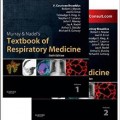
دانلود کتاب پزشکی تنفسی موری و نادل (۲ جلدی) + ویدئو
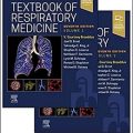
Murray & Nadel’s Textbook of Respiratory Medicine 2-Vol, 6ed + Video
Ideal for fellows and practicing pulmonologists who need an authoritative, comprehensive reference on all aspects of pulmonary medicine, Murray and Nadel’s Textbook of Respiratory Medicine offers the most definitive content on basic science, diagnosis, evaluation and treatment of the full spectrum of respiratory diseases.
- Full-color design enhances teaching points and highlights challenging concepts.
- Understand clinical applications and the scientific principles of respiratory medicine.
- Detailed explanations of each disease entity allow you to work through differential diagnoses.
- More than ۱,۰۰۰ figures and over ۲۰۰ videos and audio files.
- Key Points and Key Reading sections highlight the most useful references and resources for each chapter.
- An expanded sleep section now covers four chapters and includes control of breathing, consequences of sleep disruption, as well as obstructive and central apnea.
- New chapters in the Critical Care section cover Noninvasive Ventilation (NIV) and Extracorporeal Support of Gas Exchange (ECMO). New chapters focusing on diagnostic techniques now include Invasive Diagnostic Imaging and Image-Guided Interventions and Positron Emission Tomography, and a new chapter on Therapeutic Bronchoscopy highlights the interventional role of pulmonologists.
- Embedded videos feature thoracoscopy, therapeutic bronchoscopy, volumetric chest CT scans, and more. Brand-new audio files highlight normal and abnormal breath sounds and the separate components of cough.
- Expert Consult eBook version included with purchase. This enhanced eBook experience offers content updates, videos, review questions, and Thoracic Imaging Cases (TICs), all of which are easily navigable on any device for access on rounds or in the clinic.
Review
REVIEWS OF THE LAST EDITION:”Without a doubt, this text is a must for serious students of pulmonary medicine, whether in training, academic tenure, or mature clinical practice.” – JAMAREVIEW OF 4th EDITION:
‘It goes without saying that this is a thorough, in depth review of chest medicine and as far as I could see, leaves nothing out. I was pleased to see this is a book for active physicians rather than researchers or scientists. It is aimed sqaurely at the specialists but generalists such as GPs or specialists in other but related disciplines would also find this book a good reference source. I also liked the principal that associated non respiratory problems are covered in detail. Take for example, Sarcoidosis which has always been classified as a chest disease, but is also a multi system disease. The chapter on Sarcoidosis is an excellent review of the whole topic and not just the pulmonary aspects. In addition, there are excellent chapters on issues that you don’t always find such as air pollution, high altitude and diving medicine.
Importantly the book is readable, and releveant to physicians and adopts a well rounded approach to informing the doctor what he or she needs to know. The depth of detail acheived is just right and this book has an encylopedic feel to it. There are plenty of illusitrations in the book but in the main, this is primarily a text based reference source.’– June 2006 Dr Harry Brown www.univadis.com
About the Author
Contents
SECTION A: Anatomy and Development of the Respiratory Tract
SECTION B: Respiratory Physiology
SECTION C: Defense Mechanisms and Immunology
SECTION D: Respiratory Pathology and Inflammation
SECTION E: Diagnosis
SECTION F: Evaluation
SECTION G: Symptoms of Respiratory Disease and Their Management
SECTION H: Infectious Diseases of the Lungs
SECTION I: Obstructive Diseases
SECTION J: Neoplasms of the Lung
SECTION K: Disorders of the Pulmonary Circulation
SECTION L: Infiltrative and Interstitial Lung Diseases
SECTION M: Environmental and Occupational Hazards
SECTION N: Disorders of the Pleura
SECTION O: Disorders of the Mediastinum
SECTION P: Disorders of Sleep and Control of Breathing
SECTION Q: Respiratory Manifestations of Extrapulmonary Disorders
SECTION R: Management of Respiratory Failure
Video Contents
۱-۱. Cardiac pulsations across the lung
۱-۲. Lung hyperinlation caused by increasing positive end-expiratory pressure
۱-۳. Lung inlation determined by altering respiratory rate and peak inspiratory pressure
۱-۴. Re-expansion of collapsed lung parenchyma
۱-۵. Lung hyperinlation increases illing of the caudal vena cava
۱۱-۱. Infant withdrawal from facemask
۱۱-۲. Paciication of an unhappy patient
۱۱-۳. Mucociliary clearance measured with a gamma camera
۱۵-۱. Injury and repair of the alveolar epithelium
۱۶-۱. Normal lung sounds
۱۶-۲. Bronchial breath sounds
۱۶-۳. Tracheal breath sounds
۱۶-۴. Fine crackles
۱۶-۵. Coarse crackles
۱۶-۶A. Wheezing
۱۶-۶B. High-pitched wheeze
۱۶-۷. Rhonchi
۱۶-۸. Normal voice sounds followed by egophony
۱۶-۹. Normal voice sounds followed by bronchophony
۱۶-۱۰. Normal whispered sounds followed by whispered pectoriloquy
۱۶-۱۱. Pleural friction rub
۱۶-۱۲. Inspiratory and expiratory stridor
۱۶-۱۳. Chest hair rubbing against the diaphragm of the stethoscope
۱۶-۱۴. Subcutaneous emphysema
۱۶-۱۵. Bone crepitus
۱۸-۱. Fluoroscopic sniff test showing unilateral right diaphragmatic paralysis
۱۸-۲. Digital subtraction catheter pulmonary angiography
۱۸-۳. Normal CT angiography utilizing ECG gating (axial)
۱۸-۴. Normal CT angiography (coronal reformat)
۱۸-۵. Reduced-dose CT pulmonary angiography (CTPA)
۱۸-۶. Volumetric thin section imaging of a solitary mass detected at chest radiography
۱۸-۷A. CT features of benign causes of pulmonary nodules: rounded atelectasis (lung windows)
۱۸-۷B. CT features of benign causes of pulmonary nodules: rounded atelectasis (soft tissue windows)
۱۸-۸. Hereditary hemorrhagic telangiectasia with rteriovenous malformations
۱۸-۹. Randomly distributed small pulmonary nodules
۱۸-۱۰. Perilymphatic nodules in sarcoidosis
۱۸-۱۱A. Intrathoracic large airway neoplasia: axial CT
۱۸-۱۱B. Intrathoracic large airway neoplasia: virtual bronchoscopy
۱۸-۱۲. Air trapping on dynamic expiratory CT
۱۸-۱۳. Saddle pulmonary embolism with right ventricular strain
۱۸-۱۴. Coronal magnetic resonance angiogram of pulmonary embolism
۲۰-۱. Lung sliding sign of normal lung
۲۰-۲. Pulmonary edema with multiple B-lines seen diffusely
۲۰-۳. Pneumothorax with absence of key signs
۲۰-۴. Normal thickening of the diaphragm during inspiration
۲۰-۵. Longitudinal vascular scan with echogenic thrombus seen in the common femoral vein deep to the common femoral artery
۲۰-۶. Compression ultrasonography with no deep venous thrombosis
۲۰-۷. Hypoechoic venous thrombosis within a noncompressible vein
۲۰-۸. Pericardial effusion
۲۰-۹. Lung lapping sign of pleural effusions
۲۰-۱۰. Septations in a parapneumonic effusion
۲۰-۱۱. Swirling debris in a malignant pleural effusion
۲۰-۱۲. Heterogeneously echogenic pleural effusion with positive hematocrit sign
۲۰-۱۳. Diaphragmatic thickening and nodules in a malignant pleural effusion
۲۰-۱۴. Pleural catheter as hyperechoic linear structure
۲۲-۱. A normal bronchoscopy
۲۲-۲. Transbronchial needle aspiration of subcarinal lymph nodes via lexible bronchoscopy
۲۲-۳. Transbronchial needle aspiration via endobronchial ultrasound
۲۳-۱. Intubation using the rigid bronchoscope
۲۳-۲. Electrocautery with cautery snare
۲۳-۳. Argon plasma coagulation therapy of endobronchial papillomatosis
۲۳-۴. Neodymium:Yttrium-aluminum-garnet (Nd:YAG) laser incision of subglottic stenosis
۲۳-۵. Placement of a self-expanding stent in the right main-stem bronchus
۲۳-۶. Microdébrider used to remove granulation tissue at the tracheostomy stoma
۲۳-۷. Cryotherapy of right upper lobe squamous cell carcinoma
۲۴-۱. Thoracoscopy
۲۴-۲. Reexpansion of collapsed lung
۲۴-۳. Rigid thoracoscopy with 7-mm thoracoscope
۲۴-۴. Biopsy of parietal pleural lesion
۲۴-۵. Talc insuflation
۲۴-۶. Thoracoscopic inspection of a mesothelioma
۲۴-۷. Thoracoscopic biopsy using a rigid optical biopsy forceps via a single-port technique
۲۴-۸. Pleuroscopy in an adult with former asbestos exposure
۲۴-۹. Yellow nail syndrome
۲۴-۱۰. Empyema with septations
۲۴-۱۱. Fluorescein inhalation demonstrating apical blebs and porosity
۲۴-۱۲. Talc poudrage
۳۰-۱. Voiced or glottal phase of cough
۳۰-۲. Successive coughs
۳۰-۳. Whooping cough: child
۳۰-۴. Whooping cough: adult
۳۳-۱. S. intermedius pneumonia and empyema
۳۳-۲. Non-infectious “non-responding” pneumonia: invasive mucinous adenocarcinoma
۴۴-۱. Centriacinar emphysema with predominant apical lucency
۴۴-۲. Panacinar emphysema with predominant basilar lucency
۵۴-۱. Pulmonary artery sarcoma: imaging appearances
۵۶-۱A. Squamous papilloma of the trachea: Axial CT
۵۶-۱B. Squamous papilloma of the trachea: Coronal CT
۵۶-۲. Solitary ibrous tumor of the pleura
۵۷-۱. McConnell’s Sign, an echocardiographic sign seen with acute pulmonary embolism (PE), seen in comparison with a normal echocardiogram
۵۷-۲. Echocardiogram showing thromboemboli in right-sided chambers of the heart
۵۷-۳. CT pulmonary angiogram showing bilateral pulmonary emboli
۵۸-۱. Echocardiogram in pulmonary hypertension
۵۹-۱A. Short-axis echocardiogram of a normal heart
۵۹-۱B. Short-axis echocardiogram of a heart in a patient with pulmonary hypertension
۵۹-۲A. Apical four-chamber echocardiogram of a normal heart
۵۹-۲B. Apical four-chamber echocardiogram of a heart in a patient with pulmonary hypertension
۶۱-۱. Apical four-chamber view of an agitated saline contrast echocardiogram (Bubble Study)
۶۱-۲. Systemic-to-pulmonary vascular communication
۶۱-۳. Intralobar sequestration: anomalous systemic arterial supply
۶۲-۱. Radiographic CT images showing interstitial pulmonary edema
۶۲-۲A. Pulmonary Edema: CT at onset of symptoms
۶۲-۲B. Pulmonary Edema: CT at 2 weeks post-onset of symptoms
۶۲-۲C. Pulmonary Edema: CT at 45 days post-presentation
۶۳-۱. CT of combined pulmonary ibrosis and emphysema
۶۳-۲. CT showing peripheral consolidated opacities
۶۴-۱A. Centrilobular nodules in a patient with hypersensitivity pneumonitis
۶۴-۱B. Air trapping and small airway obstruction in a patient with hypersensitivity pneumonitis
۶۷-۱. Pulmonary alveolar microlithiasis
۶۸-۱. Idiopathic chronic eosinophilic pneumonia: chest CT indings
۷۳-۱. CT of complicated silicosis in soft tissue windows
۷۳-۲. CT of complicated silicosis in lung windows
۷۳-۳. Chest CT of silicosis and progressive massive ibrosis
۷۳-۴. Axial chest CT in a patient with asbestos exposure
۷۳-۵. Chest CT in a patient with chronic beryllium disease
۷۳-۶. Hard-metal lung disease on chest CT
۷۶-۱. Flail chest: paradoxical chest wall movement
۷۶-۲. Flail chest: post-rib ixation
۷۶-۳. Blunt thoracic traumatic injury: tracheobronchial injury
۷۷-۱. Periodic breathing at high altitude
۷۷-۲. Ataxia in high altitude cerebral edema
۷۹-۱. Pleural thickening with key features of malignancy
۷۹-۲. Pulmonary edema with interlobular septal thickening and bilateral effusions
۷۹-۳. Pericarditis with pericardial effusion and bilateral pleural effusions
۷۹-۴A. Pulmonary embolism, infarct and effusion (before resolution)
۷۹-۴B. Pulmonary embolism, infarct and effusions (after resolution)
۷۹-۵. Subdiaphragmatic abscess with associated pleural effusion
۸۰-۱. Empyema secondary to Streptococcus intermedius
۸۰-۲. Thoracoscopic surgical view of empyema
۸۱-۱. Chylothorax seen on pleuroscopy
۸۱-۲. Chest CT of chylothorax
۸۱-۳. Fibrothorax seen on pleuroscopy
۸۲-۱. Pleural tumor without effusion
۸۲-۲. Volume loss ipsilateral to pleural effusion caused by bronchial obstruction
۸۲-۳. Extrapleural clues for pleural malignancy
۸۲-۴. Mesothelioma in the setting of calciied pleural plaques (axial CT)
۸۲-۵. Mesothelioma with invasion into chest wall and diaphragm
۸۳-۱. Mediastinal mass in a patient with bronchogenic carcinoma and superior vena cava syndrome
۸۳-۲. Mediastinal mass in a patient with lymphoma and superior vena cava syndrome
۸۳-۳. Thymoma in a patient with myasthenia gravis
۸۳-۴. An anterior mediastinal teratoma
۸۳-۵. Thymolipoma
۸۳-۶. Omental fat hernia due to foramen of Morgagni hernia
۸۳-۷A. Magnetic resonance images: “Black Blood” appearance
۸۳-۷B. Magnetic resonance motion images: “White Blood” appearance
۸۳-۸. Utility of magnetic resonance for determining mediastinal involvement in bronchogenic malignancy
۸۳-۹. Seminoma
۸۳-۱۰. Embryonal cell carcinoma
۸۳-۱۱A. Schwannoma (axial CT)
۸۳-۱۱B. Schwannoma (coronal enhanced CT)
۸۳-۱۲. Extramedullary hematopoiesis containing fat
۸۳-۱۳. Mediastinal mass caused by aortic aneurysm
۸۳-۱۴. Mediastinal mass caused by enlargement of the azygos vein
۸۳-۱۵. Foramen of Morgagni hernia
۸۴-۱. Pneumomediastinum resulting from left mainstem bronchial fracture following motor vehicle collision
۸۴-۲. Venous perforation of a peripherally inserted venous catheter with entry into the mediastinum
۹۱-۱. Diffuse alveolar hemorrhage following stem cell transplantation
۹۱-۲. Peri-engraftment syndrome following stem cell transplantation
۹۴-۱. Transthoracic echocardiogram of severe pulmonary hypertension associated with sickle cell disease
۹۴-۲. Cardiac magnetic resonance image of severe pulmonary hypertension associated with sickle cell disease
۹۵-۱. Bulky goiter causing tracheal deviation
۹۵-۲. Micronodules of metastatic thyroid cancer
۹۷-۱. Diaphragmatic motion by ultrasound
۹۷-۲. Use of the mechanical insuflator-exsuflator
۹۸-۱. Pectus excavatum with right ventricular compression
۹۸-۲. Posterolateral lail chest
۱۰۰-۱. Demonstration of atelectasis from inadequate PEEP
۱۰۶-۱. Dynamic expiratory imaging in a patient with a left lung transplant for COPD
۱۰۶-۲. Inspiratory ilms of a patient with bilateral lung transplantation
۱۰۶-۳. Dynamic expiratory imaging in a patient with a bilateral lung transplant
جهت مشاهده نسخه همراه با ویدئوهای کامل ویرایش ۲۰۲۲ این کتاب کلیک کنید
لینک کوتاه : https://bookbaz.ir/?p=77415
نویسنده : V.Courtney Broaddus MD , Robert J. Mason MD
ناشر : Saunders; 6 edition
سال انتشار : 2015
زبان کتاب : انگلیسی
نوع فایل : MP4 + PDF (کیفیت اصلی)
تعداد صفحات : 2847
(ISBN) شابک : 1455733830
قیمت کتاب درآمازون : $319.07
حجم فایل : 1100 MB
