دانلود کتاب اطلس گوارش یامادا
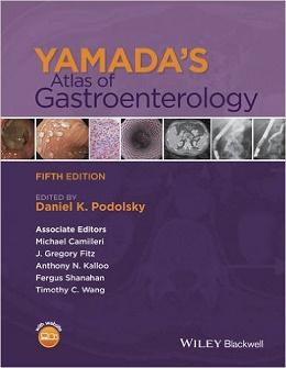
Yamada’s Atlas of Gastroenterology, 5ed
Access to accurate, high-quality images is vital for ensuring effective management of patients with GI complaints.
The fifth edition of Yamada’s Atlas of Gastroenterology sees the return of the gold-standard multi-media atlas that provides gastroenterologists with an outstanding visual tool covering all facets of the field. Every GI disorder from liver abscesses, endocrine neoplasms of the pancreas, to motility disorders of the esophagus are fully illustrated through the use of endoscopic ultrasonographs, computed tomography scans, magnetic resonance images, radionuclide images, and angiograms.
As the field of gastroenterology has evolved, a rich variety of modalities have contributed to progress in the practice of its art and science. This fifth edition of Yamada’s Atlas of Gastroenterology endeavors to provide illustrative examples that collectively provide a pictorial survey of the field. It accompanies the publication of the sixth edition of Yamada’s Textbook of Gastroenterology. With these newest editions of the original Textbook and companion Atlas, editorial responsibility has transitioned to a new set of editors; however, the goals of the books remain the same. The Textbook continues to provide comprehensive coverage of the field, incorporating significant advances since the last edition.
It is global in scope, both literally and figuratively. Figuratively in the sense that it covers all facets of gastroenterology as well as key ancillary relevant disciplines, and literally in considering the challenges encountered in the practice of gastroenterology as they may vary in different regions of the world. This newest edition of the Atlas is a companion intended to fulfill those same expectations. It is a compendium of supplementary images to the varied aspects of gastroenterology covered in the Textbook, spanning the many technologies and related fields important to the clinical practice of gastroenterology. These include gross and microscopic anatomy, and the full range of imaging modalities and endoscopies that inform the practice of gastroenterology. In addition, the Atlas includes graphics that supplement those of the Textbook to highlight key insights into many of the topics covered in the latter.
From the Back Cover
Access to accurate, high-quality images is vital for ensuring effective management of patients with GI complaints.
The fifth edition of Yamada’s Atlas of Gastroenterology sees the return of the gold-standard multi-media atlas that provides gastroenterologists with an outstanding visual tool covering all facets of the field. Every GI disorder from liver abscesses, endocrine neoplasms of the pancreas, to motility disorders of the esophagus are fully illustrated through the use of endoscopic ultrasonographs, computed tomography scans, magnetic resonance images, radionuclide images, and angiograms.
Full color throughout, it offers:
- Over 2000 exceptional images, organized by disease entity and therapeutic modality, including pathology slides, MRI and CT scans, endoscopy, EUS, and open surgery images.
- An outstanding international editorial team and 200 chapter authors, who are internationally renowned experts in their fields of expertise.
- On the companion website, www.yamadagastro.com/atlas:
- Every image as an electronic file to download – ideal for scientific presentations.
- ۲۸ downloadable high-quality surgical videos including transplantation and endoscopy clips, perfect for trainees to learn best practice techniques.
As the most comprehensive gastroenterology atlas available, Yamada’s Atlas of Gastroenterology is the perfect resource covering all GI disorders, and an essential purchase for all gastroenterologists seeking to improve their knowledge and clinical patient management.
Contents
CHAPTER 1: Esophagus: anatomy and structural anomalies
CHAPTER 2: Stomach and duodenum: anatomyand structural anomalies
CHAPTER 3: Small intestine: anatomyand structural anomalies
CHAPTER 4: Colon: anatomy and structural anomalies
CHAPTER 5: Pancreas: anatomy and structural anomalies
CHAPTER 6: Abdominal cavity: anatomy, structural anomalies, and hernias
CHAPTER 7: Gallbladder and biliary tract: anatomy and structural anomalies
CHAPTER 8: Liver: anatomy, microscopic structure, and cell types
CHAPTER 9: Motility disorders of the esophagus
CHAPTER 10: Gastroesophageal reflux disease
CHAPTER 11: Eosinophilic esophagitis
CHAPTER 12: Esophageal infections and disorders associated with acquired immunodeficiency syndrome
CHAPTER 13: Esophageal neoplasms
CHAPTER 14: Miscellaneous diseases of the esophagus: foreign bodies, physical injury, and systemic and dermatological diseases
CHAPTER 15: Disorders of gastric emptying
CHAPTER 16: Peptic ulcer disease
CHAPTER 17: Zollinger— Ellison syndrome
CHAPTER 16: Gastritis and gastropathy
CHAPTER 19: Tumors of the stomach
CHAPTER 20: Miscellaneous diseases of the stomach
CHAPTER 21: Dysmotility of the small intestine and colon
CHAPTER 22: Bacterial, viral, and toxic causes of diarrhea, gastroenteritis, and anorectal infections
CHAPTER 23: Chronic infections of the small intestine
CHAPTER 24: Disorders of epithelial transport, metabolism, and digestion in the small intestine
CHAPTER 25: Short bowel syndrome
CHAPTER 26: Tumors of the small intestine
CHAPTER 27: Miscellaneous diseases of the small intestine
CHAPTER 28: Ulcerative colitis: clinical manifestations and management
CHAPTER 29: Crohn’s disease: clinical manifestations and management
CHAPTER 30: Polyps of the colon and rectum
CHAPTER 31: Malignant tumors of the colon
CHAPTER 32: Polyposis syndromes
CHAPTER 33: Colorectal cancer screening
CHAPTER 34: Anorectal diseases
CHAPTER 35: Acute pancreatitis
CHAPTER 36: Chronic pancreatitis
CHAPTER 37: Hereditary diseases of the pancreas
CHAPTER 38: Cystic lesions of the pancreas
CHAPTER 39: Neuroendocrine tumors of the pancreas
CHAPTER 40: Gallstones
CHAPTER 41: Primary sclerosing cholangitis and other cholangiopathies
CHAPTER 42: Cystic diseases of the liver and biliary tract
CHAPTER 43: Tumors of the biliary tract
CHAPTER 44: Acute viral hepatitis
CHAPTER 45: Chronic hepatitis B viral infection
CHAPTER 46: Hepatitis C virus infection
CHAPTER 47: Drug-induced liver disease
CHAPTER 48: Autoimmune hepatitis
CHAPTER 49: Primary biliary cirrhosis
CHAPTER 50: Hemochromatosis
CHAPTER 51: Metabolic diseases of the liver
CHAPTER 52: Alcoholic liver disease
CHAPTER 53: Nonalcoholic fatty liver disease
CHAPTER 54: Hepatic fibrosis
CHAPTER 55: Approach to the patient with ascites and its complications
CHAPTER 56: Liver transplantation
CHAPTER 57: Hepatocellular carcinoma
CHAPTER 58: Liver abscess
CHAPTER 59: Vascular diseases of the liver
CHAPTER 60: Intraabdominal abscesses and fistulae
CHAPTER 61: Diseases of the peritoneum, retroperitoneum, mesentery, and omentum
CHAPTER 62: Obesity: treatment and complications
CHAPTER 63: Bariatric surgery and complications
CHAPTER 64: Complications of AIDS and other immunodeficiency states
CHAPTER 65: Gastrointestinal manifestations of immunological disorders
CHAPTER 66: Parasitic diseases: protozoa
CHAPTER 67: Helminthic infections of the gastrointestinal tract and liver
CHAPTER 68: Gastrointestinal manifestations of systemic diseases
CHAPTER 69: Skin lesions associated with gastrointestinal and liver diseases
CHAPTER 70: Oral manifestation of gastrointestinal diseases
CHAPTER 71: Intestinal ischemia and vasculitides
CHAPTER 72: Radiation injury in the gastrointestinal tract
CHAPTER 73: Upper gastrointestinal endoscopy
CHAPTER 74: Capsule and small bowel endoscopy
CHAPTER 75: Colonoscopy and flexible sigmoidoscopy
CHAPTER 76: Endoscopic retrograde cholangiopancreatography: diagnostic and therapeutic
CHAPTER 77: Gastrointestinal dilation and stent placement
CHAPTER 78: Management of upper gastrointestinal hemorrhage related to portal hypertension
CHAPTER 79: Endoscopic diagnosis and treatment of nonvariceal upper gastrointestinal hemorrhage
CHAPTER 80: Endoscopic therapy for polyps and tumors
CHAPTER 81: Laparoscopy and laparotomy
CHAPTER 82: Natural orifice translumenal endoscopic surgery [NOTES]
CHAPTER 83: Plain and contrast radiology
CHAPTER 84: Transabdominal sonography
CHAPTER 85: Endoscopic ultrasonography
CHAPTER 86: Computed tomography of the gastrointestinal tract
CHAPTER 87: Magnetic resonance imaging
CHAPTER 88: Positron emission tomography in the gastrointestinal tract
CHAPTER 89: Radionuclide imaging of the gastrointestinal tract
CHAPTER 90: Abdominal angiography
CHAPTER 91: Interventional radiology
CHAPTER 92: Liver biopsy and histopathological diagnosis
CHAPTER 93: Endoscopic mucosal biopsy: histopathological interpretation
لینک کوتاه : https://bookbaz.ir/?p=26000
نویسنده : Daniel K. Podolsky , Michael Camilleri
ناشر : Wiley-Blackwell; 5 edition
سال انتشار : 2016
زبان کتاب : انگلیسی
نوع فایل : PDF
تعداد صفحات : 979
(ISBN) شابک : 1118496434
قیمت کتاب درآمازون : $285.00
حجم فایل : $285.00
