دانلود کتاب تصویربرداری تشخیصی کودکان کافی (۲ جلدی) + ویدئو
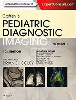
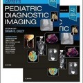
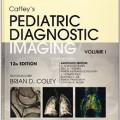
Caffey’s Pediatric Diagnostic Imaging, 2-Vol, 12ed + Video
Since 1945, radiologists have turned to Caffey’s Pediatric Diagnostic Imaging for the most comprehensive coverage and unparalleled guidance in all areas of pediatric radiology. Continuing this tradition of excellence, the completely revised 12th edition – now more concise yet still complete – focuses on the core issues you need to understand new protocols and sequences, and know what techniques are most appropriate for given clinical situations. Reviewed by: Office Journal of the European Paediatric Neurology Society Date: Jan 2014
“This is a thoroughly up-to-date text, divided into manageable topics, at a very reasonable price and I thoroughly recommend it to anyone who needs updating in the field of paediatrics or paediatric imaging.”
RAD, February 2014
- Determine the best modality for each patient with state-of-the art discussions of the latest pediatric imaging techniques.
- Quickly grasp the fundamentals you need to know through a more precise, streamlined format, reorganized by systems and disease processes, as well as “Teaching Boxes” that highlight key points in each chapter.
- Apply all the latest pediatric advances in clinical fetal neonatology techniques, technology, and pharmacology.
- Achieve accurate diagnoses as safely as possible. Increased coverage of MRI findings and newer imaging techniques for all organ systems emphasizes imaging examination appropriateness and safety.
- Reap the fullest benefit from the latest neuroimaging techniques including diffusion tensor imaging, fMRI, and susceptibility weighted imaging.
- Keep current with the latest pediatric radiological knowledge and evidence-based practices. Comprehensive updates throughout include new and revised chapters on prenatal imaging; newer anatomic and functional imaging techniques (including advances in cardiac imaging); disease classifications and insights into imaging disease processes; and advanced imaging topics in neurological, thoracoabdominal, and musculoskeletal imaging.
- Compare your findings to more than 10,000 high-quality radiology images.
- Access the full text online at Expert Consult including illustrations, videos, and bonus online-only pediatric imaging content.
Review
“Caffey’s Pediatric Diagnostic Imaging has been more than an imaging text and it has been extremely useful not only to radiologists, but also to clinicians in all medical and surgical specialties… More concist yet still complete – focuses on the core issues one needs to understand, such as new protocols and sequences, and how to know what techniques are most appropriate for given clinical situations…This text will obviously be of great interest not only to radiologists, also to those who work with children including all pediatric specialties. It is also extremely useful in countries with resource poor setting where there is shortage of well trained rdiologists in pediatric specialties.”
Journal of the European Paediatric Neurology Society, January 2014
“The writing is concise and clear, with plenty of examples covering all imaging modalities including diffusion MRI and PET/CT. It has quie a good strategic response on how to investigate a specific clinical issue. There are summaries of what the clinican needs to know and keypoints in relation to the topic which act as good revision guides and allow students to gain clear insight into the topic.The majority of the images are of high quality with clear annotation and good spatial resolution, with appropriate accompanying diagrams to add further clarity… The book is not only relevant to experts in paediatric imaging, but to anyone who has to undertake paediatric cases in their daily work. All paediatric departments should have this as a bench book. Junior doctors (in paediatrics/ radiology) and consultants whose on-call covers paediatrics would find this useful… In conclusion, this is a thoroughly up-to-date text, divided into manageable topics, at a very reasonable price and I thoroughly recommend it to anyone who needs updating in the field of paediatrics or paediatric imaging.”
RAD, February 2014
Contents
۱. Radiation Bioeffects, Risks, and Radiation Protection in Medical Imaging in Children
۲. Complications of Contrast Media
۳. Magnetic Resonance Safety
۴. Embryology, Anatomy, Normal Findings, and Imaging Techniques
۵. Prenatal, Congenital, and Neonatal Abnormalities
۶. Orbit Infection and Inflammation
۷. Orbital Neoplasia
۸. Nose and Sinonasal Cavities
۹. Embryology, Anatomy, Normal Findings, and Imaging Techniques
۱۰. Congenital and Neonatal Abnormalities
۱۱. Infection and Inflammation
۱۲. Neoplasia
۱۳. Embryology, Anatomy, Normal Findings, and Imaging Techniques
۱۴. Prenatal, Congenital, and Neonatal Abnormalities
۱۵. Infection and Inflammation
۱۶. Neoplasia
۱۷. Thyroid and Parathyroid
۱۸. Embryology, Anatomy, Normal Findings, and Imaging Techniques
۱۹. Prenatal Imaging
۲۰. Craniosynostosis, Selected Craniofacial Syndromes, and Other Abnormalities of the Skull
۲۱. Neoplasms, Neoplasm-like Lesions, and Infections of the Skull
۲۲. The Mandible
۲۳. Traumatic Lesions of the Skull and Face
۲۴. Embryology and Brain Development
۲۵. Magnetic Resonance Spectroscopy and Positron Emission Tomography
۲۶. Diffusion-Weighted Magnetic Resonance Imaging: Principles and Implementation in Clinical and Research Settings
۲۷. Functional Magnetic Resonance Imaging
۲۸. Perfusion Imaging and Magnetoencephalography
۲۹. Prenatal Imaging
۳۰. Neonatal Brain Injury
۳۱. Congenital Brain Malformations
۳۲. Hydrocephalus
۳۳. Inherited Metabolic and Neurodegenerative Disorders
۳۴. Infection and Inflammation
۳۵. Pediatric Brain Neoplasms
۳۶. Cerebrovascular Disorders
۳۷. Stroke
۳۸. Neuroimaging in Pediatric Epilepsy
۳۹. Trauma
۴۰. Embryology, Anatomy, and Normal Findings
۴۱. Spinal Cord Imaging Techniques
۴۲. Prenatal Imaging
۴۳. Congenital Abnormalities of the Spine
۴۴. Infections of the Spine and Spinal Cord
۴۵. Spinal Tumor and Tumorlike Conditions
۴۶. Vascular Lesions
۴۷. Trauma
۴۸. Embryology, Anatomy, and Normal Findings
۴۹. Imaging Techniques
۵۰. Prenatal Imaging and Intervention
۵۱. Upper Airway Disease
۵۲. Lower Large Airway Disease
۵۳. Congenital Lung Anomalies
۵۴. Pulmonary Infection
۵۵. Neoplasia
۵۶. Diffuse Lung Disease
۵۷. Systemic Conditions with Lung Involvement
۱۴۴. Child Abuse
۱۴۵. Sports Medicine
Video Contents
Video 1: 67-1 Aortic stenosis, sagittal and coronal time resolved
Video 2: 67-2 Aortic coarctation
Video 3: 67-3 Aortic stenosis
Video 4: 70-1 Cardiac MRI in a patient with previously repaired tetralogy of Fallot, view 1
Video 5: 70-2 Cardiac MRI in a patient with previously repaired tetralogy of Fallot, view 2
Video 6: 73-1 Large secundum atrial septal defect and right atrial and ventricular enlargement
Video 7: 73-2 Left-to-right ?ow across the secundum atrial septal defect
Video 8: 73-3 Flow across the large secundum atrial septal defect
Video 9: 75-1 Marginally hypoplastic left ventricle to evaluate for a two-ventricle versus single-ventricle repair
Video 10: 75-2 Bicuspid aortic valve in a 25-year-old with a history of aortic coarctation
Video 11: 75-3 Bicuspid aortic valve in a 25-year-old with a history of aortic coarctation, showing upward ballooning of the aortic valve lea?ets and turbulent poststenotic jet
Video 12: 75-4 Systolic ?ow void beyond the coarctation consistent with turbulent ?ow across the coarctation
Video 13: 76-1 Tetralogy of Fallot, status post-repair, status post–pulmonary valve replacement
Video 14: 76-2 Tetralogy of Fallot, status post-repair, status post–transcatheter pulmonary valve replacement
Video 15: 76-3 Markedly dilated right ventricle with overall mildly depressed systolic function
Video 16: 76-4 Dilated right ventricular out?ow tract with dyskinetic anterior wall in the region of the prior transannular patch
Video 17: 76-5 To-and-fro ?ow in the main pulmonary artery
Video 18: 76-6 Hypertrophy of the right ventricle with bowing of the interventricular septum into the lower pressure left ventricle
Video 19: 76-7 Anterior and leftward aorta arising from the morphologic right ventricle
Video 20: 76-8 Dephasing jet at the pulmonary valve
Video 21: 79-1 Tortuous aorta and iliac arteries with multiple stenoses
Video 22: 79-2 Diffuse arterial tortuosity and multiple stenoses
Video 23: 79-3 Thickening of the right and left ventricle myocardium with small ventricular cavities
Video 24: 79-4 Spectrum of anatomic abnormalities
Video 25: 80-1 Hypertrophic cardiomyopathy with severe asymmetric thickening of the left ventricular septal wall
Video 26: 81-1 Cardiac fbroma with Gorlin syndrome
Video 27: 86-1 Duodenal obstruction with hyperperistalsis
Video 28: 113-1 Fetal autosomal recessive polycystic kidney disease on ultrasound
Video 29: 131-1 Amniotic band syndrome
Video 30: 14-1 Normal fetal swallowing
Video 31: 98-1 Normal swallow function
Video 32: 98-2 Abnormal swallow function
Video 33: 101-1 Hypertrophied pylorus with peristalsis of the gas-flled stomach
Video 34: 101-2 Hypertrophied pylorus over time
Video 35: 103-1 Upper gastrointestinal evaluation of the duodenum
Video 36: 103-2 Midgut volvulus
Video 37: 103-3 Midgut volvulus
Video 38: 103-4 Midgut volvulus
Video 39: 108-1 Successful air enema reduction of intussusception in an infant
لینک کوتاه : https://bookbaz.ir/?p=77660
نویسنده : Brian D. Coley MD
ناشر : Saunders; 12 edition
سال انتشار : 2014
زبان کتاب : انگلیسی
نوع فایل : MP4 + PDF (کیفیت اصلی)
تعداد صفحات : 2505
(ISBN) شابک : 0323081762
قیمت کتاب درآمازون : $229.08
حجم فایل : 325 MB
