
دانلود کتاب استخوان های شکسته: اطلس رادیولوژیک شکستگی ها و دررفتگی ها
Broken Bones: The Radiologic Atlas of Fractures and Dislocations, 2ed
Broken Bones contains 434 individual cases and 1,101 radiologic images illustrating the typical and less typical appearances of fractures and dislocations throughout the body. The first chapter describes fractures and dislocations of the fingers, starting with fractures of the phalangeal tufts and progressing through the distal, middle, and proximal phalanges and the DIP and PIP joints. Subsequent chapters cover the metacarpals, the carpal bones, the radius and ulna, the elbow and upper arm, and the shoulder and thoracic cage. The cervical spine and the thoracic and lumbosacral spine are covered in separate chapters, followed by the pelvis, the femur, the knee and lower leg, the ankle, the tarsal bones, and the metatarsals and toes. The final three chapters cover the face, fractures and dislocations in children, and fractures and dislocations caused by bullets and nonmilitary blasts.
Book Description
About the Author
Contents
۱ Fractures and dislocations of the fingers
۲ Fractures and dislocations of the metacarpals
۳ Fractures and dislocations of the carpal bones
۴ Fractures and dislocations of the radius and ulna
۵ Fractures and dislocations of the elbow and arm
۶ Fractures and dislocations of the shoulder and thoracic cage
۷ Fractures and dislocations of the cervical spine
۸ Fractures and dislocations of the thoracolumbosacral spine
۹ Fractures and dislocations of the pelvis
۱۰ Fractures and dislocations of the femur
۱۱ Fractures and dislocations of the knee and leg
۱۲ Fractures and dislocations of the ankle
۱۳ Fractures and dislocations of the tarsal bones
۱۴ Fractures and dislocations of the metatarsals and toes
۱۵ Fractures and dislocations of the face
۱۶ Fractures and dislocations in children
۱۷ Fractures and dislocations caused by bullets and blasts (nonmilitary)
لینک کوتاه : https://bookbaz.ir/?p=65599
نویسنده : Felix S. Chew , Catherine Maldijan
ناشر : Cambridge University Press; 2 edition
سال انتشار : 2016
زبان کتاب : انگلیسی
نوع فایل : PDF (نسخه اصلی)
تعداد صفحات : 410
(ISBN) شابک : 1107499232
قیمت کتاب درآمازون : $89.99
حجم فایل : 95 MB
کتاب های مرتبط:
 دانلود کتاب پزشکی خانواده راکل + ویدئو
Textbook of Family Medicine, 9ed + Video
 دانلود کتاب جراحی تروما ارتوپدی: شکستگی و دررفتگی اندام فوقانی (جلد ۱)
Orthopaedic Trauma Surgery: Volume 1: Upper Extremity Fractures and Dislocations, 1ed
 دانلود کتاب بورد و بخش برای USMLE مراحل ۲ و ۳
Boards & Wards for USMLE Steps 2 & 3, 5ed
 دانلود کتاب سری کارشناسی ستون فقرات AO: ناهنجاری های ستون فقرات کودکان (جلد ۹)
AOSpine Masters Series, Volume 9: Pediatric Spinal Deformities, 1ed
 دانلود کتاب تصویربرداری ترومای اسکلتی
 Imaging Skeletal Trauma, 4ed
 دانلود کتاب بخاطر سپردن پزشکی
 Memorizing Medicine, 2ed
 دانلود کتاب ارتوپدی برای دستیاران پزشک + ویدئو
Orthopaedics for Physician Assistants, 2ed + Video
 دانلود کتاب MRCP PACES
 The MRCP PACES Handbook (MasterPass), 2ed
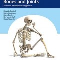 دانلود کتاب تصویربرداری از استخوان ها و مفاصل: یک روش مختصر و چند وجهی
Imaging of Bones and Joints: A Concise, Multimodality Approach, 1ed
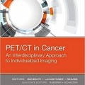 دانلود کتاب PET/CT در سرطان: رویکرد بین رشته ای به تصویربرداری فردی
PET/CT in Cancer: An Interdisciplinary Approach to Individualized Imaging, 1ed
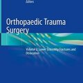 دانلود کتاب جراحی تروما ارتوپدی: شکستگی و دررفتگی اندام تحتانی (جلد ۲)
Orthopaedic Trauma Surgery: Volume 2: Lower Extremity Fractures and Dislocation, 1ed
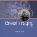 دانلود کتاب تصویربرداری پستان: ضروریات در رادیولوژی
Breast Imaging: The Requisites (Requisites in Radiology), 2ed
 دانلود کتاب تثبیت داخلی در پوکی استخوان
 Internal Fixation in Osteoporotic Bone, 1ed
 دانلود کتاب راهنمای مدیریت شکستگی AO: فیکساتور های داخلی
AO Manual of Fracture Management: Internal Fixators, 1ed
 دانلود کتاب پوکی استخوان مارکوس و فلدمن (۲ جلدی)
Osteoporosis, 2-Volume Set, Fourth Edition
 دانلود کتاب عمل جراحی اضطراری دست (ویرایش ۲۰۱۷)
Emergency Surgery of the Hand, 1ed
 دانلود کتاب تکنیک های پیشرفته در انتقال تصویر جراحی مغز و ستون فقرات
 Advanced Techniques in Image-Guided Brain and Spine Surgery, 1ed
 دانلود کتاب اندام فوقانی کودکان
 The Pediatric Upper Extremity, 2015th
 دانلود کتاب پزشکی گوش و حلق و بینی بالنگر: جراحی سر و گردن
Ballenger’s Otorhinolaryngology: Head and Neck Surgery, 18ed
 دانلود کتاب جراحی ستون فقرات حداقل تهاجمی + ویدئو
Minimally Invasive Spine Surgery, 1ed + Video
 دانلود کتاب طب اورژانس روزن: مفاهیم و تمرین بالینی (۲ جلدی)
Rosen’s Emergency Medicine: Concepts and Clinical Practice, 2-Vol, 8ed
 دانلود کتاب اطلس آسیب شناسی بافت نرم و استخوان: با بافت شناسی، سیتولوژی و رادیولوژی
Atlas of Soft Tissue and Bone Pathology: With Histologic, Cytologic, and Radiologic Correlations, 1ed
 دانلود کتاب تومورهای استخوان و بافت نرم: بررسی چند رشته با ارائه مورد
Bone and Soft Tissue Tumours: A Multidisciplinary Review With Case Presentations, 1ed
 دانلود کتاب ابزار دقیق ستون فقرات: تکنیک های جراحی
Spinal Instrumentation: Surgical Techniques, 1ed
 دانلود کتاب راهنمای ارتوپدی برای GP امروزی
An Orthopaedics Guide for Today’s GP, 1ed
 دانلود کتاب اطلس تعیین سن اسکلت: MRI دست و مچ دست در کودکان
Text-Atlas of Skeletal Age Determination: MRI of the Hand and Wrist in Children, 1ed
 دانلود کتاب جیبی ارتوپدی تروما و روماتولوژی چرچیل
Churchill’s Pocketbook of Orthopaedics, Trauma and Rheumatology, 2ed
 دانلود کتاب راهنمای آسان برای سوابق متمرکز برای OSCEs
The Easy Guide to Focused History Taking for OSCEs, 2ed
 دانلود کتاب بی ثباتی شانه در ورزشکار
 Shoulder Instability in the Athlete, 1ed
 دانلود کتاب تصویربرداری تشخیصی نقایص مادرزادی قلب
Diagnostic Imaging of Congenital Heart Defects, 1ed
 دانلود کتاب کاربردهای بالینی پرینت سه بعدی در جراحی پا و مچ پا
 Clinical Applications of 3D Printing in Foot and Ankle Surgery, 1ed
 دانلود کتاب ارتوپدی اضطراری سیمون + ویدئو
Simon’s Emergency Orthopedics, 7ed + Video
 دانلود کتاب اطلس جیبی اکوکاردیوگرافی
 Pocket Atlas of Echocardiography, 2ed
 دانلود کتاب سونوگرافی تشخیصی روماک (۲ جلدی)
Diagnostic Ultrasound, 2-Volume Set 6th Edition
 دانلود کتاب راهنمای اکوکاردیوگرافی: همگام با کتاب اکوکاردیوگرافی بالینی اوتو
Echocardiography Review Guide: Companion to the Textbook of Clinical Echocardiography, 3ed
 دانلود کتاب MRI اسکلتی عضلانی آسیف سفیودین
Asif Saifuddin’s Musculoskeletal MRI, 2ed
 دانلود کتاب سرطان پستان: هنر و علم تشخیص زود هنگام با ماموگرافی
Breast Cancer: The Art and Science of Early Detection with Mammography, 1ed
 دانلود کتاب اصول تصویربرداری کودکان
 Fundamentals of Pediatric Imaging, 2ed
 دانلود کتاب درمان جراحی و پزشکی پوکی استخوان
 Surgical and Medical Treatment of Osteoporosis, 1ed
 دانلود کتاب نورورادیولوژی: تشخیص های افتراقی کلیدی و سوالات بالینی
Neuroradiology: Key Differential Diagnoses and Clinical Questions, 2ed
 دانلود کتاب اشعه ایکس اسکلتی عضلانی برای دانشجویان و کارآموزان پزشکی
Musculoskeletal X-Rays for Medical Students and Trainees, 1ed
 دانلود کتاب رادیولوژی حوادث و اورژانس: راهنمای بقا
Accident and Emergency Radiology: A Survival Guide, 3ed
 دانلود کتاب مدیریت شکستگی برای مراقبت های اولیه
Fracture Management for Primary Care, 3ed
 دانلود کتاب مقدمه ای بر طب اورژانس بالینی
An Introduction to Clinical Emergency Medicine, 2ed
 دانلود کتاب وضعیت های اضطراری در قلب و عروق
Emergencies in Cardiology, 2ed
 دانلود کتاب تصویربرداری سر و گردن مقدماتی
Introductory Head and Neck Imaging, 1ed
 دانلود کتاب طب اورژانس تینتینالی: راهنمای جامع مطالعه + ویدئو
Tintinalli’s Emergency Medicine: A Comprehensive Study Guide, 7ed + DVD
 دانلود کتاب سونوگرافی تشخیصی: سر و گردن
Diagnostic Ultrasound: Head and Neck, 1ed
 دانلود کتاب اصول و تمرین پزشکی دیویدسون (ویرایش ۲۰۱۸)
Davidson’s Principles and Practice of Medicine, 23ed
 دانلود کتاب پزشکی گوش و حلق و بینی کودکان کامینگز
Cummings Pediatric Otolaryngology, 1ed
 دانلود کتاب راهنمای گام به گام اولتراسوند شکمی
 Abdominal Ultrasound: Step by Step, 3ed
 دانلود کتاب بیولوژیک در جراحی ارتوپدی
 Biologics in Orthopaedic Surgery, 1ed
 دانلود کتاب روش های کلارک در تصویربرداری تشخیصی
 Clark’s Procedures in Diagnostic Imaging, 1ed
 دانلود کتاب اطلس آندوسکوپی تشخیصی
Atlas of Diagnostic Endoscopy, 3ed
 دانلود کتاب اسکولیوز ایدیوپاتیک
 Idiopathic Scoliosis, 2ed
 دانلود کتاب اصول و عملکرد پزشکی دیویدسون
Davidson’s Principles and Practice of Medicine, 24ed
 دانلود کتاب هوش مصنوعی در پزشکی بالینی
AI in Clinical Medicine, 1ed
 دانلود کتاب خط مرزی و عادی اولیه پاتولوژی فین فریشمیت کوهلر/زیمر
Freyschmidt’s ,Koehler/Zimmer, Borderlands of Normal and Early Pathological Fin, 5ed
 دانلود کتاب غدد درون ریز عمومی و بالینی گرینسپن
Greenspan’s Basic and Clinical Endocrinology, 9ed
 دانلود کتاب اپیدمیولوژی بالینی تروما ارتوپدی
Clinical Epidemiology of Orthopaedic Trauma, 2ed
 دانلود کتاب فهرست تشخیص های افتراقی A تا Z فرنچ
French’s Index of Differential Diagnosis An A-Z, 16ed
 دانلود کتاب اصول رادیولوژی دهان و فک و صورت
Fundamentals of Oral and Maxillofacial Radiology, 1ed
 دانلود کتاب آنالیز تصویر رادیوگرافی
 Radiographic Image Analysis, 4ed
 دانلود کتاب بیومکانیک ستون فقرات: مفاهیم اساسی، اختلالات نخاعی و درمان
Biomechanics of the Spine: Basic Concepts, Spinal Disorders and Treatments, 1ed
 دانلود کتاب تعویض ترنس کاتتر دریچه آئورت
Transcatheter Aortic Valve Implantation, 1ed
 دانلود کتاب ۳ تفاوت برتر در پزشکی هسته ای
 Top 3 Differentials in Nuclear Medicine, 1ed
 دانلود کتاب جراحی ارتوپدی جامع چاپمن (۴ جلدی)
Chapman’s Comprehensive Orthopaedic Surgery, 4ed
 دانلود کتاب تصویربرداری تشخیصی: بیماری اسکلتی عضلانی غیر تروماتیک + ویدئو
Diagnostic Imaging: Musculoskeletal Non-Traumatic Disease, 3ed + Video
 دانلود کتاب توموگرافی پرتو مخروطی کامپیوتری در ارتودنسی: موارد مصرف، بینش و نوآوری
Cone Beam Computed Tomography in Orthodontics: Indications, Insights, and Innovations, 1ed
 دانلود کتاب ملزومات عملی شدت مدوله پرتو درمانی
Practical Essentials of Intensity Modulated Radiation Therapy, 3ed
 دانلود کتاب سونوگرافی عروقی عملی: راهنمای مصور
Practical Vascular Ultrasound: An Illustrated Guide, 1ed
 دانلود کتاب EMG آسان: راهنمای انجام مطالعات هدایت عصب و الکترومیوگرافی + ویدئو
Easy EMG: A Guide to Performing Nerve Conduction Studies and Electromyography, 2ed + Video
 دانلود کتاب رویکردهای بیولوژیکی برای ترمیم و بازسازی دیسک ستون فقرات برای پزشکان
Biological Approaches to Spinal Disc Repair and Regeneration for Clinicians, 1ed
 دانلود کتاب EKGs برای پرستار و دستیار پزشک
EKGs for the Nurse Practitioner and Physician Assistant, 2ed
 دانلود کتاب درسنامه برای Mrcog بخش ۱
Textbook for Mrcog – 1, 1ed
 دانلود کتاب تصویربرداری شکم: الزامات اصلی
Abdominal Imaging: The Core Requisites, 1ed
 دانلود کتاب اطلس روشهای ارتوپدی مداخله ای
 Atlas of Interventional Orthopedics Procedures, 1ed
 دانلود کتاب درماتولوژی برای ارائه دهنده مراقبت های اولیه
Dermatology for the Primary Care Provider, 1ed
 دانلود کتاب تصویربرداری در گوش و حلق و بینی
Imaging in Otolaryngology, 1ed
 دانلود کتاب تصویربرداری انکولوژیک: یک رویکرد چند رشته ای
Oncologic Imaging: A Multidisciplinary Approach, 2ed
 دانلود کتاب پوکی استخوان مارکوس و فلدمن (۲ جلدی)
Marcus and Feldman’s Osteoporosis, 5ed
 دانلود کتاب رادیولوژی پا و مچ پا
Foot and Ankle Radiology, 2ed
 دانلود کتاب اطلس تکنیک های جراحی تروما / اورژانس
Atlas of Trauma/Emergency Surgical Techniques, 1ed
 دانلود کتاب سری کارشناسی ستون فقرات AO: تغییر شکل ستون فقرات بزرگسالان (جلد ۴)
AOSpine Masters Series, Volume 4: Adult Spinal Deformities, 1ed
 دانلود کتاب گام به گام تا پزشکی (ویرایش ۲۰۱۶)
Stepup to Medicine, (Stepup Series), 4ed
 دانلود کتاب روشهای عملی در جراحی ارتوپدی و تروما
Operative Approaches in Orthopedic Surgery and Traumatology, 2ed
 دانلود کتاب اطلس رادیولوژی اورژانس: سیستم عروقی، قفسه سینه، شکم و لگن و سیستم تولید مثل
Atlas of Emergency Radiology: Vascular System, Chest, Abdomen and Pelvis, and Reproductive System 2015th
 دانلود کتاب آندوسونوگرافی + ویدئو
Endosonography, 3ed + Video
 دانلود کتاب سری کارشناسی ستون فقرات AO: عفونت های نخاعی (جلد ۱۰)
AOSpine Masters Series, Volume 10: Spinal Infections, 1ed
 دانلود کتاب ارزیابی فیزیکی ارتوپدی + ویدئو
Orthopedic Physical Assessment, 6ed + Video
 دانلود کتاب اصول پزشکی داخلی هریسون (۲ جلدی، ویرایش ۲۰۱۸)
Harrison’s Principles of Internal Medicine, 2-Vol, 20ed
 دانلود کتاب یک رویکرد تشریحی برای جراحی ستون فقرات کم تهاجمی
An Anatomic Approach to Minimally Invasive Spine Surgery, 2ed
 دانلود کتاب توانبخشی دست و اندام فوقانی (۲ جلدی) + ویدئو
Rehabilitation of the Hand and Upper Extremity, 2-Vol, 7ed + Video
 دانلود کتاب خلاصه ای از جراحی شانه
 Synopsis of Shoulder Surgery, 1ed
 دانلود کتاب تصویربرداری قفسه سینه (سری تشخیص مستقیم در رادیولوژی)
Thoracic Imaging (Dx-Direct), 1ed
 دانلود کتاب سونوگرافی تشخیصی: راهنما و اطلس مقدماتی
Endoscopic Ultrasound: An Introductory Manual and Atlas, 2ed
 دانلود کتاب مداخلات در پزشکی ریه
 Interventions in Pulmonary Medicine, 2ed
 دانلود کتاب اطلس بیهوشی منطقه ای هدایت اولتراسوند
Atlas of Ultrasound-Guided Regional Anesthesia, 3ed
 دانلود کتاب آلوپسی
 Alopecia, 1ed
 دانلود کتاب تصویربرداری تشخیصی پستان
Diagnostic Imaging: Breast, 3ed
 دانلود کتاب مراقبت و انتقال اورژانسی بیمار و مجروح
Emergency Care and Transportation of the Sick and Injured, 12ed
 دانلود کتاب فناوری اولتراسوند برای پزشکان بالینی
Ultrasound Technology for Clinical Practitioners, 1ed
 دانلود کتاب سونوگرافی مراقبت اورژانسی + ویدئو
Emergency Point-of-Care Ultrasound, 2ed + Video
 دانلود کتاب تصویربرداری دستگاه گوارش (سری تشخیص مستقیم در رادیولوژی)
Gastrointestinal Imaging (Direct Diagnosis in Radiology: DX-Direct!), 1ed
 دانلود کتاب آرنج تنیس: مدیریت بالینی
Tennis Elbow: Clinical Management, 2015th
 دانلود کتاب تصویربرداری رزونانس مغناطیسی: اصول فیزیکی و زیست شناختی
Magnetic Resonance Imaging: Physical and Biological Principles, 4ed
 دانلود کتاب ملزومات پزشکی بالینی کومار و کلارک
Essentials of Kumar and Clark’s Clinical Medicine, 6ed
 دانلود کتاب ارتوپدی جهانی
Global Orthopedics, 2ed
 دانلود کتاب ترمیم غضروف و حفظ مفصل زانو + ویدئو
Cartilage Repair and Joint Preservation of the Knee, 2ed + Video
 دانلود کتاب اولتراسوند تشخیصی اسکلتی عضلانی و راهنمای تزریق
Diagnostic Musculoskeletal Ultrasound and Guided Injection, 1ed
 دانلود کتاب اطلس جیبی آناتومی مقطعی: سر و گردن
Pocket Atlas of Sectional Anatomy: Head and Neck, 3ed
 دانلود کتاب اطلس آموزشی تصویربرداری شکم
Teaching Atlas of Abdominal Imaging, 1ed
 دانلود کتاب موارد رادیولوژی مغز و اعصاب
Neuroradiology Cases
 دانلود کتاب موارد اکوکاردیوگرافی عملی بالینی
Practical Bedside Echocardiography Cases, 1ed
 دانلود کتاب اطلس ویدئویی جراحی ستون فقرات
 Video Atlas of Spine Surgery, 1ed
 دانلود کتاب راهنمای روش های هیستولوژی برای استخوان و غضروف
Handbook of Histology Methods for Bone and Cartilage, 1ed
 دانلود کتاب ۱۰۱ راه حل MRI مغز
 ۱۰۱MRI Brain Solutions, 1ed
 دانلود کتاب تصویربرداری تشخیصی: بیماری غیر تروماتیک اسکلتی عضلانی
Diagnostic Imaging: Musculoskeletal Non-Traumatic Disease, 2ed
 دانلود کتاب پرتو درمانی والتر و میلر: فیزیک پرتو، درمان و انکولوژی
Walter and Miller’s Textbook of Radiotherapy: Radiation Physics, Therapy and Oncology, 7ed
 دانلود کتاب تصویربرداری استخوان گیجگاهی
 Imaging of the Temporal Bone, 4ed
 دانلود کتاب روماتولوژی کلی و فرشتین (۲ جلدی، ویرایش ۲۰۱۷)
Kelley and Firestein’s Textbook of Rheumatology, 2-Vol, 10ed
 دانلود کتاب مداخله انکولوژی (راهنمای عملی در مداخلات رادیولوژی)
Interventional Oncology (Practical Guides in Interventional Radiology), 1ed
 دانلود کتاب Vascular Ultrasound: B-Mode, Color Doppler and Duplex Ultrasound, Contrast-Enhanced Ultrasound, 1ed + Video
 دانلود کتاب پاتولوژی ارتوپدی عملی: یک روش تشخیصی
Practical Orthopedic Pathology: A Diagnostic Approach, 1ed
 دانلود کتاب تصویربرداری زنان و زایمان کوپل: سری کارشناس رادیولوژی
Obstetric Imaging: Expert Radiology Series, 1ed
 دانلود کتاب مراقبتهای ویژه سیویتا، تیلور و کربی
Civetta, Taylor and Kirby’s Critical Care, 4ed
 دانلود کتاب روش های جراحی در ارتوپدی اطفال تاچیان + ویدئو
Tachdjian’s Procedures in Pediatric Orthopaedics, 1ed + Video
 دانلود کتاب بیهوشی برای نوزادان و کودکان اسمیت
Smith’s Anesthesia for Infants and Children, 9ed
 دانلود کتاب بیماری های آئورت: اطلس تصویربرداری تشخیصی بالینی
Aortic Diseases: Clinical Diagnostic Imaging Atlas, 1ed
 دانلود کتاب تصویربرداری کودکان (سری موارد رادیولوژی)
Pediatric Imaging (RadCases), 1ed
 دانلود کتاب تسلط بر ۱۲-Lead EKG
Mastering the 12-Lead EKG, 2ed
 دانلود کتاب تفسیر رادیوگرافی قفسه سینه در بیماران قلبی کودک
Chest Radiographic Interpretation in Pediatric Cardiac Patients, 1ed
 دانلود کتاب معرفی روش های بالینی نتر + ویدئو
Netter’s Introduction to Clinical Procedures, 1ed + Video
 دانلود کتاب تشخیص و درمان پزشکی کارنت ۲۰۲۲ + ویدئو
CURRENT Medical Diagnosis and Treatment 2022, 61ed + Video
 دانلود کتاب بلوک عصب محیطی و آناتومی برای بیهوشی منطقه ای هدایت سونوگرافی هادزیک + ویدئو
Hadzic’s Peripheral Nerve Blocks and Anatomy for Ultrasound-Guided Regional Anesthesia, 2ed + Video
 دانلود کتاب جراحی ستون فقرات: ترفندهای حرفه ای + ویدئو
Spine Surgery: Tricks of the Trade, 3ed + Videos
 دانلود کتاب راهنمای مدیریت شکستگی – مچ دست
Manual of Fracture Management – Wrist, 1ed
 دانلود کتاب اختلالات زانو نویِس: جراحی، توانبخشی، نتایج بالینی
Noyes’ Knee Disorders: Surgery, Rehabilitation, Clinical Outcomes, 1ed
 دانلود کتاب موارد چالش برانگیز در تصویربرداری اسکلتی عضلانی
 Challenging Cases in Musculoskeletal Imaging, 1ed
 دانلود کتاب اولتراساوند عمومی و عروقی: مرور موردی
General and Vascular Ultrasound: Case Review, 3ed
 دانلود کتاب سندرم های حاد کرونری
 Acute Coronary Syndromes, 1ed
 دانلود کتاب نقرس
 Gout, 1ed
 دانلود کتاب مدیریت شکستگی برای مراقبت های اولیه و پزشکی اورژانس
Fracture Management for Primary Care and Emergency Medicine, 4ed
 دانلود کتاب مرور بورد انکولوژی: راهنمای مطالعه و پرسش و پاسخ
Oncology Board Review: Blueprint Study Guide and Q&A, 2ed
 دانلود کتاب بهترین آزمون فری: راهنمای عملی برای پزشکی آزمایشگاهی بالینی و تصویربرداری تشخیصی
Ferri’s Best Test: A Practical Guide to Clinical Laboratory Medicine and Diagnostic Imaging, 5ed
 دانلود کتاب ارزیابی ایمنی تجویز
 Get ahead! The Prescribing Safety Assessment, 1ed
 دانلود کتاب ماموگرافی عملی MR: امآرآی با وضوح بالا پستان
Practical MR Mammography: High-Resolution MRI of the Breast, 2ed
 دانلود کتاب درمان از دست دادن استخوان استابولوم و ناپیوستگی مزمن لگن
Treatment of Acetabular Bone Loss and Chronic Pelvic Discontinuity, 1ed
 دانلود کتاب راهنمای مراقبت اورژانس شیهی
Sheehy’s Manual of Emergency Care, 7ed
 دانلود کتاب معاینه بالینی مکلود
Macleod’s Clinical Examination, 13ed