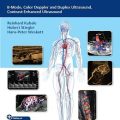
دانلود کتاب Vascular Ultrasound: B-Mode, Color Doppler and Duplex Ultrasound, Contrast-Enhanced Ultrasound, 1ed + Video
Vascular Ultrasound: B-Mode, Color Doppler and Duplex Ultrasound, Contrast-Enhanced Ultrasound An interdisciplinary guide to vascular color flow imaging and CEUS organized by anatomic region
The indications for vascular color duplex sonography (CDS) have expanded in recent years due to the availability of power Doppler, B-flow, ultrasound contrast agents, 3D reconstruction techniques, and fusion with other imaging modalities. CDS enables close-interval follow-ups after interventional procedures with improved prognoses. Edited by Reinhard Kubale, Hubert Stiegler, and Hans-Peter Weskott, Vascular Ultrasound: B-Mode, Color Doppler and Duplex Ultrasound, Contrast-Enhanced Ultrasound starts with the basic principles of diagnostic ultrasound physics and technology, followed by invaluable tips on equipment settings, possible artifacts, and limitations; hemodynamic essentials; and the use of ultrasound contrast agents. Subsequent chapters organized by anatomic region provide coverage of all peripheral and abdominal arterial and venous vascular regions; microcirculation and tumor perfusion; kidney and liver diseases; vascular malformations; non-atherosclerotic vascular diseases; the use of contrast-enhanced ultrasound (CEUS) in biliary, intestinal, splenic, and pediatric diseases; and novel/future techniques.
Key Features
- Contributions from interdisciplinary experts in angiology, neurology, radiology, vascular surgery, gastroenterology, nephrology, phlebology, rheumatology, laser medicine, and physics
- In-depth guidance on examination techniques, findings, and potential pitfalls and how to avoid them
- A wealth of comparative CT, MRI, and angiography CDS images and 34 videos enhance the understanding of impacted anatomy and the ability to master techniques and make accurate diagnoses
Video Contents
۲.۱ Increasing sound absorption by an intensely enhancing mass.
۳.۱ Jet stream behind a high grade echolucent stenosis of carotid artery.
۳.۲ Confetti signs due to high grade stenosis of celiac trunk and superior mesenteric artery
۵.۱ Floating thrombus in the carotid artery.
۵.۲ Mirror-image artifact in the carotid artery.
۵.۳ Coiling of the internal carotid artery with a 360 degrees loop.
۵.۴ Distal occlusion of the common carotid artery with a patent proximal stump.
۵.۵ Occlusion of the internal carotid artery caused by local thrombosis.
۵.۶ Occlusion of the internal carotid artery caused by embolism.
۵.۷ Distal occlusion of the internal carotid artery.
۵.۸ Dissection of the common carotid artery arising from a Stanford type A aortic dissection: transverse scan.
۵.۹ Stenosis in the V2 segment of the vertebral artery caused by a dissection with an eccentric wall hematoma.
۵.۱۰ Incomplete subclavian steal due to proximal subclavian artery stenosis, characterized by systolic deceleration
۵.۱۱ Dissection in the V2 segment of the vertebral artery with an eccentric wall hematoma.
۵.۱۲ Giant-cell arteritis of the vertebral artery, characterized by concentric, hypoechoic wall thickening.
۷.۱ Short embolic occlusion of a digital artery in the right index finger, with compensatory collateral blood flow
۷.۲ Scan of the femoral bifurcation in a 70-year-old man with Candida endocarditis and blue toe syndrome.
۷.۳ Bilocular pseudoaneurysm in the left groin after cardiac catheterization.
۷.۴ Mobile, nonocclusive thrombus in an enlarged Cockett perforator.
۷.۵ Floating thrombus in the profunda femoris vein.
۷.۶ Plantar thrombosis of the posterior tibial veins distal to the medial malleolus.
۸.۱ A 77-year-old woman with upper limb claudication and an extremely elevated erythrocyte sedimentation rate (ESR).
۸.۲ Vasculitis of the common carotid artery (CCA) in a 68-year-old woman with homogeneous wall thickening of the left CCA
۸.۳ For more than 1 year, this 62-year-old athletic diabetic male experienced severe, cramping pains in the anterior compartment of his left leg
۹.۱ Sweep from the wrist to the upper arm displays multiple arteriovenous shunts associated
۹.۲ The arteriovenous malformations in the thenar region are imaged by immersion in a warm-water bath.
۱۰.۱ Mycotic aneurysm with considerable enhancement of the inflamed surrounding tissue.
۱۰.۲ Early arterial phase after contrast agent administration (see Fig. 10.30).
۱۱.۱ Selective mesentericography in the arterial and venous phases.
۱۲.۱ Floating thrombus in the inferior vena cava.
۱۲.۲ Axial computed tomography (CT) slices show opacified, convoluted venous channels in the chest wall and mediastinum.
۱۲.۳ Coronal computed tomography (CT) slices show opacified, convoluted venous channels in the chest wall and mediastinum.
۱۲.۴ Retrograde flow in the portal vein (coded in blue) resulting from obstruction of the hepatic veins by the extracorporeal membrane…
۱۲.۵ Univentricular heart with secondary liver changes after surgical repair (see also Fig. 12.39d).
لینک کوتاه : https://bookbaz.ir/?p=377302
نویسنده : Reinhard Kubale
ناشر : Thieme; 1st edition
سال انتشار : 2023
زبان کتاب : انگلیسی
نوع فایل : MP4 + PDF (کیفیت اصلی)
تعداد صفحات : 576
(ISBN) شابک : 3132405434
قیمت کتاب درآمازون : $249.10
حجم فایل : 920 MB


