
دانلود کتاب اطلس عکاسی منطقه ای ویژگی های غیر متریک و آناتومیک اسکلت انسانی
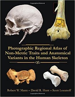
Photographic Regional Atlas of Non-Metric Traits and Anatomical Variants in the Human Skeleton, 1ed
Photographic Regional Atlas of Non-metric Traits and Anatomical Variants provides a unique collection of photographs derived from a broad array of novel skeletal specimens from across the globe. This atlas depicts skeletal features that are compiled to facilitate simple and direct access to some of the most interesting specimens currently known. This reference book is intended for clinicians, anatomists, anthropologists, forensic scientists, pathologists, biologists and other allied medical professionals who are fascinated with the expression of morphological features of the skeleton. It is particularly useful to the human biologist investigating genetic relatedness among and between skeletal samples utilizing non-metric trait analyses since this atlas provides a comprehensive visual guide for not only the identification and nomenclature of skeletal morphological features, but also for the appreciation of the range of anatomical expression. Photographic Regional Atlas of Non-metric Traits and Anatomical Variants draws from skeletal features observed from over 10,000 skeletons in collections throughout the world and provides a comprehensive yet concise presentation for rapid and reliable referral. Traits are arranged and presented based on skeletal region further facilitating ease-of-use for the reader when attempting to identify a feature of interest. Photographs are vividly displayed further enhancing the reader’s ability to compare the standard reference to a desired feature. The authors draw on their own decades of experience in skeletal anatomy to provide the best photographic atlas available for referencing daunting anatomical variations and non-metric trait morphology. As a result, Photographic Regional Atlas of Non-metric Traits and Anatomical Variants provides a one-of-a-kind reference that serves as a crucial component in the pursuit of skeletal anomaly research and education.
Review
Contents
Chapter 1 FRONTAL VIEW OF THE SKULL
Chapter 2 RIGHT LATERAL VIEW OF THE SKULL
Chapter 3 LEFT LATERAL VIEW OF THE SKULL
Chapter 4 SUPERIOR VIEW OF THE SKULL
Chapter 5 OCCIPITAL VIEW OF THE SKULL
Chapter 6 EMDOCRANIAL VIEW OF THE SKULL
Chapter 7 BASILAR (INFERIOR] VIEW OF THE SKULL
Chapter 6 MANDIBLE AND TEETH. HYOID, MAXILLA AND TEETH
Chapter 9 SHOULDER, ARM AND HAND[ CLAVICLE, SCAPULA, HUMERUS, RADIUS, ULNA, CARPAL]
Chapter 10 STERNUM, SPINE AND PELVIS( STERNUM, VERTEBRA, HIP AND SACRUM]
Chapter 11 LEG AND FOOT (FEMUR, TIBIA, FIBULA, PATELLA, TARSAL, METATARSAL, PHALANX]
Chapter 12 A METHOD FOR REMOVING SOFT TISSUE FROMA HUMAN RIB CAGE WITH BLEACH
Chapter 13 UNUSUAL COMBINATION OF SKELETAL VARIANTS INAN ADULT THAI MALE (KKU]: A PHOTOGRAPHIC CASE REPORT
لینک کوتاه : https://bookbaz.ir/?p=78697
نویسنده : Robert W. Mann , David R. Hunt
ناشر : Charles C Thomas Pub Ltd; 1 edition
سال انتشار : 2016
زبان کتاب : انگلیسی
نوع فایل : PDF
تعداد صفحات : 744
(ISBN) شابک : 039809103X
قیمت کتاب درآمازون : $99.95
حجم فایل : 115 MB
کتاب های مرتبط:
 دانلود کتاب اطلس آناتومی انسان نتر: رویکرد کلاسیک منطقه ای + ویدئو
Netter Atlas of Human Anatomy: Classic Regional Approach, 8ed + Video
 دانلود کتاب اطلس پاتولوژی مدیاستن (آناتومیک)
Atlas of Mediastinal Pathology, 2015th
 دانلود کتاب پاتوفیزیولوژی اختلالات خونی
Pathophysiology of Blood Disorders, 1ed
 دانلود کتاب بیولوژی انسان چایراس
Human Biology, 9ed
 دانلود کتاب اطلس کالبد شکافی زوبوتا
Sobotta Dissection Atlas, 3ed
 دانلود کتاب اطلس رنگ آناتومی انسان: سیستم حرکتی (جلد ۱)
Color Atlas of Human Anatomy: Vol. 1 Locomotor System, 8ed
 دانلود کتاب آناتومی سینگ (۳ جلدی)
Singh’ Textbook of Anatomy, 3-Vol, 2ed
 دانلود کتاب اطلس رنگی آناتومی انسان: اندام های داخلی (جلد ۲)
Color Atlas of Human Anatomy: Vol. 2 Internal Organs, 7ed
 دانلود کتاب پزشکی قانونی سیمپسون
Simpson’s Forensic Medicine, 14ed
 دانلود کتاب آناتومی جراحی و رادیولوژیک برای ایمپلنتولوژی دهان و دندان
Surgical and Radiologic Anatomy for Oral Implantology
 دانلود کتاب اطلس آناتومی زوبوتا: ارگان های داخلی (جلد ۲)
Sobotta Atlas of Anatomy: Internal Organs, Vol-2, 16ed
 دانلود کتاب آناتومی گری: مبنای تشریحی عمل بالینی
Gray’s Anatomy: The Anatomical Basis of Clinical Practice, 42ed
 دانلود کتاب پاسخ آناتومی و فیزیولوژی
 The Handy Anatomy Answer Book, 2ed
 دانلود کتاب ملزومات در آناتومی و فیزیولوژی انسان هول
Hole’s Essentials of Human Anatomy & Physiology, 15ed
 دانلود کتاب آناتومی و فیزیولوژی: رویکرد یکپارچه
Anatomy & Physiology: An Integrative Approach, 3ed
 دانلود کتاب فیزیولوژی گوارش: سری فیزیولوژی موزبی
Gastrointestinal Physiology: Mosby Physiology Series, 9ed
 دانلود کتاب فیزیولوژی انسان: از سلول به سیستم
Human Physiology: From Cells to Systems, 9ed
 دانلود کتاب راهنمای آزمونگاهی آناتومی بدن انسان
Human Anatomy Lab Manual, 3ed
 دانلود کتاب آناتومی بالینی منطقه ای اسنل
Snell’s Clinical Anatomy by Regions, 10ed
 دانلود کتاب آناتومی، فیزیولوژی و بیماری: مبانی برای حرفه های بهداشتی
Anatomy Physiology & Disease: Foundations for the Health Professions, 3ed
 دانلود کتاب آناتومی و فیزیولوژی سالادین: وحدت شکل و عملکرد
Anatomy & Physiology: The Unity of Form and Function, 10ed
 دانلود کتاب ملزومات آناتومی و فیزیولوژی لاپرس
Essentials of Anatomy and Physiology, 8ed
 دانلود کتاب راهنمای نوروآناتومی انسان سینگ
Inderbir Singh’s Textbook of Human Neuroanatomy, 10ed
 دانلود کتاب آناتومی بالینی مور
Moore’s Clinically Oriented Anatomy, 9ed
 دانلود کتاب آناتومی و فیزیولوژی
 Anatomy & Physiology, 5ed
 دانلود کتاب عملی آناتومی آستریون
Asterion: The Practical Handbook of Anatomy, 2ed
 دانلود کتاب آناتومی حالت صورت
Anatomy of Facial Expression, 1ed
 دانلود کتاب اطلس رنگی هیستولوژی گارتنر
Color Atlas and Text of Histology, 7ed
 دانلود کتاب ساختار و عملکرد بدن انسان مملر
Memmler’s Structure and Function of the Human Body, 11ed
 دانلود کتاب جداول عضلات، مفاصل و اعصاب زوبوتا
Sobotta Tables of Muscles, Joints and Nerves, 2ed
 دانلود کتاب تعلیمات آناتومی برای درمانگران دستی و متخصصان حرکتی + ویدئو
Anatomy Trains: Myofascial Meridians for Manual Therapists and Movement Professionals, 4ed + Video
 دانلود کتاب آناتومی: اطلس عکسی (اطلس رنگی آناتومی مطالعه عکاسی از بدن انسان)
Anatomy: A Photographic Atlas (Color Atlas of Anatomy a Photographic Study of the Human Body), 8ed
 دانلود کتاب اندام فوقانی کودکان
 The Pediatric Upper Extremity, 2015th
 دانلود کتاب آناتومی گری برای دانشجویان
Gray’s Anatomy for Students, 3ed
 دانلود کتاب تکنیک های اجرایی ضروری و آناتومی اسکات-کانر و داوسون
Scott-Conner & Dawson: Essential Operative Techniques and Anatomy, 4ed
 دانلود کتاب آناتومی و فیزیولوژی ماریئب
Anatomy & Physiology, 6ed
 دانلود کتاب پاتوفیزیولوژی تنفسی کاربردی
Applied Respiratory Pathophysiology, 1ed
 دانلود کتاب تاریخچه آناتومی: چشم انداز بین المللی
History of Anatomy: An International Perspective, 1ed
 دانلود کتاب مبانی حرکت شناسی
Foundations of Kinesiology, 2ed
 دانلود کتاب آناتومی برای دندانپزشکی
Anatomy for Dental Medicine, 3ed
 دانلود کتاب آسیب شناسی کالبد شکافی کودکان
Handbook of Pediatric Autopsy Pathology
 دانلود کتاب آناتومی جنین انسان
The Anatomy of the Human Embryo, 1ed
 دانلود کتاب آناتومی و فیزیولوژی برای پرستاران در یک نگاه
Anatomy and Physiology for Nurses at a Glance, 1ed
 دانلود کتاب آپوپتوز و فراتر از آن: راه های بسیار مرگ سلول ها (۲ جلدی)
Apoptosis and Beyond: The Many Ways Cells Die, 2-Vol, 1ed
 دانلود کتاب آناتومی تفصیلی بالینی مور
Moore’s Clinically Oriented Anatomy, 8ed
 دانلود کتاب اطلس آناتومی زوبوتا: سر، گردن و نوروآناتومی (جلد ۳)
Sobotta Atlas of Anatomy: Head, Neck and Neuroanatomy, Vol-3, 16ed
 دانلود کتاب آناتومی مصور سر و گردن
 Illustrated Anatomy of the Head and Neck, 5ed
 دانلود کتاب سیستم قلبی عروقی در یک نگاه
The Cardiovascular System at a Glance, 5ed
 دانلود کتاب اطلس آناتومی زوبوتا: آناتومی عمومی و سیستم اسکلتی عضلانی (جلد ۱)
Sobotta Atlas of Anatomy, Vol.1: General Anatomy and Musculoskeletal System, 17ed
 دانلود کتاب پاتولوژی پزشکی قانونی دی مایو
DiMaio’s Forensic Pathology, 3ed
 دانلود کتاب فیزیولوژی کلیه وَندرز
Vanders Renal Physiology, 8ed
 دانلود کتاب آناتومی فوری
Instant Anatomy, 5ed
 دانلود کتاب اطلس رنگی کالبد شکافی واگنر (ویرایش ۲۰۱۷)
Color Atlas of the Autopsy, 2ed
 دانلود کتاب اطلس آناتومی زوبوتا: آناتومی عمومی و سیستم عضلانی اسکلتی (جلد ۱)
Sobotta Atlas of Anatomy: General Anatomy and Musculoskeletal System, Vol-1, 16ed
 دانلود کتاب اطلس آناتومی زوبوتا: اندام های داخلی (جلد ۲)
Sobotta Atlas of Anatomy, Vol. 2: Internal Organs, 17ed
 دانلود کتاب پایه آناتومیک عصب شناسی تشخیصی
Anatomic Basis of Neurologic Diagnosis, 1ed
 دانلود کتاب آناتومی انسان برای دانشجویان دندانپزشکی آناند
Anand’s Human Anatomy for Dental Students, 3ed
 دانلود کتاب مغز انسان در عکس ها و نمودارها نولته
Nolte’s The Human Brain in Photographs and Diagrams, 5ed
 دانلود کتاب مقدمه ای بر بدن انسان
Introduction to the Human Body, 10ed
 دانلود کتاب اصول آناتومی و فیزیولوژی مارتینی (ویرایش ۲۰۱۸)
Fundamentals of Anatomy & Physiology, 11ed
 دانلود کتاب اطلس جیبی آناتومی مقطعی: سر و گردن
Pocket Atlas of Sectional Anatomy: Head and Neck, 3ed
 دانلود کتاب دانشنامه جامع تنوع آناتومیک انسانی برگمن
Bergman’s Comprehensive Encyclopedia of Human Anatomic Variation, 1ed
 دانلود کتاب نوروپاتولوژی قانونی
Forensic Neuropathology, 2ed
 دانلود کتاب جداول یادگیری عضلات، مفاصل و اعصاب زوبوتا
Sobotta Learning Tables of Muscles, Joints and Nerves, 3ed
 دانلود کتاب اطلس جیبی آناتومی مقطعی: سر و گردن، توموگرافی کامپیوتری و تصویربرداری رزونانس مغناطیسی
Pocket Atlas of Sectional Anatomy: Head and Neck, Computed Tomography and Magnetic Resonance Imaging, 4ed
 دانلود کتاب خفگی، اختناق و مرگ ناشی از فشار گردن
Asphyxiation, Suffocation, and Neck Pressure Deaths, 1ed
 دانلود کتاب پزشکی قانونی ضروری
Essential Forensic Medicine, 1ed
 دانلود کتاب میکروسکوپی پزشکی قانونی: راهنمای آزمایشگاه
Practical Forensic Microscopy: A Laboratory Manual, 2ed
 دانلود کتاب راهنمای تافونومی پزشکی قانونی
Manual of Forensic Taphonomy, 2ed
 دانلود کتاب هندبوک پزشکی قانونی
 Handbook of Forensic Medicine, 1ed
 دانلود کتاب آسیب مصور: چگونه تصاویر پزشکی در پرونده های حقوقی پیروز می شوند Injury Illustrated: How Medical Images Win Legal Cases, 1ed
 دانلود کتاب ابزارها و دستگاه های پزشکی قانونی مدرن: روندها در تحقیقات جنایی
Modern Forensic Tools and Devices: Trends in Criminal Investigation, 1ed
 دانلود کتاب پزشکی قانونی و سم شناسی
Textbook of Forensic Medicine and Toxicology
 دانلود کتاب آنالیز DNA پزشکی قانونی ساده شده: راهنمای افراد کنجکاو
Forensic DNA Analyses Made Simple: A Guide for the Curious, 1ed
 دانلود کتاب اطلس ناهنجاریهای میدان انکشاف اسکلت انسانی
Atlas of Developmental Field Anomalies of the Human Skeleton, 1ed
 دانلود کتاب بدن انسان! (دانشنامه دانش)
Human Body! (Knowledge Encyclopedias), 1ed
 دانلود کتاب آناتومی سر و گردن آکسفورد
Oxford Handbook of Head and Neck Anatomy, 1ed
 دانلود کتاب اطلس ترومای اسکلتی در زمینه پزشکی قانونی
 An Atlas of Skeletal Trauma in Medico-Legal Contexts, 1ed
 دانلود کتاب راهنمای آمادگی آزمون برای دانشجویان: پزشکی قانونی و سم شناسی
Exam Preparatory Manual for Undergraduates: Forensic Medicine & Toxicology, 1ed
 دانلود کتاب آناتومی بالینی مور
Moore Clinically Oriented Anatomy, 7ed
 دانلود کتاب Advances in Paleoimaging: Applications for Paleoanthropology, Bioarchaeology, Forensics, and Cultural Artifacts, 1ed
 دانلود کتاب پزشکی قانونی: اصول و دیدگاه
Forensic Medicine: Fundamentals and Perspectives, 2014th
 دانلود کتاب اطلس رنگی آناتومی انسان: سیستم عصبی و اندام حسی (جلد ۳)
Color Atlas of Human Anatomy, Vol. 3: Nervous System and Sensory Organs, 7ed
 دانلود کتاب روانپزشکی قانونی آکسفورد
Forensic Psychiatry, 2ed
 دانلود کتاب تخمین زمان پس از مرگ
Estimation of the Time Since Death, 4ed
 داناود کتاب فیزیولوژی BRS
BRS Physiology, 7ed
 دانلود کتاب اطلس رنگی آسیب شناسی پزشکی قانونی
 Color Atlas of Forensic Medicine and Pathology
 دانلود کتاب راهنمای پزشکی قانونی (۳ جلدی)
Handbook of Forensic Medicine, 3-Vol, 2ed
 دانلود کتاب هندبوک پزشکی قانونی آکسفورد
Oxford Handbook of Forensic Medicine
 دانلود کتاب نقد و بررسی فیزیولوژی پزشکی گانونگ
Ganong’s Review of Medical Physiology, 25ed
 دانلود کتاب نوروآناتومی BRS
BRS Neuroanatomy, 6ed
 دانلود کتاب پاتوفیزیولوژی
Pathophysiology (Copstead), 5ed
 دانلود کتاب پاتوفیزیولوژی فوق العاده آسان!
Pathophysiology Made Incredibly Easy!, 5ed
 دانلود کتاب درسی فیزیولوژی
Textbook of Physiology, 3ed
 دانلود کتاب کار برای آناتومی مقطعی برای متخصصان تصویربرداری
 Workbook for Sectional Anatomy for Imaging Professionals, 3ed
 دانلود کتاب آناتومی انسان ماریب (ویرایش ۲۰۱۷)
Human Anatomy, 8ed
 دانلود کتاب آناتومی مقطعی برای متخصصان تصویربرداری
Sectional Anatomy for Imaging Professionals, 4ed
 دانلود کتاب رنگ آمیزی آناتومی
 The Anatomy Coloring Book, 4ed
 دانلود کتاب ملزومات آناتومی و فیزیولوژی انسان ماریئب (ویرایش جهانی)
Essentials of Human Anatomy & Physiology (Global Edition), 12ed
 دانلود کتاب عجایب بدن انسان: سیستم قلبی عروقی و تنفسی (جلد ۲)
Wonders of the Human Body: Cardiovascular & Respiratory Systems, 1ed
 دانلود کتاب سیستم های بدن (۲ جلدی)
Body Systems: 2 Volume Set, 1ed
 دانلود کتاب فیزیولوژی در یک نگاه
Physiology at a Glance, 4ed
 دانلود کتاب مرور فیزیولوژی
 Review of Physiology, 2ed
 دانلود کتاب خون: مقدمه بسیار کوتاه
Blood: A Very Short Introduction, 1ed
 دانلود کتاب ویتامین ها
 Handbook of Vitamins, 5ed
 دانلود مجموعه ویدئویی مدیریت تشخیص ارتوپدی مخصوص دست و مچ دست
 Diagnosis-Specific Orthopedic Management of the Wrist and Hand
 دانلود کتاب مصور رنگی آناتومی اعصاب
 Neuroanatomy: An Illustrated Colour Text, 5ed
 دانلود کتاب آناتومی انسان مصور لوگان
Logan’s Illustrated Human Anatomy, 1ed
 دانلود کتاب آناتومی بالینی برای مبتدیان
Clinical Anatomy For Dummies, 1ed
 دانلود کتاب نوروآناتومی مختصر نتر
Netter’s Concise Neuroanatomy, 2ed
 دانلود کتاب کار رنگی آناتومی و فیزیولوژی راس و ویلسون
Ross & Wilson Anatomy and Physiology Colouring and Workbook, 5ed
 دانلود کتاب درک آناتومی و فیزیولوژی
 Understanding Anatomy & Physiology, 3ed
 دانلود کتاب آناتومی جراحی گری
Gray’s Surgical Anatomy, 1ed
 دانلود کتاب ملزومات آناتومی و فیزیولوژی سیلی
Seeley’s Essentials of Anatomy and Physiology, 11ed
 دانلود کتاب آناتومی و فیزیولوژی
Anatomy and Physiology, 2ed
 دانلود کتاب طرح تشخیص: راهنمای تصویری برای تشخیص پزشکی برای مخاطبان غیرپزشکی
Diagnosketch: A Visual Guide to Medical Diagnosis for the Non-Medical Audience, 1ed
 دانلود کتاب LATER: تصمیم گیری فیزیولوژی عصبی
LATER: The Neurophysiology of Decision-Making, 1ed
 دانلود کتاب پاتوفیزیولوژی: اساس بیولوژیک بیماری در کودکان و بزرگسالان
Pathophysiology: The Biologic Basis for Disease in Adults and Children, 7ed
 دانلود کتاب پاتوفیزیولوژی برای حرفه بهداشت
Pathophysiology for the Health Professions, 4ed
 دانلود کتاب مبانی پزشکی قانونی دیجیتال
The Basics of Digital Forensics: The Primer for Getting Started in Digital Forensics, 2ed
 دانلود کتاب فیزیولوژی انسان فاکس
Fox’ Human Physiology, 14ed
 دانلود کتاب آناتومی گری: اساس تشریحی طب بالینی (ویرایش ۲۰۱۶)
Gray’s Anatomy: The Anatomical Basis of Clinical Practice, 41ed
 دانلود کتاب اصول اولیه آناتومی بدن انسان: برای دوره های کارشناسی پیراپزشکی
Basics in Human Anatomy: For Bsc Paramedical Courses
 دانلود کتاب اطلس آناتومی انسان نتر
Atlas of Human Anatomy, 6ed
 دانلود کتاب اطلس آناتومی گیلروی
Atlas of Anatomy (Gilroy), 2ed
 دانلود کتاب اصول آناتومی و فیزیولوژی کودکان
Fundamentals of Children’s Anatomy and Physiology, 1ed
 دانلود کتاب فیزیولوژی غدد درون ریز
 Endocrine Physiology, 4ed
 دانلود کتاب آناتومی اجمالی رادیولوژیک نتر
Netter’s Concise Radiologic Anatomy, 2ed
 دانلود کتاب پاتولوژی پزشکی قانونی نایت
Knight’s Forensic Pathology, 4ed
 دانلود کتاب اصطلاحات آناتومی اعصاب: واژگان از ریشه کلاسیک و مبانی تاریخی
Neuroanatomical Terminology: A Lexicon of Classical Origins and Historical Foundations, 1ed
 دانلود کتاب اطلس پاتولوژی کالبد شکافی بزرگسالان
Atlas of Adult Autopsy Pathology, 1ed
 دانلود کتاب آناتومی برای تصویربرداری تشخیصی
Anatomy for Diagnostic Imaging, 3ed
 دانلود کتاب بیولوژی انسانی مِیدر
Human Biology, 14th Edition
 دانلود کتاب بیولوژی انسان: مفاهیم، کاربردها و مسائل
Biology of Humans: Concepts, Applications, and Issues, 6ed
 دانلود کتاب آناتومی پایه گری (ویرایش ۲۰۱۸)
Gray’s Basic Anatomy, 2ed
 دانلود کتاب اطلس آناتومی گری
Gray’s Atlas of Anatomy, 2ed
 دانلود کتاب فلش کارت های اسکلتی عضلانی نتر
Netter’s Musculoskeletal Flash Cards Updated Edition, 1ed
 دانلود کتاب مبانی آناتومی مقطعی: روش تصویربرداری
Fundamentals of Sectional Anatomy: An Imaging Approach, 2ed
 دانلود کتاب ملزومات بیماری های عفونی بالینی
Essentials of Clinical Infectious Diseases, 2ed
 دانلود کتاب آناتومی سینگ: قفسه سینه، شکم و لگن (جلد ۲)
Inderbir Singh’s Textbook of Anatomy: Thorax, Abdomen and Pelvis, 6ed
 دانلود کتاب فیزیولوژی قلبی عروقی
Cardiovascular Physiology, 1ed
 دانلود کتاب راهنمای آناتومی عملی سر، گردن و مغز کانینگهام (جلد ۳)
Cunningham’s Manual of Practical Anatomy: Head, Neck and Brain, Vol-3, 16ed
 دانلود کتاب بررسی و مرور بورد فیزیولوژی گانونگ
 Ganong’s Physiology Examination and Board Review, 1ed
 دانلود کتاب آناتومی عملکردی لگن و مفصل ساکروایلیاک
 Functional Anatomy of the Pelvis and the Sacroiliac Joint, 1ed
 دانلود کتاب آناتومی بالینی: آناتومی کاربردی برای دانشجویان و پزشکان
Clinical Anatomy: Applied Anatomy for Students and Junior Doctors, 14ed
 دانلود کتاب ملزومات آناتومی برای مبتدیان
Anatomy Essentials For Dummies, 1ed
 دانلود کتاب جراحی زیر پوستی مجاری ادراری فوقانی
Percutaneous Surgery of the Upper Urinary Tract, 1ed
 دانلود کتاب راهنمای حرکت شناسی ساختاری
Manual of Structural Kinesiology, 20ed
 دانلود کتاب مرور مصور فیزیولوژی لیپینکات
Lippincott® Illustrated Reviews: Physiology, 2ed
 دانلود کتاب آناتومی و فیزیولوژی قلبی ریوی: موارد ضروری مراقبت های تنفسی
Cardiopulmonary Anatomy & Physiology: Essentials of Respiratory Care, 7ed