
دانلود کتاب آناتومی جنین انسان
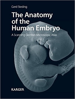
The Anatomy of the Human Embryo, 1ed
The present anatomical atlas concentrates on the early weeks of prenatal development of the human embryo. It comprises more than 800 scanning electron-microscopic pictures of specimens of exclusively human embryos. The three-dimensional appearing illustrations show the development of the external form of the face, neck, trunk and limbs. Besides, the brain and the viscera of the head, neck, thorax, abdomen and pelvis – all dissected into layers – are represented in their position and spatial form. The juxtaposition of pictures of temporally close developmental stages reveals the changes in the form of the organs. Photographs of the same organic system are usually shown at the same magnification and clearly demonstrate the growth process. Simple outline drawings provided with the principal nomenclature facilitate the orientation within the specimens. A brief introduction to each chapter explains the most significant developmental steps depicted. This atlas is of great interest not only to anatomists, embryologists, histologists and developmental biologists, but also to biologists, biochemists and geneticists. Moreover, it serves as a valuable reference book for clinicians such as gynecologists, obstetricians, pediatric surgeons and pediatric cardiologists.
In this atlas, the forms of the major human organs during the early weeks of prenatal development are depicted. Illustrations of the external form of human embryos have been published in great numbers and are to be found in numerous textbooks dealing with human embryology. However, we predominantly fi nd a description of organ development with pictures of two-dimensional histological sections or with more or less schematic drawings often made according to historical originals. It is not rare to fi nd in textbooks, even today, pictures of animal embryos as material for human development. Since it has been shown that results for animal embryos cannot always be simply and uncritically applied to human development, in this atlas of human embryology, only original images of the major organs of human embryos during the fi rst few weeks of prenatal development are shown.
Contents
۱ External Aspects
۲ Endodermal Organs in the Head and Neck
۳ Organs in the Thorax
۴ Organs in the Abdomen
۵ Urogenital Organs
۶ Brain and Sensory Organs
لینک کوتاه : https://bookbaz.ir/?p=37448
نویسنده : G. Steding
ناشر : S. Karger; 1 edition
سال انتشار : 2011
زبان کتاب : انگلیسی
نوع فایل : PDF
تعداد صفحات : 516 Double
(ISBN) شابک : 3805597053
قیمت کتاب درآمازون : $246.00
حجم فایل : 16 MB
کتاب های مرتبط:
 دانلود کتاب رشد رفتاری کودکان لِوین
Levine’s Developmental-Behavioral Pediatrics, 4ed
 دانلود کتاب یادداشت های آموزشی USMLE مرحله ۲ CK 2018 (5 کتاب)
USMLE Step 2 CK Lecture Notes 2018: 5-Book Set, 1ed
 دانلود کتاب فیزیولوژی انسان: از سلول به سیستم
Human Physiology: From Cells to Systems, 9ed
 دانلود کتاب پاتوفیزیولوژی اختلالات خونی
Pathophysiology of Blood Disorders, 1ed
 دانلود کتاب تشخیص مبتنی بر نشانه کودکان نلسون
Nelson Pediatric Symptom-Based Diagnosis, 1ed
 دانلود کتاب بیولوژی انسان چایراس
Human Biology, 9ed
 دانلود کتاب اطلس کالبد شکافی زوبوتا
Sobotta Dissection Atlas, 3ed
 دانلود کتاب آناتومی بالینی مور
Moore’s Clinically Oriented Anatomy, 9ed
 دانلود کتاب اطلس رنگ آناتومی انسان: سیستم حرکتی (جلد ۱)
Color Atlas of Human Anatomy: Vol. 1 Locomotor System, 8ed
 دانلود کتاب پوست کودکان هارپر (۲ جلدی)
Harper’s Textbook of Pediatric Dermatology, 2-Vol, 3ed
 دانلود کتاب اطلس آناتومی انسان نتر: رویکرد کلاسیک منطقه ای + ویدئو
Netter Atlas of Human Anatomy: Classic Regional Approach, 8ed + Video
 دانلود کتاب جداول یادگیری عضلات، مفاصل و اعصاب زوبوتا
Sobotta Learning Tables of Muscles, Joints and Nerves, 3ed
 دانلود کتاب اطلس رنگی آناتومی انسان: اندام های داخلی (جلد ۲)
Color Atlas of Human Anatomy: Vol. 2 Internal Organs, 7ed
 دانلود کتاب خود ارزیابی و مرور آناتومی
 Self Assessment and Review of Anatomy, 1ed
 دانلود کتاب راهنمای نوروآناتومی انسان سینگ
Inderbir Singh’s Textbook of Human Neuroanatomy, 10ed
 دانلود کتاب جنین شناسی انسانی لارسن
Larsen’s Human Embryology, 6ed
 دانلود کتاب آناتومی سر و گردن آکسفورد
Oxford Handbook of Head and Neck Anatomy, 1ed
 دانلود کتاب بیولوژی انسان: مفاهیم، کاربردها و مسائل
Biology of Humans: Concepts, Applications, and Issues, 6ed
 دانلود کتاب پزشکی کودکان بلوپرینت
Blueprints Pediatrics, 7ed
 دانلود کتاب فیزیولوژی کاستانزو
Costanzo Physiology, 7ed
 دانلود کتاب برنامه اصلی برای مراقبت شیردهی بین رشته ای
Core Curriculum for Interdisciplinary Lactation Care, 1ed
 دانلود کتاب پزشکی کودکان برکوویتز: روش مراقبت های اولیه
Berkowitz’s Pediatrics: A Primary Care Approach, 5ed
 دانلود کتاب رویکرد عملی برای بیهوشی کودکان
A Practical Approach to Pediatric Anesthesia, 2ed
 دانلود کتاب اطلس عکاسی منطقه ای ویژگی های غیر متریک و آناتومیک اسکلت انسانی
Photographic Regional Atlas of Non-Metric Traits and Anatomical Variants in the Human Skeleton, 1ed
 دانلود کتاب آسان سازی معاینه بالینی کودکان
 Paediatric Clinical Examination Made Easy, 6ed
 دانلود کتاب کاوش آناتومی و فیزیولوژی در آزمایشگاه
Exploring Anatomy & Physiology in the Laboratory, 3ed
 دانلود کتاب آناتومی تصویربرداری: سر و گردن
Imaging Anatomy: Head and Neck, 1ed
 دانلود کتاب جراحی کودکان: تشخیص و مدیریت
Pediatric Surgery: Diagnosis and Management, 2ed
 دانلود کتاب آناتومی و فیزیولوژی: یک رویکرد یکپارچه
Anatomy and Physiology: An Integrated Approach, 1ed
 دانلود کتاب فیزیولوژی کاستانزو
Physiology Costanzo, 6ed
 دانلود کتاب بیماری های سیستم عصبی در دوران کودکی ایکاردی
Aicardi’s Diseases of the Nervous System in Childhood, 4ed
 دانلود کتاب مرور بورد نوزادشناسی اوری
Avery’s Neonatology Board Review, 1ed
 دانلود کتاب مراقبتهای قبل از زایمان و پس از زایمان: یک رویکرد زنانه محور
Prenatal and Postnatal Care: A Woman-Centered Approach, 2ed
 دانلود کتاب درمان کنونی کان (ویرایش ۲۰۱۶)
Conn’s Current Therapy 2016, 1ed
 دانلود کتاب آناتومی و فیزیولوژی بصری
Visual Anatomy & Physiology, 2ed
 دانلود کتاب پزشکی مادر-جنین کریزی و رزنیک: اصول و عمل
Creasy and Resnik’s Maternal-Fetal Medicine: Principles and Practice, 7ed
 دانلود کتاب راهنمای مطالعه برای ساختار و عملکرد بدن انسان مملر
Study Guide for Memmler’s Structure and Function of the Human Body, 11ed
 دانلود کتاب بیهوشی کودکان گرگوری
Gregory’s Pediatric Anesthesia, 5ed
 دانلود کتاب آناتومی و فیزیولوژی برای پرستاران در یک نگاه
Anatomy and Physiology for Nurses at a Glance, 1ed
 دانلود کتاب همراه جیبی کتاب فیزیولوژی پزشکی گایتون و هال
Pocket Companion to Guyton and Hall Textbook of Medical Physiology, 13ed
 دانلود کتاب محدودیت رشد جنین و جفت جنین
 Placental-Fetal Growth Restriction, 1ed
 دانلود کتاب اطلس آناتومی زوبوتا: ارگان های داخلی (جلد ۲)
Sobotta Atlas of Anatomy: Internal Organs, Vol-2, 16ed
 دانلود کتاب آناتومی و رویکردهای جراحی نتر
Netter’s Surgical Anatomy and Approaches, 2ed
 دانلود کتاب سناریوهای موردی در تمرین کودکان و نوجوانان
Case Scenarios in Pediatric and Adolescent Practice, 1ed
 دانلود کتاب سیستم عصبی انسان بار: یک دیدگاه تشریحی
Barr’s The Human Nervous System: An Anatomical Viewpoint, 10ed
 دانلود کتاب ارزیابی خود در آناتومی و فیزیولوژی در سلامت و بیماری راس و ویلسون
Ross & Wilson Self-Assessment in Anatomy and Physiology in Health and Illness, 1ed
 دانلود کتاب پرسش و پاسخ مبتنی بر شایستگی در آناتومی برای آزمون حرفهای اول Mbbs
Competency Based Q&A In Anatomy For First Mbbs Professional Examination, 1ed
 دانلود کتاب جراحی بالینی اطفال جونز
Jones’ Clinical Paediatric Surgery, 7ed
 دانلود کتاب ملزومات تصویری آناتومی و فیزیولوژی
 Visual Essentials of Anatomy & Physiology, 1ed
 دانلود کتاب ملزومات آناتومی و فیزیولوژی سالدین
Essentials of Anatomy & Physiology, 2ed
 دانلود کتاب اطلس آناتومی زوبوتا: سر، گردن و نوروآناتومی (جلد ۳)
Sobotta Atlas of Anatomy: Head, Neck and Neuroanatomy, Vol-3, 16ed
 دانلود کتاب آناتومی مصور سر و گردن
 Illustrated Anatomy of the Head and Neck, 5ed
 دانلود کتاب سونوگرافی کودکان
 Pediatric Ultrasound, 1ed
 دانلود کتاب اطلس آناتومی زوبوتا: آناتومی عمومی و سیستم اسکلتی عضلانی (جلد ۱)
Sobotta Atlas of Anatomy, Vol.1: General Anatomy and Musculoskeletal System, 17ed
 دانلود کتاب آناتومی و تکنیک جراحی
 Surgical Anatomy and Technique: A Pocket Manual, 4ed
 دانلود کتاب اطلس آناتومی زوبوتا: آناتومی عمومی و سیستم عضلانی اسکلتی (جلد ۱)
Sobotta Atlas of Anatomy: General Anatomy and Musculoskeletal System, Vol-1, 16ed
 دانلود کتاب اطلس آناتومی زوبوتا: اندام های داخلی (جلد ۲)
Sobotta Atlas of Anatomy, Vol. 2: Internal Organs, 17ed
 دانلود کتاب کار درک آناتومی و فیزیولوژی: بینایی، شنوایی، رویکرد تعاملی
Workbook to Accompany Understanding Anatomy & Physiology: A Visual, Auditory, Interactive Approach, 2ed
 دانلود کتاب آناتومی انسان برای دانشجویان دندانپزشکی آناند
Anand’s Human Anatomy for Dental Students, 3ed
 دانلود کتاب راهنمای آناتومی عملی اندام فوقانی و تحتانی کانینگهام (جلد ۱)
Cunningham’s Manual of Practical Anatomy Upper and Lower limbs, Vol-1, 16ed
 دانلود کتاب راهنمای آناتومی عملی قفسه سینه و شکم کانینگهام (جلد ۲)
Cunningham’s Manual of Practical Anatomy: Thorax and Abdomen, Vol-2, 16ed
 دانلود کتاب راهنمای تغذیه، رژیم غذایی و چشم
Handbook of Nutrition, Diet, and the Eye, 2ed
 دانلود کتاب آناتومی گری: مبنای تشریحی عمل بالینی
Gray’s Anatomy: The Anatomical Basis of Clinical Practice, 42ed
 دانلود کتاب آناتومی و فیزیولوژی انسان هول (ویرایش ۲۰۱۶)
Hole’s Human Anatomy & Physiology, 14ed
 دانلود کتاب اورولوژی بالینی کودکان کلالیس – کینگ – بلمن
The Kelalis-King-Belman Textbook of Clinical Pediatric Urology, 6ed
 دانلود کتاب اطلس جیبی آناتومی مقطعی: سر و گردن
Pocket Atlas of Sectional Anatomy: Head and Neck, 3ed
 دانلود کتاب راهنمای بالینی اورژانس کودکان
 Clinical Manual of Emergency Pediatrics, 6ed
 دانلود کتاب آناتومی مقطعی برای متخصصان تصویربرداری
 Sectional Anatomy for Imaging Professionals, 3ed
 دانلود کتاب تشخیص و درمان پزشکی کارنت ۲۰۲۲
CURRENT Medical Diagnosis and Treatment 2022, 61ed
 دانلود کتاب راهنمای ارتوپدی اطفال
 Handbook of Pediatric Orthopedics, 2ed
 دانلود کتاب اطلس جیبی آناتومی مقطعی: سر و گردن، توموگرافی کامپیوتری و تصویربرداری رزونانس مغناطیسی
Pocket Atlas of Sectional Anatomy: Head and Neck, Computed Tomography and Magnetic Resonance Imaging, 4ed
 دانلود کتاب دستورالعمل مبتنی بر شواهد پزشکی زایمان
Obstetric Evidence Based Guidelines, 3ed
 دانلود کتاب اطلس آناتومی لگن و جراحی زنان باگیش + ویدئو
Atlas of Pelvic Anatomy and Gynecologic Surgery, 5ed + Video
 دانلود کتاب مراقبتهای ویژه کودکان فورمن و زیمرمن
Fuhrman & Zimmerman’s Pediatric Critical Care, 5ed
 دانلود کتاب پزشکی مادر-جنین کریزی و رزنیک: اصول و عمل + ویدئو
Creasy and Resnik’s Maternal-Fetal Medicine: Principles and Practice, 7ed + Video
 دانلود کتاب پزشکی جنین: علوم پایه و تمرین بالینی
Fetal Medicine: Basic Science and Clinical Practice, 3ed
 دانلود کتاب PCEP I: ارزیابی مادر و جنین و مراقبت فوری از نوزادان
 PCEP Book I: Maternal and Fetal Evaluation and Immediate Newborn Care, 3ed
 دانلود کتاب آتالارینژولوژی کودکان: مدیریت بالینی عملی
Pediatric Otolaryngology: Practical Clinical Management, 1ed
 دانلود کتاب پاسخ آناتومی و فیزیولوژی
 The Handy Anatomy Answer Book, 2ed
 دانلود کتاب غددشناسی کودکان اسپرلینگ
Sperling Pediatric Endocrinology, 5ed
 دانلود کتاب ملزومات در آناتومی و فیزیولوژی انسان هول
Hole’s Essentials of Human Anatomy & Physiology, 15ed
 دانلود کتاب آناتومی و فیزیولوژی: رویکرد یکپارچه
Anatomy & Physiology: An Integrative Approach, 3ed
 دانلود کتاب فیزیولوژی گوارش: سری فیزیولوژی موزبی
Gastrointestinal Physiology: Mosby Physiology Series, 9ed
 دانلود کتاب اختلالات رشد مادرزادی اپستین: اساس مولکولی اختلالات بالینی مورفوژنز
Epstein’s Inborn Errors of Development: The Molecular Basis of Clinical Disorders of Morphogenesis, 3ed
 دانلود کتاب غددشناسی کودکان
Pediatric Endocrinology, 4ed
 دانلود کتاب راهنمای آزمونگاهی آناتومی بدن انسان
Human Anatomy Lab Manual, 3ed
 دانلود کتاب آناتومی بالینی منطقه ای اسنل
Snell’s Clinical Anatomy by Regions, 10ed
 دانلود کتاب آناتومی، فیزیولوژی و بیماری: مبانی برای حرفه های بهداشتی
Anatomy Physiology & Disease: Foundations for the Health Professions, 3ed
 دانلود کتاب آناتومی و فیزیولوژی سالادین: وحدت شکل و عملکرد
Anatomy & Physiology: The Unity of Form and Function, 10ed
 دانلود کتاب ملزومات آناتومی و فیزیولوژی لاپرس
Essentials of Anatomy and Physiology, 8ed
 دانلود کتاب جنین شناسی انسانی لارسن
Larsen’s Human Embryology, 5ed
 دانلود کتاب الگوهای قابل تشخیص تغییر شکل انسان اسمیت + ویدئو
Smith’s Recognizable Patterns of Human Deformation, 4ed + Video
 دانلود کتاب بیماری قلبی بحرانی در نوزادان و کودکان + ویدئو
Critical Heart Disease in Infants and Children, 3ed + Video
 دانلود کتاب آناتومی سینگ (۳ جلدی)
Singh’ Textbook of Anatomy, 3-Vol, 2ed
 دانلود کتاب ساختار و عملکرد بدن انسان مملر
Memmler’s Structure and Function of the Human Body, 11ed
 دانلود کتاب پزشکی موبایل هریت لین
The Harriet Lane Handbook: Mobile Medicine Series, 20ed
 دانلود کتاب بیماری صرع کودکان پیلاک: تشخیص و درمان
Pellock’s Pediatric Epilepsy: Diagnosis and Therapy, 4ed
 دانلود کتاب فیزیوتراپی کودکان
 Pediatric Physical Therapy, 5ed
 دانلود کتاب بدن انسان! (دانشنامه دانش)
Human Body! (Knowledge Encyclopedias), 1ed
 دانلود کتاب آناتومی بالینی مور
Moore Clinically Oriented Anatomy, 7ed
 دانلود کتاب اطلس رنگی آناتومی انسان: سیستم عصبی و اندام حسی (جلد ۳)
Color Atlas of Human Anatomy, Vol. 3: Nervous System and Sensory Organs, 7ed
 دانلود کتاب جنین انسان در شرایط آزمایشگاهی
The Human Embryo In Vitro, 1ed
 دانلود کتاب هماتولوژی-انکولوژی کودکان در کشورهای با منابع محدود
Pediatric Hematology-Oncology in Countries with Limited Resources, 2014th
 داناود کتاب فیزیولوژی BRS
BRS Physiology, 7ed
 دانلود کتاب نقد و بررسی فیزیولوژی پزشکی گانونگ
Ganong’s Review of Medical Physiology, 25ed
 دانلود کتاب تروما اسکلتی در کودکان گرین
Green’s Skeletal Trauma in Children, 5ed
 دانلود کتاب جنین شناسی دندانی، بافت شناسی و آناتومی
Illustrated Dental Embryology, Histology, and Anatomy, 4ed
 دانلود کتاب درمان فلج مغزی و تاخیر حرکتی
Treatment of Cerebral Palsy and Motor Delay, 6ed
 دانلود کتاب تشخیص افتراقی در تصویربرداری کودکان
Differential Diagnosis in Pediatric Imaging, 1ed
 دانلود کتاب نوروآناتومی BRS
BRS Neuroanatomy, 6ed
 دانلود کتاب صدمات اعصاب و عصب: تاریخچه، جنین شناسی، آناتومی، تصویربرداری و تشخیص (جلد اول)
Nerves and Nerve Injuries: Vol 1: History, Embryology, Anatomy, Imaging, and Diagnostics
 دانلود کتاب پزشکی تورنتو نوت (نسخه ۲۰۱۵)
Toronto Notes 2015 (Essential Med Notes), 31ed
 دانلود کتاب آناتومی بالینی، بافت شناسی، جنین شناسی و نوروآناتومی
Clinical Anatomy Histology Embryology and Neuroanatomy, 1ed
 دانلود کتاب پاتوفیزیولوژی
Pathophysiology (Copstead), 5ed
 دانلود کتاب پاتوفیزیولوژی فوق العاده آسان!
Pathophysiology Made Incredibly Easy!, 5ed
 دانلود کتاب درسی فیزیولوژی
Textbook of Physiology, 3ed
 دانلود کتاب کار برای آناتومی مقطعی برای متخصصان تصویربرداری
 Workbook for Sectional Anatomy for Imaging Professionals, 3ed
 دانلود کتاب آناتومی انسان ماریب (ویرایش ۲۰۱۷)
Human Anatomy, 8ed
 دانلود کتاب درمان ضد میکروبی کودکان نلسون ۲۰۱۶
۲۰۱۶Nelson’s Pediatric Antimicrobial Therapy, 22ed
 دانلود کتاب تخصصی جامعه کودکان آکسفورد
The Oxford Specialist Handbook of Community Paediatrics, 1ed
 دانلود کتاب آناتومی مقطعی برای متخصصان تصویربرداری
Sectional Anatomy for Imaging Professionals, 4ed
 دانلود کتاب اصول پزشکی داخلی هریسون ، چاپ نوزدهم (۲ جلدی)
Harrison’s Principles of Internal Medicine 2Vol, 19ed
 دانلود کتاب اندام فوقانی کودکان
 The Pediatric Upper Extremity, 2015th
 دانلود کتاب فیزیولوژی جنینی و نوزادی (۲ جلدی)
Fetal and Neonatal Physiology, 2-Vol, 5ed
 دانلود کتاب اصول پاتوفیزیولوژی کاربردی کودکان
Fundamentals of Children’s Applied Pathophysiology, 1ed
 دانلود کتاب فیزیولوژی جنین و نوزاد (۲ جلدی)
Fetal and Neonatal Physiology, 2-Vol, 6ed
 دانلود کتاب اصول و تمرین انکولوژی اطفال
Principles and Practice of Pediatric Oncology, 5ed
 دانلود کتاب بیهوشی اطفال
 Handbook of Pediatric Anesthesia
 دانلود کتاب اورولوژی اطفال: استراتژی معاصر از زندگی جنینی به نوجوانی
Pediatric Urology: Contemporary Strategies from Fetal Life to Adolescence
 دانلود کتاب موارد بالینی در طب گرمسیری روث
Clinical Cases in Tropical Medicine, 1ed
 دانلود کتاب پزشکی نوزادان ضروری
Essential Neonatal Medicine, 5ed
 دانلود کتاب جراحی مغز و اعصاب کودکان کوهن: ترفندهای حرفه ای + ویدئو
Pediatric Neurosurgery: Tricks of the Trade + Video, 1ed
 دانلود کتاب طب اطفال در یک نگاه
Paediatrics at a Glance, 4ed
 دانلود کتاب رنگ آمیزی آناتومی
 The Anatomy Coloring Book, 4ed
 دانلود کتاب رادیولوژی EXPERTddx: کودکان
EXPERTddx: Pediatrics, 1ed
 دانلود کتاب پزشکی کودکان نتر
Netter’s Pediatrics, 1ed
 دانلود کتاب اطلس تومورهای مغزی کودکان
Atlas of Pediatric Brain Tumors, 2ed
 دانلود کتاب پرستاری کودکان دیمستیفاید
Pediatric Nursing Demystified, 1ed
 دانلود کتاب دستورالعمل نوزادان: استفاده از دارو در دوران بارداری و سال اول زندگی
Neonatal Formulary: Drug Use in Pregnancy and the First Year of Life, 7ed
 دانلود کتاب ملزومات آناتومی و فیزیولوژی انسان ماریئب (ویرایش جهانی)
Essentials of Human Anatomy & Physiology (Global Edition), 12ed
 دانلود کتاب موارد و مشکلات در تصویربرداری کودکان
Pearls and Pitfalls in Pediatric Imaging, 1ed
 دانلود کتاب کمک های اولیه برای نوزادان و کودکان
First Aid Fast for Babies and Children, 1ed
 دانلود کتاب عجایب بدن انسان: سیستم قلبی عروقی و تنفسی (جلد ۲)
Wonders of the Human Body: Cardiovascular & Respiratory Systems, 1ed
 دانلود کتاب پزشکی نوزاد ضروری
Essential Neonatal Medicine, 6ed
 دانلود کتاب سیستم های بدن (۲ جلدی)
Body Systems: 2 Volume Set, 1ed
 دانلود کتاب فیزیولوژی در یک نگاه
Physiology at a Glance, 4ed
 دانلود کتاب مرور فیزیولوژی
 Review of Physiology, 2ed
 دانلود کتاب خون: مقدمه بسیار کوتاه
Blood: A Very Short Introduction, 1ed
 دانلود کتاب ویتامین ها
 Handbook of Vitamins, 5ed
 دانلود مجموعه ویدئویی مدیریت تشخیص ارتوپدی مخصوص دست و مچ دست
 Diagnosis-Specific Orthopedic Management of the Wrist and Hand