دانلود کتاب سکته مغزی: پاتوفیزیولوژی، تشخیص و مدیریت + ویدئو
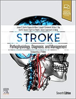
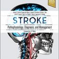
Stroke: Pathophysiology, Diagnosis, and Management, 7ed + Video
Authored by the world’s foremost stroke experts, this classic text brings you fully up to date with current research findings and management approaches for cerebrovascular disease. Stroke: Pathophysiology, Diagnosis, and Management, 7th Edition, covers every aspect of this fast-moving field, and is an ideal resource for stroke specialists, general neurologists, and other medical professionals with an interest in stroke. You’ll find expert clinical guidance, comprehensive pathophysiology coverage, data from recent trials, advances in diagnostic tests, full-color CT images, pathology slides, and much more, for a complete picture of today’s stroke medicine.
- Helps you recognize the clinical manifestations of stroke, use the latest laboratory and imaging studies to arrive at a diagnosis, and generate an effective medical and surgical treatment plan.
- Keeps you abreast of the overwhelming volume of studies and guidelines in this dynamic field, providing clear summaries and practical evaluations of all relevant data.
- Contains updates throughout, including the latest clinical trials (thrombectomy, DAWN, DEFUSE), genetics research, prevention research, new therapies, and the new guidelines from the ASA.
- Includes new slides for lectures, covering basic science, case studies, and interventional treatment overviews.
- Features a Key Points summary at the beginning of each chapter so you can quickly find important information.
- Provides abundant full-color CT images and pathology slides that help you make efficient and accurate diagnoses.
Review
“The book covers the basic gross anatomy with histopathology and etiopathogenesis of different types of strokes. The best aspect is the very well organized elaboration of disease characteristics with pathophysiological changes through to treatment.”
-© Doody’s Review Service,2021,Ram K Saha, M.D. (Thomas Jefferson University) Score: 92-4 Stars!
Contents
SECTION I Pathophysiology
SECTION II Epidemiology and Risk Factors
SECTION III Clinical Manifestations
SECTION IV Specific Conditions and Stroke
SECTION V Diagnostic Studies
SECTION VI Therapy
Video Contents
۱. Video 30.1 Important concepts regarding angioarchitecture, classifcation, and risk factors for arteriovenous malformations.
۲. Video 30.2 Endovascular embolization of a frontal arteriovenous malformation.
۳. Video 30.3 Endovascular and surgical techniques in the treatment of dural arteriovenous fstulae.
۴. Video 32.1 Mobile atheroma: An intraoperative video 3D TEE image of a mobile atheroma in the aortic arch, visualized during a transcatheter aortic valve replacement (TAVR), consistent with grade 5 atheromatous disease.
۵. Video 32.2 (A) Positive bubble study: Markedly positive bubble study consistent with a large atrial level shunt.
۶. Video 32.2 (B) Negative bubble study: Agitated saline contrast (“bubble”) study showing no evidence of an interatrial communication.
۷. Video 32.3 TEE bubble study with intrapulmonary shunt: A bubble study on TEE with delayed passage of bubbles into the left atrium via the right upper pulmonary vein, suggesting an intrapulmonary shunt, such as a pulmonary atriovenous malformation.
۸. Video 32.4 Takotsubo cardiomyopathy: TTE revealing akinesis of mid- and apical left ventricular segments with hypercontractile basal segments, consistent with a stress-mediated cardiomyopathy (Takotsubo cardiomyopathy).
۹. Video 32.5 Mobile atrial septum with positive bubble study: Mobile atrial septum, consistent with an atrial septal aneurysm.
۱۰. Video 32.6 TTE of a large left atrial myxoma, flling the entire left atrium and resulting in obstruction to left ventricular flling.
۱۱. Video 32.7 Papillary fbroelastoma: TEE of the aortic valve revealing a linear echo density on the ventricular surface of the valve, consistent with a papillary fbroelastoma.
۱۲. Video 32.8 (A) 2D images of a large burden of clot in the left atrium and left atrial appendage in a patient with a cardiomyopathy and atrial flutter
۱۳. Video 32.8 (B) 3D images of a large burden of clot in the left atrium and left atrial appendage in a patient with a cardiomyopathy and atrial flutter
۱۴. Video 32.9 TTE of large posterior mitral annular calcifcation (MAC) with central echo lucency, suggesting caseous MAC.
۱۵. Video 33.1 Large atherosclerotic plaque in the mid-portion of the aortic arch with large superimposed, mobile thrombus
۱۶. Video 33.2 Mobile aortic plaque in the superior aspect of the aortic arch, just proximal to the takeoff of the innominate artery
۱۷. Video 35.1 3D reconstruction of carotid angiography in a 40-year-old woman with right-sided numbness. Anterior, posterior, and left lateral rotational video of the left internal carotid artery demonstrates a web.
۱۸. Video 60.1 Knee-ankle-foot-orthosis: A knee-ankle-foot orthosis enabled this hemiplegic person withsensorimotor loss to prevent the knee from snapping back in the mid- to late stance phase of gait, prevent catching the forefoot when initiating swing, and improved control of the stability of foot placement during the stance phase.
۱۹. Video 72.1 Minimally invasive endoscopic ICH evacuation. The procedure begins with a 1.5-cm incision, a 1-cm craniectomy, and stereotactic placement of a 19F (6.3-mm) sheath into the hematoma.
۲۰. Video 72.2 Minimally invasive endoport-mediated ICH evacuation. The procedure begins with a 4-cm incision, a 2.5-cm craniotomy, and stereotactic placement of a 13.5-cm endoport into the hematoma.
۲۱. Video 75.1 Surgical management of a cerebral cavernous malformation.
۲۲. Video 77.1 Superfcial temporal artery to middle cerebral artery bypass.
۲۳. Video 78.1 Decompressive craniectomy in a patient with malignant middle cerebral artery infarction.
لینک کوتاه : https://bookbaz.ir/?p=339793
نویسنده : James C. Grotta MD
ناشر : Elsevier; 7th edition
سال انتشار : 2022
زبان کتاب : انگلیسی
نوع فایل : MP4 + PDF (کیفیت اصلی) 380 MB
تعداد صفحات : 1542
(ISBN) شابک : 0323694241
قیمت کتاب درآمازون : $194.99
حجم فایل : 630 MB
