
دانلود کتاب اطلس مغز انسان (ویرایش ۲۰۱۶)
Atlas of the Human Brain, 4ed
The fourth edition of Atlas of the Human Brain presents the anatomy of the brain at macroscopic and microscopic levels, featuring different aspects of brain morphology and topography. This greatly enlarged new edition provides the most detailed and accurate delineations of brain structure available. It includes features which assist in the new fields of neuroscience – functional imaging, resting state imaging and tractography. Atlas of the Human Brain is an essential guide to those working with human brain imaging or attempting to relate their observations on experimental animals to humans. Totally new in this edition is the inclusion of Nissl plates with delineation of cortical areas (Brodmann’s areas), the first time that these areas have been presented in serial histological sections.
- The contents of the Atlas of the brain in MNI stereotaxic space has been extensively expanded from 143 pages, showing 69 levels through the hemisphere, to 314 pages representing 99 levels.
- In addition to the fiber-stained (myelin) plates, we now provide fifty new (Nissl) plates covering cytoarchitecture. These are interdigitated within the existing myelin plates of the stereotaxic atlas.
- All photographic plates now represent the complete hemisphere.
- All photographs of the cell- and fiber-stained sections have been transformed to fit the MNI-space.
- Major fiber tracts are identified in the fiber-stained sections.
- In the Nissl plates cortical delineations (Brodmann’s areas) are provided for the first time.
- The number of diagrams increased to 99. They were now generated from the 3D reconstruction of the hemisphere registered to the MNI- stereotaxic space. They can be used for immediate comparison between our atlas and experimental and clinical imaging results.
- Parts of cortical areas are displayed at high magnification on the facing page of full page Nissl sections. Images selected highlight those areas which are thought to correspond with those published by von Economo and Koskinas (1925).
- A novel way of depicting cortical areal pattern is used: The cortical cytoarchitectonic ribbon is unfolded and presented linearly. This linear representation of the cortex enables the comparison of different interpretations of cortecal areas and allows mapping of activation sites.
- Low magnification diagrams in the horizontal (axial) and sagittal planes are included, calculated from the 3D model of the atlas brain.
About the Author
The main research interests are (i) the structural and molecular anatomy of the mammalian brain, especially of the human brain and (ii) expression patterns and regulation of terminal carbohydrates in development, cell activation and disease (III) operation planning in stereotactic neurosurgery. He works on a “Digital Brain Atlas for Planning and Interindividual Registration of Targets in Deep Brain Stimulation” and on a “Spatial Information Management Resource for the Human Brain”. J. K. Mai has edited the catalogue of human brain sections from the Vogt collection; he is author and editor of several books, e.g. the awarded “Atlas of the Human Brain” with CD-ROM (Academic Press/Elsevier, San Diego), “The Human Nervous System” (Academic Press/Elsevier, San Diego, Amsterdam, 3rd ed. 2012), Funktionelle Anatomie für Zahnmediziner (Quintessenz, Berlin, 2nd. ed. 2008; Sensi Divini (ital., engl., ger, russ. eds). J. K. Mai is CEO of MR-X-Brain GmbH.Milan Majtanik received his diploma in neuropsychology and training in neuroinformatics from the University of Bochum. He completed his diploma in mathematics and his PhD in psychology at the University of Düsseldorf. In his research at the Research Center Jülich he combined advanced analysis techniques in magnetoencephalography (synchronization tomography) with computational modelling in order to measure the impact of desynchronizing sensory stimulation on brain functions. His work on neural plasticity and desynchronizing neural stimulation provided framework for the developent of novel therapeutic techniques. He is currently focusing on the development of novel algorithms for high precision mapping and analysis of individual MRI scans.Professor George Paxinos, AO (BA, MA, PhD, DSc) completed his BA at The University of California at Berkeley, his PhD at McGill University, and spent a postdoctoral year at Yale University. He is the author of almost 50 books on the structure of the brain of humans and experimental animals, including The Rat Brain in Stereotaxic Coordinates, now in its 7th Edition, which is ranked by Thomson ISI as one of the 50 most cited items in the Web of Science. Dr. Paxinos paved the way for future neuroscience research by being the first to produce a three-dimensional (stereotaxic) framework for placement of electrodes and injections in the brain of experimental animals, which is now used as an international standard. He was a member of the first International Consortium for Brain Mapping, a UCLA based consortium that received the top ranking and was funded by the NIMH led Human Brain Project. Dr. Paxinos has been honored with more than nine distinguished awards throughout his years of research, including: The Warner Brown Memorial Prize (University of California at Berkeley, 1968), The Walter Burfitt Prize (1992), The Award for Excellence in Publishing in Medical Science (Assoc Amer Publishers, 1999), The Ramaciotti Medal for Excellence in Biomedical Research (2001), The Alexander von Humbolt Foundation Prize (Germany 2004), and more.
Contents
Part 1: Three Atlases of the Brain in the Head
۱.۱ Materials and Methods
۱.۱.۱ Anatomical Preparations
۱.۱.۲ Magnetic Resonance Imaging (MRI)
۱.۱.۳ Preparation and Photography of the Anatomical Slices
۱.۱.۴ Preparation of 100 µm Thick Frozen Histological Brain Sections
۱.۱.۵ Presentation of the Images for the Atlases of the Brain in the Head
۱.۱.۶ References
۱.۲ Horizontal Atlas
۱.۳ Coronal Atlas
۱.۴ Sagittal Atlas
Part 2: Myelo- and Cytoarchitectonic Atlas of the Human Brain in Stereotaxic (MNI) Space
۲.۱ Materials and Methods
۲.۱.۱ The Brain
۲.۱.۲ Methods
۲.۱.۳ Earlier Histological, Morphometric, Immunohistochemical Studies
۲.۱.۴ Nomenclature
۲.۱.۵ Photographic Plates and Corresponding Diagrams
۲.۱.۶ Three-Dimensional Reconstructions
۲.۱.۷ Standardization
۲.۱.۸ Mapping of the Atlas Space to the Talairach Space
۲.۱.۹ Mapping of the Atlas Space to the MNI/ICBM2009b Template
۲.۱.۱۰ Atlas of the Human Brain (AHB) Reconstruction with MNI/ICBM2009b Shape Constrain
۲.۱.۱۱ Registration of the Histological Sections to the Reconstructed Volume
۲.۱.۱۲ Use of the Atlas for the Interpretation of Individual in vivo/in vitro Brains
۲.۱.۱۳ Mapping of the Cortex Areas
۲.۱.۱۴ Generation of the Linear Representation of Cortex „Stripes“
۲.۱.۱۵ Layout of the Myelo- and Cytoarchitectonic Atlas of the Human Brain in Stereotaxic (MNI) Space
۲.۱.۱۶ References
۲.۲ Surface Views
۲.۳ Plates, Figures and Diagrams
۲.۴ Diagrams with Reduced Detail
۲.۴.۱ Horizontal (axial) Diagrams
۲.۴.۲ Sagittal Diagrams
۲.۵ Maps of Subcortical Areas
۲.۵.۱ Thalamus
۲.۵.۲ Hypothalamus
۲.۶ Published Studies Referring to the Brain Represented in the Myelo- and Cytoarchitectonic
Atlas of the Human Brain in Stereotaxic (MNI) Space
۲.۶.۱ Histological, Morphometric and Histochemical Studies
۲.۶.۲ References
لینک کوتاه : https://bookbaz.ir/?p=34757
نویسنده : Juergen K. Mai , Milan Majtanik
ناشر : Academic Press; 4 edition
سال انتشار : 2016
زبان کتاب : انگلیسی
نوع فایل : PDF
تعداد صفحات : 458
(ISBN) شابک : 0128028009
قیمت کتاب درآمازون : $215.35
حجم فایل : 200 MB

کتاب های مرتبط:
 دانلود کتاب جراحی آندوسکوپی کرانیوسینوستوز
Endoscopic Craniosynostosis Surgery, 1ed
 دانلود کتاب پاتوفیزیولوژی اختلالات خونی
Pathophysiology of Blood Disorders, 1ed
 دانلود کتاب بیولوژی انسان چایراس
Human Biology, 9ed
 دانلود کتاب آناتومی انسان برای دانشجویان دندانپزشکی آناند
Anand’s Human Anatomy for Dental Students, 3ed
 دانلود کتاب اطلس کالبد شکافی زوبوتا
Sobotta Dissection Atlas, 3ed
 دانلود کتاب سری کارشناسی ستون فقرات AO: تغییر شکل ستون فقرات بزرگسالان (جلد ۴)
AOSpine Masters Series, Volume 4: Adult Spinal Deformities, 1ed
 دانلود کتاب معاینه عصبی دژانگ
DeJong’s The Neurologic Examination, 8ed
 دانلود کتاب اطلس رنگ آناتومی انسان: سیستم حرکتی (جلد ۱)
Color Atlas of Human Anatomy: Vol. 1 Locomotor System, 8ed
 دانلود کتاب آناتومی سینگ (۳ جلدی)
Singh’ Textbook of Anatomy, 3-Vol, 2ed
 دانلود کتاب آنژیوگرافی مغزی کاربردی: آناتومی و پاتولوژی عروقی نرمال
Applied Cerebral Angiography: Normal Anatomy and Vascular Pathology, 1ed
 دانلود کتاب اطلس آناتومی انسان نتر: رویکرد کلاسیک منطقه ای + ویدئو
Netter Atlas of Human Anatomy: Classic Regional Approach, 8ed + Video
 دانلود کتاب اطلس رنگی آناتومی انسان: اندام های داخلی (جلد ۲)
Color Atlas of Human Anatomy: Vol. 2 Internal Organs, 7ed
 دانلود کتاب پاسخ آناتومی و فیزیولوژی
 The Handy Anatomy Answer Book, 2ed
 دانلود کتاب فیزیولوژی انسان: از سلول به سیستم
Human Physiology: From Cells to Systems, 9ed
 دانلود کتاب تعادل ساژیتال ستون فقرات: از عادی به پاتولوژی
Sagittal Balance of the Spine: From Normal to Pathology, 1ed
 دانلود کتاب آناتومی بالینی مور
Moore’s Clinically Oriented Anatomy, 9ed
 دانلود کتاب اطلس علوم اعصاب نتر + ویدئو
Netter’s Atlas of Neuroscience, 3ed + Video
 دانلود کتاب توانبخشی سکته مغزی: رویکرد مبتنی بر عملکرد
Stroke Rehabilitation: A Function-Based Approach, 4ed
 دانلود کتاب دانشنامه جامع تنوع آناتومیک انسانی برگمن
Bergman’s Comprehensive Encyclopedia of Human Anatomic Variation, 1ed
 دانلود کتاب بیماری صرع کودکان پیلاک: تشخیص و درمان
Pellock’s Pediatric Epilepsy: Diagnosis and Therapy, 4ed
 دانلود کتاب اطلس ویدیویی پایش نوروفیزیولوژیک در جراحی تومورهای نفوذی مغز
Video Atlas of Neurophysiological Monitoring in Surgery of Infiltrating Brain Tumors, 1ed
 دانلود کتاب اختلالات مزمن: راهنمای جیبی فوق العاده آسان
Chronic Disorders: An Incredibly Easy Pocket Guide
 دانلود کتاب نورولوژی بالینی تاراسکون
Tarascon Clinical Neurology Pocketbook, 1ed
 دانلود کتاب متاستازهای مغز از تومورهای اولیه (۳ جلدی)
Brain Metastases from Primary Tumors, 3-Vol, 1ed
 دانلود کتاب آناتومی و فیزیولوژی: رویکرد یکپارچه
Anatomy & Physiology: An Integrative Approach, 3ed
 دانلود کتاب مغز و اعصاب در عمل بالینی برادلی (۲ جلدی)
Bradley’s Neurology in Clinical Practice, 2-Vol, 7ed
 دانلود کتاب اطلس آناتومی و جراحی پایین تنه زنان باگیش (ویرایش ۲۰۱۶) + ویدئو
Baggish’s Atlas of Pelvic Anatomy and Gyncecologic Surgery, 4ed + Video
 دانلود کتاب فیزیولوژی انسان وندر
Vander’s Human Physiology, 14ed
 دانلود کتاب آناتومی و فیزیولوژی سالادین: وحدت شکل و عملکرد
Anatomy & Physiology: The Unity of Form and Function, 10ed
 دانلود کتاب راهنمای نوروآناتومی انسان سینگ
Inderbir Singh’s Textbook of Human Neuroanatomy, 10ed
 دانلود کتاب ۵ دقیقه مشورت مغز و اعصاب
 The 5-Minute Neurology Consult, 2ed
 دانلود کتاب بیماری عصبی در بارداری: اصول و تمرین
Neurological Illness in Pregnancy: Principles and Practice, 1ed
 دانلود کتاب تصویربرداری در جراحی ستون فقرات
 Imaging in Spine Surgery, 1ed
 دانلود کتاب ساختار و عملکرد بدن انسان مملر
Memmler’s Structure and Function of the Human Body, 11ed
 دانلود کتاب اطلس توسعه سیستم عصبی مرکزی انسان (۵ جلدی)
Atlas of Human Central Nervous System Development
 دانلود کتاب آناتومی و فیزیولوژی
 Anatomy & Physiology, 5ed
 دانلود کتاب جراحی ستون فقرات حداقل تهاجمی + ویدئو
Minimally Invasive Spine Surgery, 1ed + Video
 دانلود کتاب حفاظت مغز در اسکیزوفرنی ، اختلالات خلقی و شناختی
Brain Protection in Schizophrenia, Mood and Cognitive Disorders
 دانلود کتاب عملی آناتومی آستریون
Asterion: The Practical Handbook of Anatomy, 2ed
 دانلود کتاب مغز و اعصاب بالینی هانکی
Clinical Neurology Hankey, 1ed
 دانلود کتاب ابزار دقیق ستون فقرات: تکنیک های جراحی
Spinal Instrumentation: Surgical Techniques, 1ed
 دانلود کتاب آناتومی حالت صورت
Anatomy of Facial Expression, 1ed
 دانلود کتاب اطلس رنگی هیستولوژی گارتنر
Color Atlas and Text of Histology, 7ed
 دانلود کتاب تکنیک های پیشرفته در انتقال تصویر جراحی مغز و ستون فقرات
 Advanced Techniques in Image-Guided Brain and Spine Surgery, 1ed
 دانلود کتاب الکترومیوگرافی و اختلالات عصبی عضلانی
Electromyography and Neuromuscular Disorders, 4ed
 دانلود کتاب تفسیر نوار مغز
 Handbook of EEG Interpretation, 3ed
 دانلود کتاب چشم پزشکی عصبی لیو، والپ و گالتا: تشخیص و مدیریت + ویدئو
Liu, Volpe, and Galetta’s Neuro-Ophthalmology: Diagnosis and Management, 3ed + Video
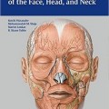 دانلود کتاب آناتومی برای جراحی پلاستیک صورت، سر و گردن
Anatomy for Plastic Surgery of the Face, Head and Neck, 1ed
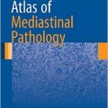 دانلود کتاب اطلس پاتولوژی مدیاستن (آناتومیک)
Atlas of Mediastinal Pathology, 2015th
 دانلود کتاب اطلس عمل جراحی مغز و اعصاب (۸ جلدی)
AANS’ Neurosurgical Operative Atlas, 8-Vol
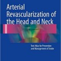 دانلود کتاب بازسازی عروق شریانی سر و گردن
Arterial Revascularization of the Head and Neck, 1ed
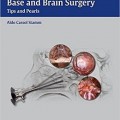 دانلود کتاب اندوسکوپی ترانس نازال قاعده جمجمه و جراحی مغز
Transnasal Endoscopic Skull Base and Brain Surgery, 1ed
 دانلود کتاب سری کارشناسی ستون فقرات AO: ناهنجاری های ستون فقرات کودکان (جلد ۹)
AOSpine Masters Series, Volume 9: Pediatric Spinal Deformities, 1ed
 دانلود کتاب جداول عضلات، مفاصل و اعصاب زوبوتا
Sobotta Tables of Muscles, Joints and Nerves, 2ed
 دانلود کتاب تعلیمات آناتومی برای درمانگران دستی و متخصصان حرکتی + ویدئو
Anatomy Trains: Myofascial Meridians for Manual Therapists and Movement Professionals, 4ed + Video
 دانلود کتاب نورورادیولوژی: تشخیص های افتراقی کلیدی و سوالات بالینی
Neuroradiology: Key Differential Diagnoses and Clinical Questions, 2ed
 دانلود کتاب آناتومی: اطلس عکسی (اطلس رنگی آناتومی مطالعه عکاسی از بدن انسان)
Anatomy: A Photographic Atlas (Color Atlas of Anatomy a Photographic Study of the Human Body), 8ed
 دانلود کتاب اندام فوقانی کودکان
 The Pediatric Upper Extremity, 2015th
 دانلود کتاب آناتومی گری برای دانشجویان
Gray’s Anatomy for Students, 3ed
 دانلود کتاب تکنیک های اجرایی ضروری و آناتومی اسکات-کانر و داوسون
Scott-Conner & Dawson: Essential Operative Techniques and Anatomy, 4ed
 دانلود کتاب تصویربرداری سر و گردن مقدماتی
Introductory Head and Neck Imaging, 1ed
 دانلود کتاب مغز و اعصاب شناختی و زوال عقل آکسفورد
Oxford Textbook of Cognitive Neurology and Dementia, 1ed
 دانلود کتاب آناتومی و فیزیولوژی ماریئب
Anatomy & Physiology, 6ed
 دانلود کتاب پاتوفیزیولوژی تنفسی کاربردی
Applied Respiratory Pathophysiology, 1ed
 دانلود کتاب اپیدوروسکوپی: اطلس روشها
Epiduroscopy: Atlas of Procedures, 1ed
 دانلود کتاب ضروریات بیهوشی اعصاب و مراقبت ویژه اعصاب گوپتا و گلب
Gupta and Gelb’s Essentials of Neuroanesthesia and Neurointensive Care, 2ed
 دانلود کتاب اختلالات مرتبط با ورزش: تشخیص و مدیریت
Sports-Related Concussion: Diagnosis and Management, 2ed
 دانلود کتاب تاریخچه آناتومی: چشم انداز بین المللی
History of Anatomy: An International Perspective, 1ed
 دانلود کتاب نقشه برداری مغز: نشانه ها و تکنیک ها + ویدئو
Brain Mapping: Indications and Techniques, 1ed + Video
دانلود کتاب الکترومیوگرافی و اختلالات عصبی عضلانی + ویدئوElectromyography and Neuromuscular Disorders, 4ed + Video
 دانلود کتاب سکته مغزی: پاتوفیزیولوژی، تشخیص و مدیریت
Stroke: Pathophysiology, Diagnosis, and Management, 7ed
 دانلود کتاب نورولوژی نتر
Netter’s Neurology, 3ed
 دانلود کتاب اسکولیوز ایدیوپاتیک
 Idiopathic Scoliosis, 2ed
 دانلود کتاب مبانی حرکت شناسی
Foundations of Kinesiology, 2ed
 دانلود کتاب آناتومی برای دندانپزشکی
Anatomy for Dental Medicine, 3ed
 دانلود کتاب اطلس آناتومی زوبوتا: سر، گردن و نوروآناتومی (جلد ۳)
Sobotta Atlas of Anatomy: Head, Neck and Neuroanatomy, Vol-3, 16ed
 دانلود کتاب راهنمای نوروپاتولوژی عمومی اسکرول و پوریِر
Escourolle & Poirier’s Manual of Basic Neuropathology, 5ed
 دانلود کتاب علوم اعصاب شناختی سیستم های بشری: کار و زندگی روزمره
Cognitive Neuroscience of Human Systems: Work and Everyday Life
 دانلود کتاب کالبدشناسی اعصاب بالینی گری: اساس آناتومی علوم اعصاب بالینی
Gray’s Clinical Neuroanatomy: The Anatomic Basis for Clinical Neuroscience, 1ed
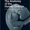 دانلود کتاب آناتومی جنین انسان
The Anatomy of the Human Embryo, 1ed
 دانلود کتاب غددشناسی عصبی رفتاری
Behavioral Neuroendocrinology, 1ed
 دانلود کتاب آناتومی و فیزیولوژی برای پرستاران در یک نگاه
Anatomy and Physiology for Nurses at a Glance, 1ed
 دانلود کتاب بیومکانیک ستون فقرات: مفاهیم اساسی، اختلالات نخاعی و درمان
Biomechanics of the Spine: Basic Concepts, Spinal Disorders and Treatments, 1ed
 دانلود کتاب آپوپتوز و فراتر از آن: راه های بسیار مرگ سلول ها (۲ جلدی)
Apoptosis and Beyond: The Many Ways Cells Die, 2-Vol, 1ed
 دانلود کتاب مغز و اعصاب بالینی لانگه (امینف، نسخه اصلی)
Aminoff Clinical Neurology, 8ed
 دانلود کتاب اطلس عصب صورت و ساختارهای مرتبط + آلبوم تصویری
Atlas of the Facial Nerve and Related Structures, 1ed + Image Album
 دانلود کتاب مراقبت زوال عقل: رویکرد عملی
Dementia Care: A Practical Approach, 1ed
 دانلود کتاب جراحی زاویه سربلوپونتین
 Surgery of the Cerebellopontine Angle, 1ed
 دانلود کتاب رویکردهای بیولوژیکی برای ترمیم و بازسازی دیسک ستون فقرات برای پزشکان
Biological Approaches to Spinal Disc Repair and Regeneration for Clinicians, 1ed
 دانلود کتاب اطلس رنگی جراحی ساقه مغز
 Color Atlas of Brainstem Surgery, 1ed
 دانلود کتاب علوم اعصاب رفتاری
Behavioral Neuroscience, 8ed
 دانلود کتاب آناتومی تفصیلی بالینی مور
Moore’s Clinically Oriented Anatomy, 8ed
 دانلود کتاب از دست دادن حافظه، آلزایمر و دمانس: راهنمای عملی برای پزشکان + ویدئو
Memory Loss, Alzheimer’s Disease, and Dementia: A Practical Guide for Clinicians, 2ed + Video
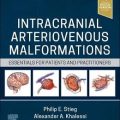 دانلود کتاب ناهنجاری های شریانی وریدی داخل جمجمه: موارد ضروری برای بیماران و پزشکان
Intracranial Arteriovenous Malformations: Essentials for Patients and Practitioners, 1ed
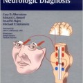 دانلود کتاب پایه آناتومیک عصب شناسی تشخیصی
Anatomic Basis of Neurologic Diagnosis, 1ed
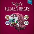 دانلود کتاب مغز انسان در عکس ها و نمودارها نولته
Nolte’s The Human Brain in Photographs and Diagrams, 5ed
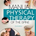 دانلود کتاب راهنمای درمان فیزیکی ستون فقرات اولسون
Manual Physical Therapy of the Spine, 2ed
 دانلود کتاب یک رویکرد تشریحی برای جراحی ستون فقرات کم تهاجمی
An Anatomic Approach to Minimally Invasive Spine Surgery, 2ed
 دانلود کتاب جراحی مغز و اعصاب کودکان کوهن
Pediatric Neurosurgery: Tricks of the Trade, 1ed
 دانلود کتاب آناتومی مصور سر و گردن
 Illustrated Anatomy of the Head and Neck, 5ed
 دانلود کتاب اصول علوم اعصاب برای علوم و اختلالات ارتباطاتی
Neuroscience Fundamentals for Communication Sciences and Disorders, 1ed
 دانلود کتاب سیستم قلبی عروقی در یک نگاه
The Cardiovascular System at a Glance, 5ed
 دانلود کتاب نورولوژی بالینی کودکان فنیچل: رویکرد علائم و نشانه ها
Fenichel’s Clinical Pediatric Neurology: A Signs and Symptoms Approach, 8ed
 دانلود کتاب اطلس آناتومی زوبوتا: آناتومی عمومی و سیستم اسکلتی عضلانی (جلد ۱)
Sobotta Atlas of Anatomy, Vol.1: General Anatomy and Musculoskeletal System, 17ed
 دانلود کتاب تب مغزی: چگونه واکسن ها از مننژیت و سایر بیماری های کشنده جلوگیری می کنند
Brain Fever: How Vaccines Prevent Meningitis And Other Killer Diseases, 1ed
 دانلود کتاب تصویربرداری از مغز (سری کارشناس رادیولوژی)
Imaging of the Brain (Expert Radiology Series), 1ed
 دانلود کتاب فیزیولوژی کلیه وَندرز
Vanders Renal Physiology, 8ed
 دانلود کتاب آناتومی فوری
Instant Anatomy, 5ed
 دانلود کتاب مانیتورینگ مراقبت حاد مغز و اعصاب
Neurocritical Care Monitoring, 1ed
 دانلود کتاب اطلس آناتومی زوبوتا: آناتومی عمومی و سیستم عضلانی اسکلتی (جلد ۱)
Sobotta Atlas of Anatomy: General Anatomy and Musculoskeletal System, Vol-1, 16ed
 دانلود کتاب اطلس ضایعات سلار، سوپراسلار و پاراسلار
Atlas of Sellar, Suprasellar, and Parasellar Lesions, 1ed
 دانلود کتاب راهنمای بالینی عصب شناسی
 Neurology: A Clinical Handbook, 1ed
 دانلود کتاب اطلس آناتومی زوبوتا: اندام های داخلی (جلد ۲)
Sobotta Atlas of Anatomy, Vol. 2: Internal Organs, 17ed
 دانلود کتاب مغز و اعصاب بین المللی
International Neurology, 2ed
 دانلود کتاب راهنمای ابزار جراحی مغز و اعصاب
 The Neurosurgical Instrument Guide, 1ed
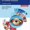 دانلود کتاب اطلس مدیریت چند رشته ای بیماری های حدقه ای
Interdisciplinary Management of Orbital Diseases: Textbook and Atlas, 1ed
 دانلود کتاب مقدمه ای بر بدن انسان
Introduction to the Human Body, 10ed
 دانلود کتاب اصول آناتومی و فیزیولوژی مارتینی (ویرایش ۲۰۱۸)
Fundamentals of Anatomy & Physiology, 11ed
 دانلود کتاب مغز و اعصاب بالینی امینوف
Lange Clinical Neurology, 10ed
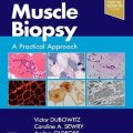 دانلود کتاب بیوپسی عضلانی: یک رویکرد عملی
Muscle Biopsy: A Practical Approach, 5ed
 دانلود کتاب اطلس جیبی آناتومی مقطعی: سر و گردن
Pocket Atlas of Sectional Anatomy: Head and Neck, 3ed
 دانلود کتاب موارد رادیولوژی مغز و اعصاب
Neuroradiology Cases
 دانلود کتاب اطلس ویدئویی روشهای نورواندوواسکولار
 Video Atlas of Neuroendovascular Procedures, 1ed
 دانلود کتاب اطلس ویدئویی جراحی ستون فقرات
 Video Atlas of Spine Surgery, 1ed
 دانلود کتاب جراحی قاعده جمجمه جانبی: اطلس کلینیک هاوس
Lateral Skull Base Surgery: The House Clinic Atlas, 1ed
 دانلود کتاب ۱۰۱ راه حل MRI مغز
 ۱۰۱MRI Brain Solutions, 1ed
 دانلود کتاب آزمون USMLE مرحله ۱ ۲۰۱۶: آناتومی
USMLE Step 1 Lecture Notes 2016: Anatomy, 1ed
 دانلود کتاب اختلالات خواب در نورولوژی: یک رویکرد عملی
Sleep Disorders in Neurology: A Practical Approach, 2ed
 دانلود کتاب اطلس ویدئویی جراحی آنوریسم داخل جمجمه
Video Atlas of Intracranial Aneurysm Surgery, 1ed
 دانلود کتاب تکنیک های جراحی مغز و اعصاب اشمیدک و سویت (۲ جلدی) + ویدئو
Schmidek and Sweet Operative Neurosurgical Techniques, 2-Vol, 7ed + Video
 دانلود کتاب اطلس جیبی آناتومی مقطعی: سر و گردن، توموگرافی کامپیوتری و تصویربرداری رزونانس مغناطیسی
Pocket Atlas of Sectional Anatomy: Head and Neck, Computed Tomography and Magnetic Resonance Imaging, 4ed
 دانلود کتاب جداول یادگیری عضلات، مفاصل و اعصاب زوبوتا
Sobotta Learning Tables of Muscles, Joints and Nerves, 3ed
 دانلود کتاب بورد و بخش برای USMLE مراحل ۲ و ۳
Boards & Wards for USMLE Steps 2 & 3, 5ed
 دانلود کتاب الکترودیاگنوز در مغز و اعصاب بالینی امینف
Aminoff’s Electrodiagnosis in Clinical Neurology, 6ed
 دانلود کتاب اطلس تکنیک های مغز و اعصاب: مغز (دو جلدی)
Atlas of Neurosurgical Techniques: Brain, 2-Vol, 2ed
 دانلود کتاب آناتومی سینگ: ژنتیک آناتومی عصبی سر و گردن (جلد ۳)
Inderbir Singh’s Textbook of Anatomy: Head and Neck Neuroanatomy Genetics, 6ed
 دانلود کتاب مراقبت سکته مغزی حاد
Acute Stroke Care, 3ed
 دانلود کتاب درک پس از ضربه مغزی
 Understanding Traumatic Brain Injury, 1ed
 دانلود کتاب بررسی جامع در مغز و اعصاب بالینی
Comprehensive Review in Clinical Neurology, 2ed
 دانلود کتاب اطلس توپوگرافی و پاتوتوپوگرافی آناتومی سر و گردن
Atlas of Topographical and Pathotopographical Anatomy of the Head and Neck, 1ed
 دانلود کتاب شیمی اعصاب پایه: اصول مولکولی، سلولی و نوروبیولوژی پزشکی
Basic Neurochemistry: Principles of Molecular, Cellular, and Medical Neurobiology, 8ed
 دانلود کتاب اطلس جراحی اضطراری مغز و اعصاب
Atlas of Emergency Neurosurgery, 1ed
 دانلود کتاب فیزیولوژی کاستانزو
Physiology Costanzo, 6ed
 دانلود کتاب MKSAP 17 : عصب شناسی
 MKSAP (R) 17 Neurology, 17ed
 دانلود کتاب اطلس مصنوعات در فیزیولوژی عصبی بالینی
Atlas of Artifacts in Clinical Neurophysiology, 1ed
 دانلود کتاب سیستم عصبی انسان بار: یک دیدگاه تشریحی
Barr’s The Human Nervous System: An Anatomical Viewpoint, 10ed
 دانلود کتاب تغذیه و عضلات اسکلتی
Nutrition and Skeletal Muscle, 1ed
 دانلود کتاب فلش کارت آناتومی بالینی مور
Moore’s Clinical Anatomy Flash Cards, 2ed
 دانلود کتاب اصول نوروسایکولوژی انسان
Fundamentals of Human Neuropsychology, 7ed
 دانلود کتاب های زیست شناسی: علوم برای زندگی با فیزیولوژی و کارت مطالعه
Books a la Carte for Biology: Science for Life with Physiology & Study Card, 3ed