دانلود کتاب تصویربرداری اسکلتی عضلانی
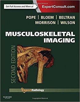
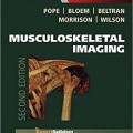
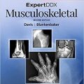
Musculoskeletal Imaging, 2ed
In its fully revised and updated second edition, Musculoskeletal Imaging covers every aspect of musculoskeletal radiology. This medical reference book incorporates the latest diagnostic modalities and interventional techniques, as well as must-read topics such as hip, groin and cartilage imaging; newly described impingements; and new concepts in the hip including teres ligament pathology. Accessibility in print, online and across portable devices makesMusculoskeletal Imaging a fully searchable and dependable sourcefor both reading and reference. This publication is a key title in the popular Expert Radiology Series, which delivers evidence-based expert guidance from around the globe.
“This is an excellent benchbook and accompanying electronic resource which will be of value to trainee radiologists and established consultants.”Reviewed by: Dr Steve Amerasekara, Consultant Radiologist on behalf of journal RAD Magazine Date: July 2015
“This outstanding text is now an acclaimed primary resource and therefore belongs in the libraries and at the work stations of all general and orthopedic hospital departments of radiology and, indeed, at any and all imaging facilities involved in musculoskeletal imaging.” Foreword by: Lee F. Rogers, June 2015
- Fully understand each topic with a format that delivers essential background information.
- Streamline the decision-making process with integrated protocols, classic signs, and ACR guidelines, as well as a design that structures every chapter consistently to include pathophysiology, imaging techniques, imaging findings, differential diagnosis, and treatment options.
- Write the most comprehensive reports possible with help from boxes highlighting what the referring physician needs to know, as well as suggestions for treatment and future imaging studies.
- Access in-depth case studies, valuable appendices, and additional chapters covering all of the most important musculoskeletal procedures performed today.
- Quickly locate important information with a full-color design that includes color-coded tables and bulleted lists highlighting key concepts, as well as color artwork that lets you easily find critical anatomic views of diseases and injuries.
- Engage with more than 40 brand-new videos, including arthroscopic videos.
- Easily comprehend complicated material with over 5,000 images and new animations.
- Explore integrated clinical perspectives on the newest modalities such as PET-CT in cancer, diffusion MR, as well as ultrasonography, fusion imaging, multi-slice CT and nuclear medicine.
- Learn from team of international experts provides a variety of evidence-based guidance, including the pros and cons of each modality, to help you overcome difficult challenges.
- Expert Consult eBook version included with purchase. This enhanced eBook experience allows you to search all of the text, figures, references, and videos from the book on a variety of devices.
Review
Contents
۱. General Imaging Principles
۲. General Principles of Osseous Injury
۳. Imaging of Facial and Skull Trauma
۴. Cervical Spine Injuries
۵. Injury of the Thoracic Cage and Thoracolumbar Spine
۶. Normal Shoulder
۷. Osseous Injuries of the Shoulder Girdle
۸. Shoulder Impingement Syndromes
۹. Glenohumeral Instability
۱۰. Normal Elbow
۱۱. Acute Osseous Injury of the Elbow and Forearm
۱۲. Soft Tissue Injury to the Elbow
۱۳. Normal Wrist
۱۴. Acute Osseous Injury to the Wrist
۱۵. Internal Derangement of the Wrist
۱۶. Acute Osseous Trauma to the Hand
۱۷. Compressive and Entrapment Neuropathies of the Upper Extremities
۱۸. Soft Tissue Injuries of the Hand and Wrist
۱۹. Normal Pelvis and Hip
۲۰. Acute Osseous Injury to the Pelvis and Acetabulum
۲۱. Athletic Pubalgia
۲۲. Acute Osseous Injury to the Hip and Proximal Femur
۲۳. Internal Derangement of the Hip and Proximal Femur
۲۴. Normal Knee
۲۵. Acute Osseous Injury to the Knee
۲۶. Internal Derangement of the Knee: Meniscal Injuries
۲۷. Internal Derangement of the Knee: Ligament Injuries
۲۸. Internal Derangement of the Knee: Tendon Injuries
۲۹. Internal Derangement of the Knee: Cartilage and Osteochondral Injuries
۳۰. Normal Ankle and Foot
More…
۹۰. The Patient with a Tumor or a Tumor-Like Lesion of Bone
۹۱. The Patient with a Soft Tissue Lump
۹۲. Primary Bone Tumors
۹۳. Myeloma
۹۴. Tumor-Like Lesions of Bone
۹۵. Soft Tissue Tumors
۹۶. Tumor-Like Soft Tissue Lesions
۹۷. Metastatic Disease
۹۸. Treatment Strategies for Musculoskeletal Tumors and Tumor-Like Lesions
۹۹. Staging Bone and Soft Tissue Tumors
۱۰۰. Monitoring Therapy in Bone and Soft Tissue Tumors
۱۰۱. Focal Growth Disturbances
۱۰۲. Developmental Dysplasia of the Hip
۱۰۳. Coalitions
۱۰۴. Dysplasias
۱۰۵. Spinal Deformity
۱۰۶. Principles and Complications of Orthopedic Hardware
۱۰۷. Postoperative Shoulder
۱۰۸. Postoperative Elbow, Wrist, and Hand
۱۰۹. Postoperative Hip
۱۱۰. Postoperative Knee
۱۱۱. Postoperative Ankle and Foot
۱۱۲. Imaging of the Residual Limb after Amputation
۱۱۳. Postoperative Infections
۱۱۴. Temporomandibular Joint
۱۱۵. Dental Imaging
۱۱۶. Normal Variants
۱۱۷. Biopsy: Soft Tissue
۱۱۸. Percutaneous Biopsy of the Appendicular Skeleton
۱۱۹. Percutaneous Biopsy of the Spine
۱۲۰. Tumor Ablation
۱۲۱. Spinal Injections
۱۲۲. Discography
۱۲۳. Vertebroplasty and Kyphoplasty
۱۲۴. Percutaneous Intradiskal Therapies
۱۲۵. Ultrasound Procedures
جهت مشاهده نسخه همراه با ویدئوهای کامل کتاب کلیک کنید
لینک کوتاه : https://bookbaz.ir/?p=78764
نویسنده : Thomas Pope MD FACR , Hans L. Bloem MD PhD
ناشر : Saunders; 2 edition
سال انتشار : 2015
زبان کتاب : انگلیسی
نوع فایل : PDF
تعداد صفحات : 2884
(ISBN) شابک : 1455708135
قیمت کتاب درآمازون : $209.88
حجم فایل : 217 MB


