
دانلود کتاب تصویربرداری حساسیت وزنی در MRI
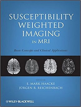
Susceptibility Weighted Imaging in MRI, 1ed
This book presents the first in-depth reference to discuss Susceptibility Weighted Imaging for students and professionals
Within the pages of this book, the reader will find comprehensive coverage of the major concepts that underlie the origins of susceptibility weighted imaging (SWI) in magnetic resonance imaging (MRI) and its wide range of applications. This book provides readers a thorough understanding of the technique now commonly called SWI, a major enhancement of traditional MRI with the power to produce high-resolution images that are exquisitely sensitive to blood products and iron. Since its inception, SWI has become a powerful tool for investigating a number of important clinical aspects of neuro-imaging, especially the diagnosis and pathophysiology of traumatic brain injury, the detection of acute hemorrhagic stroke, and the detection of microbleeds in dementia.
Edited by the originators of SWI, this groundbreaking text is the definitive resource on this critically important technology. Featuring contributions from the top leaders in the science and clinical use of the modality, the book:
- Introduces the fundamentals of SWI
- Presents an even balance between the technical aspects and its clinical applications
- Explains how to image brain tumors, cerebral microbleeds, and hemorrhage as well as many other clinical applications
- Explains how to quantify iron content for diseases such as multiple sclerosis, Parkinson’s disease, and other neurodegenerative diseases
- Introduces the use of SWI in visualizing the vessel wall
- Covers the use of SWI at ultra-high magnetic fields
- Introduces the important concept of susceptibility mapping as the next generation of SWI
- Includes over 100 high-quality images and tables
This reference also covers more advanced topics, from improved contrast in MRI of the midbrain using SWI to functional susceptibility weighted MRI, automated vein segmentation and lesion detection, rapid acquisition methods, and more.
Suitable for all levels of experience, Susceptibility Weighted Imaging in MRI is the ideal source for neuroradiologists, radiologists, imaging and medical physicists, cardiologists, oncologists, biochemists, and students who want authoritative information on the basic elements and practical applications of this exciting new medical imaging technique.
Review
Contents
۱ Introduction to Susceptibility Weighted Imaging
۲ Magnetic Susceptibility
۳ Gradient Echo Imaging
۴ Phase and Its Relationship to Imaging Parameters and Susceptibility
۵ Understanding T2*-Related Signal Loss
۶ Processing Concepts and SWI Filtered Phase Images
۷ MR Angiography and Venography of the Brain
۸ Brain Anatomy with Phase
۹ SWI Venographic Anatomy of the Cerebrum
۱۰ Novel Approaches to Imaging Brain Tumors
۱۱ Traumatic Brain Injury
۱۲ Imaging Cerebral Microbleeds with SWI
۱۳ Imaging Ischemic Stroke and Hemorrhage with SWI
۱۴ Visualizing Deep Medullary Veins with SWI in Newborn and Young Infants
۱۵ Susceptibility Weighted Imaging in Multiple Sclerosis
۱۶ Cerebral Venous Diseases and Occult Intracranial Vascular Malformations
۱۷ Sturge–Weber Syndrome
۱۸ Visualizing the Vessel Wall Using Susceptibility Weighted Imaging
۱۹ Imaging Breast Calcification Using SWI
۲۰ Susceptibility Weighted Imaging at Ultrahigh Magnetic Fields
۲۱ Improved Contrast in MR Imaging of the Midbrain Using SWI
۲۲ Measuring Iron Content with Phase
۲۳ Validation of Phase Iron Detection with Synchrotron X-Ray Fluorescence
۲۴ Rapid Calculation of Magnetic Field Perturbations from Biological Tissue in Magnetic Resonance Imaging
۲۵ SWIM: Susceptibility Mapping as a Means to Visualize Veins and Quantify Oxygen Saturation
۲۶ Effects of Contrast Agents in Susceptibility Weighted Imaging
۲۷ Oxygen Saturation: Quantification
۲۸ Quantification of Oxygen Saturation of Single Cerebral Veins, the Blood Capillary Network, and Its Dependency on Perfusion
۲۹ Integrating Perfusion Weighted Imaging, MR Angiography, and Susceptibility Weighted Imaging
۳۰ Functional Susceptibility Weighted Magnetic Resonance Imaging
۳۱ Complex Thresholding Methods for Eliminating Voxels That Contain Predominantly Noise in Magnetic Resonance Images
۳۲ Automatic Vein Segmentation and Lesion Detection: from SWI-MIPs to MR Venograms
۳۳ Rapid Acquisition Methods
۳۴ High-Resolution Venographic BOLD MRI of Animal Brain at 9.4 T: Implications for BOLD fMRI
۳۵ Susceptibility Weighted Imaging in Rodents
۳۶ Ultrashort TE Imaging: Phase and Frequency Mapping of Susceptibility Effects in Short T2 Tissues of the Musculoskeletal System
لینک کوتاه : https://bookbaz.ir/?p=81257
نویسنده : E. Mark Haacke , Jürgen R. Reichenbach
ناشر : Wiley-Blackwell; 1 edition
سال انتشار : 2011
زبان کتاب : انگلیسی
نوع فایل : PDF
تعداد صفحات : 752
(ISBN) شابک : 0470043431
قیمت کتاب درآمازون : $119.81
حجم فایل : 25 MB
کتاب های مرتبط:
 دانلود کتاب آناتومی تصویربرداری: سر و گردن
Imaging Anatomy: Head and Neck, 1ed
 دانلود کتاب تصویربرداری اسکلتی عضلانی
Musculoskeletal Imaging, 2ed
 دانلود کتاب پرسش و پاسخ تصویربرداری گوارشی رادکِیس
RadCases Plus Q&A Gastrointestinal Imaging, 2ed
 دانلود کتاب تصویربرداری بیماری های بینابینی ریه (سری رادیولوژی کلینیکو)
Clinico Radiological Series: Imaging of Interstitial Lung Diseases, 1ed
 دانلود کتاب موارد تصویربرداری پستان
Breast Imaging Cases
 دانلود کتاب تصویربرداری تشخیصی کودکان کافی (ویرایش ۲۰۱۹، ۲ جلدی)
Caffey’s Pediatric Diagnostic Imaging, 2-Volume Set, 13ed
 دانلود کتاب تصویربرداری تومور قفسه سینه (سری رادیولوژی کلینیکو)
Clinico Radiological Series: Imaging Of Chest Tumors, 1ed
 دانلود کتاب MRI در یک نگاه
MRI at a Glance, 2ed
 دانلود کتاب تصویربرداری قفسه سینه و قلب و عروق
Chest and Cardiovascular Imaging, 3ed
 دانلود کتاب تصویربرداری در جراحی شکم
Imaging in Abdominal Surgery, 1ed
 دانلود کتاب تصویربرداری در گوارش
Imaging in Gastroenterology, 1ed
 دانلود کتاب اصول سی تی اسکن بدن
Fundamentals of Body CT, 5ed
 دانلود کتاب هدایت حین عمل MRI جراحی مغز و اعصاب
Intraoperative MRI-Guided Neurosurgery, 1ed
 دانلود کتاب اصول و تمرین پزشکی خواب (۲ جلدی) + ویدئو
Principles and Practice of Sleep Medicine, 2-Vol, 7ed + Video
 دانلود کتاب تصویربرداری تشخیصی: مغز
Diagnostic Imaging: Brain, 3ed
 دانلود کتاب تصویربرداری تشخیصی: پزشکی زایمان
Diagnostic Imaging: Obstetrics, 3ed
 دانلود کتاب تفسیر رادیوگرافی قفسه سینه در بیماران قلبی کودک
Chest Radiographic Interpretation in Pediatric Cardiac Patients, 1ed
 دانلود کتاب اطلس بیهوشی منطقه ای هدایت اولتراسوند + ویدئو
Atlas of Ultrasound-Guided Regional Anesthesia, 3ed + Video
 دانلود کتاب تصویربرداری تشخیصی دستگاه گوارش
Diagnostic Imaging: Gastrointestinal, 4ed
 دانلود کتاب تصویربرداری تشخیصی زایمان
Diagnostic Imaging: Obstetrics, 4ed
 دانلود کتاب تصویربرداری تشخیصی سر و گردن
Diagnostic Imaging: Head and Neck, 4ed
 دانلود کتاب تصویربرداری تشخیصی انکولوژی
Diagnostic Imaging: Oncology, 2ed
 دانلود کتاب تصویربرداری از مغز با MRI و CT: رویکرد الگوی تصویری
Brain Imaging with MRI and CT: An Image Pattern Approach, 1ed
 دانلود کتاب صدمات عصب کلاین و هادسون: صدمات عصب، انترپمنت و تومور
Kline and Hudson’s Nerve Injuries: Operative Results for Major Nerve Injuries, Entrapments and Tumors, 2ed
 دانلود کتاب سونوگرافی مداخله ای: راهنمای عملی و اطلس
Interventional Ultrasound: A Practical Guide and Atlas, 1ed
 دانلود کتاب فیزیک ضروری در تصویربرداری برای پرتونگار کلارک
Clark’s Essential Physics in Imaging for Radiographers, 1ed
 دانلود کتاب تصویربرداری ادراری تناسلی (سری موارد رادیولوژی)
Genitourinary Imaging (Radcase), 1ed
 دانلود کتاب تشخیص افتراقی در تصویربرداری سونوگرافی
 Differential Diagnosis in Ultrasound Imaging, 2ed
 دانلود کتاب اطلس درمان عصبی با بیحسی موضعی
 Atlas of Neural Therapy With Local Anesthetics, 3ed
 دانلود کتاب به دام افتادن اعصاب محیطی: تشخیص و مدیریت بالینی
Peripheral Nerve Entrapments: Clinical Diagnosis and Management, 1ed
 دانلود کتاب تصویربرداری تشخیصی: زنان و زایمان
Diagnostic Imaging: Gynecology, 2ed
 دانلود کتاب تصویربرداری زنان و زایمان: تشخیص و مراقبت از جنین
Obstetric Imaging: Fetal Diagnosis and Care, 2ed
 دانلود کتاب اندوسکوپی ترانس نازال قاعده جمجمه و جراحی مغز
Transnasal Endoscopic Skull Base and Brain Surgery, 1ed
 دانلود کتاب نوروپاتولوژی تکاملی
Developmental Neuropathology, 2ed
 دانلود کتاب موقعیت در رادیوگرافی کلارک
Clark’s Positioning in Radiography, 13ed
 دانلود کتاب مدیریت جامع شوانوما وستیبولار
 Comprehensive Management of Vestibular Schwannoma, 1ed
 دانلود کتاب تصویربرداری تشخیصی قفسه سینه
Diagnostic Imaging: Chest, 3ed
 دانلود کتاب تصویربرداری تشخیصی زنان
Diagnostic Imaging: Gynecology, 3ed
 دانلود کتاب لندمارک پیشرفته
Landmark Advanced: Students Book Advanced level
 دانلود کتاب کالبدشناسی اعصاب بالینی واکسمن
Clinical Neuroanatomy, 27ed
 دانلود کتاب روش های افتراقی در میکروجراحی مغز سیگر
Differential Approaches in Microsurgery of the Brain, 1ed
 دانلود کتاب تشخیص افتراقی در توموگرافی کامپیوتری
Differential Diagnosis in Computed Tomography, 2ed
 دانلود کتاب آناتومی تصویربرداری: قفسه سینه، شکم، لگن
Imaging Anatomy: Chest, Abdomen, Pelvis, 2ed
 دانلود کتاب رادیولوژی اضطراری (سری موارد رادیولوژی)
Radcases Emergency Radiology, 1ed
 دانلود کتاب اطلس سر، گردن و مغز روتون: تصاویر دو بعدی و سه بعدی
Rhoton’s Atlas of Head, Neck, and Brain: 2D and 3D Images, 1ed
 دانلود کتاب اطلس توپوگرافی و پاتوتوپوگرافی آناتومی سر و گردن
Atlas of Topographical and Pathotopographical Anatomy of the Head and Neck, 1ed
 دانلود کتاب رادیولوژی اعصاب تشخیصی و مداخله ای
Diagnostic and Interventional Neuroradiology, 1ed
 دانلود کتاب اولتراسوند: مرور کلی
Ultrasound: A Core Review, 1ed
 دانلود کتاب اختلالات مرتبط با ورزش: تشخیص و مدیریت
Sports-Related Concussion: Diagnosis and Management, 2ed
 دانلود کتاب درمان های مولکولی و سلولی برای بیماری های حرکتی نورون
Molecular and Cellular Therapies for Motor Neuron Diseases, 1ed
 دانلود کتاب ترومای فک و صورت: یک راهنمای بالینی
Maxillofacial Trauma: A Clinical Guide, 1ed
 دانلود کتاب مداخلات ساختاری قلب + ویدئو
Handbook of Structural Heart Interventions, 1ed + Video
 دانلود کتاب یادگیری رادیولوژی هِرینگ: شناخت مبانی
Learning Radiology: Recognizing the Basics, 3ed
 دانلود کتاب مغز و اعصاب کودکان
 Handbook of Pediatric Neurology, 1ed
 دانلود کتاب رادیولوژی ۱۰۱: مبانی و اصول تصویربرداری
Radiology 101: The Basics & Fundamentals of Imaging, 4ed
 دانلود کتاب تصویربرداری عمومی اسکلتی عضلانی
Basic Musculoskeletal Imaging, 1ed
 دانلود کتاب داروها در عصب شناسی
 Drugs in Neurology, 1ed
 دانلود کتاب اندوسونوگرافی
 Endosonography, 4ed
 دانلود کتاب تعویض ترنس کاتتر دریچه آئورت
Transcatheter Aortic Valve Implantation, 1ed
 دانلود کتاب تکنیک های اصلی در جراحی مغز و اعصاب عملی
Core Techniques in Operative Neurosurgery, 2ed
 دانلود کتاب پرسش و پاسخ پزشکی هسته ای رادکِیس
RadCases Plus Q&A Nuclear Medicine, 2ed
 دانلود کتاب اطلس تشخیصی پاتولوژی کلیه
 Diagnostic Atlas of Renal Pathology, 4ed
 دانلود کتاب اطلس جیبی آناتومی مقطعی: ستون فقرات، اندام، مفاصل
 Pocket Atlas of Sectional Anatomy: Spine Extremities Joints, 3ed
 دانلود کتاب اطلس رنگی خون رسانی مجدد مغز: آناتومی، تکنیک، موارد بالینی
Color Atlas of Cerebral Revascularization: Anatomy, Techniques, Clinical Cases, 1ed
 دانلود کتاب توموگرافی پرتو مخروطی کامپیوتری در ارتودنسی: موارد مصرف، بینش و نوآوری
Cone Beam Computed Tomography in Orthodontics: Indications, Insights, and Innovations, 1ed
 دانلود کتاب تصویربرداری پزشکی آکسفورد
Oxford Handbook of Medical Imaging, 1ed
 دانلود کتاب رژیم غذایی کتوژنیک و متابولیک درمانی: گسترش نقش در سلامت و بیماری
Ketogenic Diet and Metabolic Therapies: Expanded Roles in Health and Disease, 1ed
 دانلود کتاب سری مرور موردی رادیولوژی: تصویربرداری قفسه سینه
 Radiology Case Review Series: Thoracic Imaging, 1ed
 دانلود کتاب راهنمای رادیو مابولیزاسیون: فیزیک، بیولوژی، پزشکی هسته ای و تصویربرداری
Handbook of Radioembolization: Physics, Biology, Nuclear Medicine, and Imaging, 1ed
 دانلود کتاب تعمیرات X-Ray: راهنمای جامع نصب و سرویس تجهیزات رادیوگرافی
X-Ray Repair: A Comprehensive Guide to the Installation and Servicing of Radiographic Equipment, 3ed
 دانلود کتاب دوز، فواید و خطر در تصویربرداری پزشکی
 Dose, Benefit, and Risk in Medical Imaging, 1ed
 دانلود کتاب چشم انداز محاسباتی و پردازش تصویری پزشکی
 Computational Vision and Medical Image Processing V: VipIMAGE 2015
 دانلود کتاب سری بررسی موردی رادیولوژی: تصویربرداری پستان
 Radiology Case Review Series: Breast Imaging, 1ed
 دانلود کتاب تمرین مراقبت نورولوژیک
 The Practice of Neurocritical Care, 1ed
 دانلود کتاب رادیوگرافی PREP (مرور برنامه و آمادگی آزمون)
Radiography PREP (Program Review and Exam Preparation), 8ed
 دانلود کتاب CT قلبی بالینی: آناتومی و عملکرد + ویدئو
Clinical Cardiac CT: Anatomy and Function, 2ed + Video
 دانلود کتاب اصول علوم اعصاب برای علوم و اختلالات ارتباطاتی
Neuroscience Fundamentals for Communication Sciences and Disorders, 1ed
 دانلود کتاب سونوگرافی کودکان
 Pediatric Ultrasound, 1ed
 دانلود کتاب روشهای اسکلتی عضلانی هدایت شده با سونوگرافی در پزشکی ورزشی
Ultrasound Guided Musculoskeletal Procedures in Sports Medicine, 1ed
 دانلود کتاب ۳ تفاوت برتر در رادیولوژی مغز و اعصاب
 Top 3 Differentials in Neuroradiology, 1ed
 دانلود کتاب مرور بالینی مغز و اعصاب مبتنی بر تصویر
Neurology Image-Based Clinical Review, 1ed
 دانلود کتاب مانیتورینگ مراقبت حاد مغز و اعصاب
Neurocritical Care Monitoring, 1ed
 دانلود کتاب MRI بافتها همراه با T2 یا T2*s کوتاه
MRI of Tissues with Short T2s or T2*s, 1ed
 دانلود کتاب توسعه دیجیتال ریه: از اولین سی تی ریه تا هوش مصنوعی بالینی
Developing the Digital Lung: From First Lung CT to Clinical AI, 1ed
 دانلود کتاب سونوگرافی مراقبت اورژانسی + ویدئو
Emergency Point-of-Care Ultrasound, 2ed + Video
 دانلود کتاب تصویربرداری از استخوان ها و مفاصل: یک روش مختصر و چند وجهی
Imaging of Bones and Joints: A Concise, Multimodality Approach, 1ed
 دانلود کتاب تصویربرداری و تجسم در اتاق عمل مدرن: راهنمای جامع برای پزشکان
Imaging and Visualization in The Modern Operating Room: A Comprehensive Guide for Physicians, 2015th
 دانلود کتاب آناتومی مقطعی برای متخصصان تصویربرداری
 Sectional Anatomy for Imaging Professionals, 3ed
 دانلود کتاب تصویربرداری پستان (سری تشخیص مستقیم در رادیولوژی)
Breast Imaging (Direct Diagnosis in Radiology: DX-Direct!), 1ed
 دانلود کتاب صدمات اعصاب و عصب: درد، درمان، آسیب بیماری و دستورالعمل های آینده (جلد دوم)
Nerves and Nerve Injuries: Vol 2: Pain, Treatment, Injury, Disease and Future Directions
 دانلود کتاب تصویربرداری زنان و زایمان کوپل: سری کارشناس رادیولوژی
Obstetric Imaging: Expert Radiology Series, 1ed
 دانلود کتاب تصویربرداری قلبی (سری تشخیص مستقیم در رادیولوژی)
Cardiac Imaging (Direct Diagnosis in Radiology: DX-Direct!), 1ed
 دانلود کتاب تصمیم گیری در مراقبت نورولوژی
 Decision Making in Neurocritical Care, 1ed
 دانلود کتاب مداخله انکولوژی (راهنمای عملی در مداخلات رادیولوژی)
Interventional Oncology (Practical Guides in Interventional Radiology), 1ed
 دانلود کتاب مغز انسان نولته: مقدمه ای بر آناتومی عملکردی آن
Nolte’s The Human Brain: An Introduction to its Functional Anatomy, 8ed
 دانلود کتاب اطلس جیبی آناتومی مقطعی: سر و گردن، توموگرافی کامپیوتری و تصویربرداری رزونانس مغناطیسی
Pocket Atlas of Sectional Anatomy: Head and Neck, Computed Tomography and Magnetic Resonance Imaging, 4ed
 دانلود کتاب تشخیص افتراقی در تصویربرداری عصبی: ستون فقرات
Differential Diagnosis in Neuroimaging: Spine, 1ed
 دانلود کتاب نورونولوژی و تصویربرداری عصبی سکته مغزی
Neurosonology and Neuroimaging of Stroke, 2ed
 دانلود کتاب مشکلات رایج در بیماریهای مولتیپل اسکلروزیس و میلین زدا CNS
Common Pitfalls in Multiple Sclerosis and CNS Demyelinating Diseases, 1ed
 دانلود کتاب سرطان پستان: تشخیص زودرس با ماموگرافی
Breast Cancer: Early Detection with Mammography: Casting Type Calcifications Sign of a Subtype with Deceptive Features, 1ed
 دانلود کتاب SPECT و SPECT/CT: راهنمای بالینی
SPECT and SPECT/CT: A Clinical Guide, 1ed
 دانلود کتاب CT ریه با وضوح بالا
High-Resolution CT of the Lung, 5ed
 دانلود کتاب تصویربرداری تشخیصی پستان
Diagnostic Imaging: Breast, 3ed
 دانلود کتاب تصویربرداری بالینی تروما ستون فقرات
Clinical Imaging of Spinal Trauma, 1ed
 دانلود کتاب تصویربرداری هیبرید در پزشکی قلب و عروق
Hybrid Imaging in Cardiovascular Medicine, 1ed
 دانلود کتاب آناتومی مقطعی برای متخصصان تصویربرداری
Sectional Anatomy for Imaging Professionals, 4ed
 دانلود کتاب تصویربرداری قفسه سینه مولر
Muller’s Imaging of the Chest, 2ed
 دانلود کتاب پرسش و پاسخ تصویربرداری عصبی رادکِیس
RadCases Plus Q&A Neuro Imaging, 2ed
 دانلود کتاب تصویربرداری در بیماریهای عصبی عروقی: رویکرد مبتنی بر مورد
Imaging in Neurovascular Disease: A Case-Based Approach, 1ed
 دانلود کتاب تصویربرداری رزونانس مغناطیسی: اصول فیزیکی و طراحی
Magnetic Resonance Imaging: Physical Principles and Sequence Design, 2ed
 دانلود کتاب تصویربرداری پستان: ضروریات در رادیولوژی
Breast Imaging: The Requisites (Requisites in Radiology), 2ed
 دانلود کتاب تصویربرداری اشباع وزنی و اشباع تانسور
Diffusion Weighted and Diffusion Tensor Imaging, 1ed
 دانلود کتاب تصویربرداری عروقی و مداخله ای: سری بررسی موارد
Vascular and Interventional Imaging: Case Review Series, 3ed
 دانلود کتاب آناتومی تصویربرداری: سونوگرافی
Imaging Anatomy: Ultrasound, 2ed
 دانلود کتاب روش های تصویربرداری پزشکی: نظریه و کاربردها
Medical Imaging Methods: Theory and Applications, 1ed
 دانلود کتاب اطلس تعیین سن اسکلت: MRI دست و مچ دست در کودکان
Text-Atlas of Skeletal Age Determination: MRI of the Hand and Wrist in Children, 1ed
 دانلود کتاب اولتراسونوگرافی سر و گردن: اطلس تصویربرداری
Ultrasonography of the Head and Neck: An Imaging Atlas, 1ed
 دانلود کتاب هندبوک تکنیک ام آر آی
Handbook of MRI Technique, 4ed
 دانلود کتاب تکنیک های ام آر آی
MRI Techniques
 دانلود کتاب تصویربرداری اسکلتی عضلانی: موارد ضروری
Musculoskeletal Imaging: The Requisites, 4ed
 دانلود کتاب ۵۰ مطالعه تصویربرداری که هر پزشک باید بداند
۵۰Imaging Studies Every Doctor Should Know, 1ed
 دانلود کتاب ام آر آی: ملزومات برای فن آوری های نوآورانه
MRI: Essentials for Innovative Technologies
 دانلود کتاب استراتژی های تصویربرداری برای شانه
Imaging Strategies for the Shoulder, 1ed
 دانلود کتاب تصویربرداری تخصصی: مشکلات و علائم کلاسیک شکم و لگن
Specialty Imaging: Pitfalls and Classic Signs of the Abdomen and Pelvis, 1ed
 دانلود کتاب تصویربرداری پزشکی برای متخصصین سلامت
Medical Imaging for Health Professionals, 1ed
 دانلود کتاب بررسی آناتومی مقطعی CT و MRI
 Cross Sectional Anatomy CT & MRI, 1ed
 دانلود کتاب MRI اسکلتی عضلانی
Musculoskeletal MRI, 3ed
 دانلود کتاب تفسیر ام آر آی پستان
Breast MRI Interpretation, 1ed
 دانلود کتاب پرسش و پاسخ تصویربرداری قفسه سینه رادکِیس
RadCases Plus Q&A Thoracic Imaging, 2ed
 دانلود کتاب رادیولوژی تشخیصی: تصویربرداری دستگاه گوارش و کبد
Diagnostic Radiology. Gastrointestinal and Hepatobiliary Imaging, 3ed
 دانلود کتاب مبانی MRI بدن
 Fundamentals of Body MRI, 2ed
 دانلود کتاب تصویربرداری تشخیصی: بیماری اسکلتی عضلانی غیر تروماتیک + ویدئو
Diagnostic Imaging: Musculoskeletal Non-Traumatic Disease, 3ed + Video
 دانلود کتاب تصویربرداری و قرار گرفتن در معرض رادیوگرافی
Radiographic Imaging and Exposure, 5ed
 دانلود کتاب تصویربرداری رزونانس مغناطیسی از مغز و ستون فقرات
Magnetic Resonance Imaging of the Brain and Spine, 5ed
 دانلود کتاب اصول تصویربرداری رادیوگرافی: هنر و علم
Principles of Radiographic Imaging: An Art and A Science, 6ed
 دانلود کتاب تصویربرداری سینوازال (سری رادیولوژی کلینیکو)
Clinico Radiological Series: Sinonasal Imaging, 1ed
 دانلود کتاب ۱۰۱ راه حل MRI مغز
 ۱۰۱MRI Brain Solutions, 1ed
 دانلود کتاب ام آر آی لگن خاصره زن و مرد
MRI of the Female and Male Pelvis
 دانلود کتاب آناتومی تصویربرداری: ریه ها، مدیاستن و قلب (جلد ۱)
Imaging Anatomy: Text and Atlas Volume 1, Lungs, Mediastinum, and Heart, 1ed
 دانلود کتاب MRI در عمل
MRI in Practice, 5ed
 دانلود کتاب تمرین پیشرفته پرستاری
Advanced Practice Nursing Procedures
 دانلود کتاب راهنمای روش های آئین نامه CDM 2015
CDM Regulations 2015 Procedures Manual, 4ed
 دانلود کتاب بیماری های عصبی: پیامدها در اقدامات پزشکی و دندانپزشکی
Neurological Diseases: Implications in Medical and Dental Practices, 1ed
 دانلود کتاب ماموگرافی عملی MR: امآرآی با وضوح بالا پستان
Practical MR Mammography: High-Resolution MRI of the Breast, 2ed
 دانلود کتاب PACS ، RIS و انفورماتیک تصویربرداری ضروری کلارک
Clark’s Essential PACS, RIS and Imaging Informatics, 1ed
دانلود کتاب تصویربرداری اسکلتی عضلانی + ویدئوMusculoskeletal Imaging, 2ed + Video
 دانلود کتاب راهنمای عملی MRI مختصر از پستان
Abbreviated MRI of the Breast: A Practical Guide, 1ed
 دانلود کتاب سونوگرافی تشخیصی بالینی: شکم و ساختار ظاهری + کتاب کار
Diagnostic Medical Sonography: Abdomen and Superficial Structures + Workbook, 3ed
 دانلود کتاب راهنمای جیبی برای رادیوگراف های کلارک
Clark’s Pocket Handbook for Radiographers, 2ed
 دانلود کتاب اطلس MRI پاتولوژی هیپوفیز
MRI Atlas of Pituitary Pathology