
دانلود کتاب تصویربرداری از مغز با MRI و CT: رویکرد الگوی تصویری
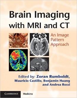
Brain Imaging with MRI and CT: An Image Pattern Approach, 1ed
Most imaging books are ordered according to underlying etiology. However, in real life clinical practice, radiologists usually make their differential diagnoses according to the image patterns, as the etiology is often unknown. Brain Imaging with CT and MRI presents over 180 disease processes and normal variants, grouping entities by these basic patterns to accentuate differential diagnostic features. High quality CT and MRI scans show multiple typical and distinguishing images for each entity. Common and unusual clinical scenarios are described, including dilated perivascular spaces, capillary teleangiectasia, Susac’s syndrome and desmoplastic infantile ganglioglioma. Both basic and advanced imaging techniques are used, reflecting the reality of clinical practice. This image-focused book emphasises the most pertinent clinical information relevant to the diagnostic process. Trainee and practising radiologists will find Brain Imaging with CT and MRI an invaluable and clinically relevant tool for learning and teaching.
Book Description
About the Author
Mauricio Castillo is Professor of Radiology and Section Chief of Neuroradiology, University of North Carolina School of Medicine, New Chapel, North Carolina, USA.
Benjamin Huang is Clinical Assistant Professor of Radiology in the Division of Neuroradiology, University of North Carolina School of Medicine, New Chapel, North Carolina, USA.
Andrea Rossi is Senior Staff Neuroradiologist in the Department of Pediatric Neuroradiology, G. Gaslini Children’s Research Hospital, Genoa, Italy.
Contents
Section 1 Bilateral Predominantly Symmetric Abnormalities
Section 2 Sellar, Perisellar and Midline Lesions
Section 3 Parenchymal Defects or Abnormal Volume
Section 4 Abnormalities Without Significant Mass Effect
Section 5 Primarily Extra-Axial Focal Space-Occupying Lesions
Section 6 Primarily Intra-Axial Masses
Section 7 Intracranial Calcifications
لینک کوتاه : https://bookbaz.ir/?p=22357
نویسنده : Zoran Rumboldt MD , Mauricio Castillo
ناشر : Cambridge University Press; 1 edition
سال انتشار : 2012
زبان کتاب : انگلیسی
نوع فایل : PDF
تعداد صفحات : 434
(ISBN) شابک : 0521119448
قیمت کتاب درآمازون : $129.43
حجم فایل : 35 MB
کتاب های مرتبط:
 دانلود کتاب توسعه دیجیتال ریه: از اولین سی تی ریه تا هوش مصنوعی بالینی
Developing the Digital Lung: From First Lung CT to Clinical AI, 1ed
 دانلود کتاب جراحی ستون فقرات سینه ای: اصول و تکنیک ها
Surgery of the Thoracic Spine: Principles and Techniques, 1ed
 دانلود کتاب صدمات عصب کلاین و هادسون: صدمات عصب، انترپمنت و تومور
Kline and Hudson’s Nerve Injuries: Operative Results for Major Nerve Injuries, Entrapments and Tumors, 2ed
 دانلود کتاب تصویربرداری از استخوان ها و مفاصل: یک روش مختصر و چند وجهی
Imaging of Bones and Joints: A Concise, Multimodality Approach, 1ed
 دانلود کتاب اطلس رنگی جراحی ساقه مغز + ویدئو
Color Atlas of Brainstem Surgery, 1ed + Video
 دانلود کتاب قدرت فرا صوت: کاربرد فرا صوت شدت بالا
Power Ultrasonics: Applications of High-Intensity Ultrasound, 1ed
 دانلود کتاب جراحی و انکولوژی سر و گردن جاتین شاه
Jatin Shah’s Head and Neck Surgery and Oncology, 4ed
 دانلود کتاب مثانه نوروژنیک
 Textbook of the Neurogenic Bladder, 3ed
 دانلود کتاب مبانی اولیه EEG روآن
Rowan’s Primer of EEG, 2ed
 دانلود کتاب اطلس سر، گردن و مغز روتون: تصاویر دو بعدی و سه بعدی + تصاویر
Rhoton’s Atlas of Head, Neck, and Brain: 2D and 3D Images, 1ed + Images
 دانلود کتاب پزشکی آسیب مغزی: اصول و تمرین
Brain Injury Medicine: Principles and Practice, 2ed
 دانلود کتاب علوم اعصاب رفتاری
 Behavioral Neuroscience, 9ed
 دانلود کتاب ترموگرافی پزشکی انسانی
Human Medical Thermography, 1ed
 دانلود کتاب تصویربرداری در جراحی شکم
Imaging in Abdominal Surgery, 1ed
 دانلود کتاب تصویربرداری در گوارش
Imaging in Gastroenterology, 1ed
 دانلود کتاب اصول سی تی اسکن بدن
Fundamentals of Body CT, 5ed
 دانلود کتاب هدایت حین عمل MRI جراحی مغز و اعصاب
Intraoperative MRI-Guided Neurosurgery, 1ed
 دانلود کتاب آناتومی جراحی سر و گردن پرویز جانفزا
Janfaza Surgical Anatomy of the Head and Neck, 1ed
 دانلود کتاب مجتمع علوم اعصاب و عصب شناسی
 Integrated Neuroscience and Neurology, 2ed
 دانلود کتاب CT بدن: ضروریات
Body CT: The Essentials, 1ed
 دانلود کتاب آسیب مغزی جنین و نوزاد
Fetal and Neonatal Brain Injury, 5ed
 دانلود کتاب اصول و تمرین پزشکی خواب (۲ جلدی) + ویدئو
Principles and Practice of Sleep Medicine, 2-Vol, 7ed + Video
 دانلود کتاب تصویربرداری تشخیصی: مغز
Diagnostic Imaging: Brain, 3ed
 دانلود کتاب نورو دیجنریشن: آسیب شناسی مولکولی اختلالات دمانس و حرکتی
Neurodegeneration: The Molecular Pathology of Dementia and Movement Disorders, 2ed
 دانلود کتاب فیزیک ضروری تصویربرداری پزشکی
 The Essential Physics of Medical Imaging, 3ed
 دانلود کتاب نوروپاتولوژی جراحی عملی: یک رویکرد تشخیصی
Practical Surgical Neuropathology: A Diagnostic Approach, 2ed
 دانلود کتاب PET و PET/CT: یک راهنمای بالینی
PET and PET/CT: A Clinical Guide, 3ed
 دانلود کتاب بیماری پارکینسون
 Parkinson’s Disease, 2ed
 دانلود کتاب راهنمای جراحی مغز و اعصاب گرینبرگ
Greenberg’s Handbook of Neurosurgery, 10ed
 دانلود کتاب اطلس رنگی آناتومی اولتراسوند
Color Atlas of Ultrasound Anatomy, 3ed
 دانلود کتاب موارد تصویربرداری پستان
Breast Imaging Cases
 دانلود کتاب تصویربرداری تشخیصی: پزشکی زایمان
Diagnostic Imaging: Obstetrics, 3ed
 دانلود کتاب ملزومات سونوگرافی شکمی و لگنی
Essentials of Abdomino-Pelvic Sonography, 1ed
 دانلود کتاب تفسیر رادیوگرافی قفسه سینه در بیماران قلبی کودک
Chest Radiographic Interpretation in Pediatric Cardiac Patients, 1ed
 دانلود کتاب تصویربرداری تشخیصی کودکان کافی (ویرایش ۲۰۱۹، ۲ جلدی)
Caffey’s Pediatric Diagnostic Imaging, 2-Volume Set, 13ed
 دانلود کتاب اطلس بیهوشی منطقه ای هدایت اولتراسوند + ویدئو
Atlas of Ultrasound-Guided Regional Anesthesia, 3ed + Video
 دانلود کتاب آناتومی تصویربرداری: سر و گردن
Imaging Anatomy: Head and Neck, 1ed
 دانلود کتاب تصویربرداری تشخیصی دستگاه گوارش
Diagnostic Imaging: Gastrointestinal, 4ed
 دانلود کتاب تصویربرداری تشخیصی زایمان
Diagnostic Imaging: Obstetrics, 4ed
 دانلود کتاب تصویربرداری تشخیصی سر و گردن
Diagnostic Imaging: Head and Neck, 4ed
 دانلود کتاب تصویربرداری تشخیصی انکولوژی
Diagnostic Imaging: Oncology, 2ed
 دانلود کتاب صرع کاربردی
Practical Epilepsy, 1ed
 دانلود کتاب اطلس ویدئویی جراحی آنوریسم داخل جمجمه + ویدئو
Video Atlas of Intracranial Aneurysm Surgery, 1ed + Video
 دانلود کتاب MRI در یک نگاه
MRI at a Glance, 2ed
 دانلود کتاب سونوگرافی مداخله ای: راهنمای عملی و اطلس
Interventional Ultrasound: A Practical Guide and Atlas, 1ed
 دانلود کتاب فیزیک ضروری در تصویربرداری برای پرتونگار کلارک
Clark’s Essential Physics in Imaging for Radiographers, 1ed
 دانلود کتاب تصویربرداری ادراری تناسلی (سری موارد رادیولوژی)
Genitourinary Imaging (Radcase), 1ed
 دانلود کتاب تشخیص افتراقی در تصویربرداری سونوگرافی
 Differential Diagnosis in Ultrasound Imaging, 2ed
 دانلود کتاب اطلس درمان عصبی با بیحسی موضعی
 Atlas of Neural Therapy With Local Anesthetics, 3ed
 دانلود کتاب به دام افتادن اعصاب محیطی: تشخیص و مدیریت بالینی
Peripheral Nerve Entrapments: Clinical Diagnosis and Management, 1ed
 دانلود کتاب تصویربرداری تشخیصی: زنان و زایمان
Diagnostic Imaging: Gynecology, 2ed
 دانلود کتاب تصویربرداری زنان و زایمان: تشخیص و مراقبت از جنین
Obstetric Imaging: Fetal Diagnosis and Care, 2ed
 دانلود کتاب تصویربرداری اسکلتی عضلانی
Musculoskeletal Imaging, 2ed
 دانلود کتاب اندوسکوپی ترانس نازال قاعده جمجمه و جراحی مغز
Transnasal Endoscopic Skull Base and Brain Surgery, 1ed
 دانلود کتاب نوروپاتولوژی تکاملی
Developmental Neuropathology, 2ed
 دانلود کتاب موقعیت در رادیوگرافی کلارک
Clark’s Positioning in Radiography, 13ed
 دانلود کتاب مدیریت جامع شوانوما وستیبولار
 Comprehensive Management of Vestibular Schwannoma, 1ed
 دانلود کتاب تصویربرداری تشخیصی قفسه سینه
Diagnostic Imaging: Chest, 3ed
 دانلود کتاب تصویربرداری تشخیصی زنان
Diagnostic Imaging: Gynecology, 3ed
 دانلود کتاب تصویربرداری قفسه سینه و قلب و عروق
Chest and Cardiovascular Imaging, 3ed
 دانلود کتاب کالبدشناسی اعصاب بالینی واکسمن
Clinical Neuroanatomy, 27ed
 دانلود کتاب روش های افتراقی در میکروجراحی مغز سیگر
Differential Approaches in Microsurgery of the Brain, 1ed
 دانلود کتاب مغز و اعصاب کودکان
 Handbook of Pediatric Neurology, 1ed
 دانلود کتاب تشخیص افتراقی در توموگرافی کامپیوتری
Differential Diagnosis in Computed Tomography, 2ed
 دانلود کتاب آناتومی تصویربرداری: قفسه سینه، شکم، لگن
Imaging Anatomy: Chest, Abdomen, Pelvis, 2ed
 دانلود کتاب رادیولوژی اضطراری (سری موارد رادیولوژی)
Radcases Emergency Radiology, 1ed
 دانلود کتاب اطلس سر، گردن و مغز روتون: تصاویر دو بعدی و سه بعدی
Rhoton’s Atlas of Head, Neck, and Brain: 2D and 3D Images, 1ed
 دانلود کتاب اطلس توپوگرافی و پاتوتوپوگرافی آناتومی سر و گردن
Atlas of Topographical and Pathotopographical Anatomy of the Head and Neck, 1ed
 دانلود کتاب رادیولوژی اعصاب تشخیصی و مداخله ای
Diagnostic and Interventional Neuroradiology, 1ed
 دانلود کتاب اولتراسوند: مرور کلی
Ultrasound: A Core Review, 1ed
 دانلود کتاب اختلالات مرتبط با ورزش: تشخیص و مدیریت
Sports-Related Concussion: Diagnosis and Management, 2ed
 دانلود کتاب درمان های مولکولی و سلولی برای بیماری های حرکتی نورون
Molecular and Cellular Therapies for Motor Neuron Diseases, 1ed
 دانلود کتاب پرسش و پاسخ تصویربرداری گوارشی رادکِیس
RadCases Plus Q&A Gastrointestinal Imaging, 2ed
 دانلود کتاب علم رادیولوژیک برای تکنولوژیست ها: فیزیک، بیولوژی و حفاظت
Radiologic Science for Technologists: Physics, Biology, and Protection, 11ed
 دانلود کتاب یادگیری رادیولوژی هِرینگ: شناخت مبانی
Learning Radiology: Recognizing the Basics, 3ed
 دانلود کتاب رادیولوژی ۱۰۱: مبانی و اصول تصویربرداری
Radiology 101: The Basics & Fundamentals of Imaging, 4ed
 دانلود کتاب تصویربرداری عمومی اسکلتی عضلانی
Basic Musculoskeletal Imaging, 1ed
 دانلود کتاب داروها در عصب شناسی
 Drugs in Neurology, 1ed
 دانلود کتاب اندوسونوگرافی
 Endosonography, 4ed
 دانلود کتاب تعویض ترنس کاتتر دریچه آئورت
Transcatheter Aortic Valve Implantation, 1ed
 دانلود کتاب تکنیک های اصلی در جراحی مغز و اعصاب عملی
Core Techniques in Operative Neurosurgery, 2ed
 دانلود کتاب پرسش و پاسخ پزشکی هسته ای رادکِیس
RadCases Plus Q&A Nuclear Medicine, 2ed
 دانلود کتاب تصویربرداری بیماری های بینابینی ریه (سری رادیولوژی کلینیکو)
Clinico Radiological Series: Imaging of Interstitial Lung Diseases, 1ed
 دانلود کتاب اطلس جیبی آناتومی مقطعی: ستون فقرات، اندام، مفاصل
 Pocket Atlas of Sectional Anatomy: Spine Extremities Joints, 3ed
 دانلود کتاب درمان سلولی برای آسیب مغز و اعصاب
Cellular Therapy for Neurological Injury, 1ed
 دانلود کتاب اطلس جراحی آندوسکوپی سینوس و قاعده جمجمه + ویدئو
Atlas of Endoscopic Sinus and Skull Base Surgery, 2ed + Video
 دانلود کتاب اطلس رنگی خون رسانی مجدد مغز: آناتومی، تکنیک، موارد بالینی
Color Atlas of Cerebral Revascularization: Anatomy, Techniques, Clinical Cases, 1ed
 دانلود کتاب توموگرافی پرتو مخروطی کامپیوتری در ارتودنسی: موارد مصرف، بینش و نوآوری
Cone Beam Computed Tomography in Orthodontics: Indications, Insights, and Innovations, 1ed
 دانلود کتاب تصویربرداری پزشکی آکسفورد
Oxford Handbook of Medical Imaging, 1ed
 دانلود کتاب رژیم غذایی کتوژنیک و متابولیک درمانی: گسترش نقش در سلامت و بیماری
Ketogenic Diet and Metabolic Therapies: Expanded Roles in Health and Disease, 1ed
 دانلود کتاب سری مرور موردی رادیولوژی: تصویربرداری قفسه سینه
 Radiology Case Review Series: Thoracic Imaging, 1ed
 دانلود کتاب راهنمای رادیو مابولیزاسیون: فیزیک، بیولوژی، پزشکی هسته ای و تصویربرداری
Handbook of Radioembolization: Physics, Biology, Nuclear Medicine, and Imaging, 1ed
 دانلود کتاب تعمیرات X-Ray: راهنمای جامع نصب و سرویس تجهیزات رادیوگرافی
X-Ray Repair: A Comprehensive Guide to the Installation and Servicing of Radiographic Equipment, 3ed
 دانلود کتاب دوز، فواید و خطر در تصویربرداری پزشکی
 Dose, Benefit, and Risk in Medical Imaging, 1ed
 دانلود کتاب استراتژی های جراحی قاعده جمجمه + ویدئو
Skull Base Surgery: Strategies, 1ed + Video
 دانلود کتاب چشم انداز محاسباتی و پردازش تصویری پزشکی
 Computational Vision and Medical Image Processing V: VipIMAGE 2015
 دانلود کتاب سری بررسی موردی رادیولوژی: تصویربرداری پستان
 Radiology Case Review Series: Breast Imaging, 1ed
 دانلود کتاب تمرین مراقبت نورولوژیک
 The Practice of Neurocritical Care, 1ed
 دانلود کتاب رادیوگرافی PREP (مرور برنامه و آمادگی آزمون)
Radiography PREP (Program Review and Exam Preparation), 8ed
 دانلود کتاب CT قلبی بالینی: آناتومی و عملکرد + ویدئو
Clinical Cardiac CT: Anatomy and Function, 2ed + Video
 دانلود کتاب اصول علوم اعصاب برای علوم و اختلالات ارتباطاتی
Neuroscience Fundamentals for Communication Sciences and Disorders, 1ed
 دانلود کتاب سونوگرافی کودکان
 Pediatric Ultrasound, 1ed
 دانلود کتاب روشهای اسکلتی عضلانی هدایت شده با سونوگرافی در پزشکی ورزشی
Ultrasound Guided Musculoskeletal Procedures in Sports Medicine, 1ed
 دانلود کتاب ۳ تفاوت برتر در رادیولوژی مغز و اعصاب
 Top 3 Differentials in Neuroradiology, 1ed
 دانلود کتاب مرور بالینی مغز و اعصاب مبتنی بر تصویر
Neurology Image-Based Clinical Review, 1ed
 دانلود کتاب پزشکی هسته ای (سری موارد رادیولوژی)
Nuclear Medicine (RadCases), 1ed
 دانلود کتاب مانیتورینگ مراقبت حاد مغز و اعصاب
Neurocritical Care Monitoring, 1ed
 دانلود کتاب MRI بافتها همراه با T2 یا T2*s کوتاه
MRI of Tissues with Short T2s or T2*s, 1ed
 دانلود کتاب تصویربرداری تومور قفسه سینه (سری رادیولوژی کلینیکو)
Clinico Radiological Series: Imaging Of Chest Tumors, 1ed
 دانلود کتاب CT ریه با وضوح بالا
High-Resolution CT of the Lung, 5ed
 دانلود کتاب سونوگرافی مراقبت اورژانسی + ویدئو
Emergency Point-of-Care Ultrasound, 2ed + Video
 دانلود کتاب تصویربرداری و تجسم در اتاق عمل مدرن: راهنمای جامع برای پزشکان
Imaging and Visualization in The Modern Operating Room: A Comprehensive Guide for Physicians, 2015th
 دانلود کتاب آناتومی مقطعی برای متخصصان تصویربرداری
 Sectional Anatomy for Imaging Professionals, 3ed
 دانلود کتاب تصویربرداری پستان (سری تشخیص مستقیم در رادیولوژی)
Breast Imaging (Direct Diagnosis in Radiology: DX-Direct!), 1ed
 دانلود کتاب صدمات اعصاب و عصب: درد، درمان، آسیب بیماری و دستورالعمل های آینده (جلد دوم)
Nerves and Nerve Injuries: Vol 2: Pain, Treatment, Injury, Disease and Future Directions
 دانلود کتاب تصویربرداری قلبی (سری تشخیص مستقیم در رادیولوژی)
Cardiac Imaging (Direct Diagnosis in Radiology: DX-Direct!), 1ed
 دانلود کتاب بررسی سوالات ام آر آی
Review Questions for MRI, 2ed
 دانلود کتاب تصمیم گیری در مراقبت نورولوژی
 Decision Making in Neurocritical Care, 1ed
 دانلود کتاب مداخله انکولوژی (راهنمای عملی در مداخلات رادیولوژی)
Interventional Oncology (Practical Guides in Interventional Radiology), 1ed
 دانلود کتاب تصویربرداری زنان و زایمان کوپل: سری کارشناس رادیولوژی
Obstetric Imaging: Expert Radiology Series, 1ed
 دانلود کتاب مغز انسان نولته: مقدمه ای بر آناتومی عملکردی آن
Nolte’s The Human Brain: An Introduction to its Functional Anatomy, 8ed
 دانلود کتاب اطلس جیبی آناتومی مقطعی: سر و گردن، توموگرافی کامپیوتری و تصویربرداری رزونانس مغناطیسی
Pocket Atlas of Sectional Anatomy: Head and Neck, Computed Tomography and Magnetic Resonance Imaging, 4ed
 دانلود کتاب عصب شناسی، روانپزشکی و اختلالات سیستمیک مرتبط اجمالی
Synopsis of Neurology, Psychiatry and Related Systemic Disorders, 1ed
 دانلود کتاب پرتودرمانی عملی: فیزیک و تجهیزات
Practical Radiotherapy: Physics and Equipment, 3ed
 دانلود کتاب مداخلات با راهنمای تصویری
 Image-Guided Interventions, 3ed
 دانلود کتاب نشانگرهای زیستی برای آسیب مغزی تروماتیک
Biomarkers for Traumatic Brain Injury, 1ed
 دانلود کتاب رادیولوژی مداخله ای: راهنمای بقا
Interventional Radiology: A Survival Guide, 4ed
 دانلود کتاب بیماری های جراحی مغز و اعصاب: یک رویکرد مبتنی بر شواهد برای هدایت عمل
Neurosurgical Diseases: An Evidence-Based Approach to Guide Practice, 1ed
 دانلود کتاب راهنمای ابزار جراحی مغز و اعصاب
 The Neurosurgical Instrument Guide, 1ed
 دانلود کتاب تشخیص افتراقی در تصویربرداری عصبی: ستون فقرات
Differential Diagnosis in Neuroimaging: Spine, 1ed
 دانلود کتاب نورونولوژی و تصویربرداری عصبی سکته مغزی
Neurosonology and Neuroimaging of Stroke, 2ed
 دانلود کتاب مشکلات رایج در بیماریهای مولتیپل اسکلروزیس و میلین زدا CNS
Common Pitfalls in Multiple Sclerosis and CNS Demyelinating Diseases, 1ed
 دانلود کتاب جراحی مغز و اعصاب: راهنمای ضروری آزمون جراحی دهان و مغز و اعصاب بالینی
Neurosurgery: The Essential Guide to the Oral and Clinical Neurosurgical Exam
 دانلود کتاب سرطان پستان: تشخیص زودرس با ماموگرافی
Breast Cancer: Early Detection with Mammography: Casting Type Calcifications Sign of a Subtype with Deceptive Features, 1ed
 دانلود کتاب SPECT و SPECT/CT: راهنمای بالینی
SPECT and SPECT/CT: A Clinical Guide, 1ed
 دانلود کتاب بیماری های عفونی مغز و اعصاب: مدیریت جراحی و غیرجراحی
Neurosurgical Infectious Disease: Surgical and Nonsurgical Management, 1ed
 دانلود کتاب کمک های اولیه برای بورد مغز و اعصاب
 First Aid for the Neurology Boards, 2ed
 دانلود کتاب اشعه ایکس قفسه سینه برای دانشجویان پزشکی
Chest X-rays for Medical Students, 1ed
 دانلود کتاب رینوره مایع مغزی نخاعی: راهنمای جامع ارزیابی و مدیریت + ویدئو
Cerebrospinal Fluid Rhinorrhea: Comprehensive Guide to Evaluation and Management, 1ed + Video
 دانلود کتاب نوروسایکولوژی کودکان: پژوهش، نظریه و تمرین
Pediatric Neuropsychology: Research, Theory, and Practice 2ed
 دانلود کتاب انکولوژی تابشی بالینی گاندرسون و تِپِر + ویدئو
Gunderson & Tepper’s Clinical Radiation Oncology, 4ed + Video
 دانلود کتاب تصویربرداری عفونت قفسه سینه (سری رادیولوژی کلینیکو)
Clinico Radiological Series: Imaging of Chest Infections, 1ed
 دانلود کتاب فناوری اولتراسوند برای پزشکان بالینی
Ultrasound Technology for Clinical Practitioners, 1ed
 دانلود کتاب اطلس تصویربرداری تشخیصی خاله مینی
Aunt Minnie’s Atlas and Imaging-Specific Diagnosis, 4ed
 دانلود کتاب تصویربرداری تشخیصی پستان
Diagnostic Imaging: Breast, 3ed
 دانلود کتاب پرتو درمانی والتر و میلر: فیزیک پرتو، درمان و انکولوژی
Walter and Miller’s Textbook of Radiotherapy: Radiation Physics, Therapy and Oncology, 7ed
 دانلود کتاب بیماری های عصبی: پیامدها در اقدامات پزشکی و دندانپزشکی
Neurological Diseases: Implications in Medical and Dental Practices, 1ed
 دانلود کتاب سونوگرافی تشخیصی بالینی: شکم و ساختار ظاهری + کتاب کار
Diagnostic Medical Sonography: Abdomen and Superficial Structures + Workbook, 3ed
 دانلود کتاب راهنمای جیبی برای رادیوگراف های کلارک
Clark’s Pocket Handbook for Radiographers, 2ed
دانلود کتاب نورونولوژی و تصویربرداری عصبی سکته مغزی + ویدئوNeurosonology and Neuroimaging of Stroke, 2ed + Video