
دانلود کتاب آناتومی تصویربرداری از مغز انسان
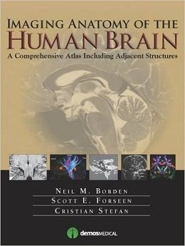
Imaging Anatomy of the Human Brain, 1ed
This volume presents a detailed and beautifully illustrated tour of neuroanatomy, employing…standard and advanced modalities… Ample use of color illustrates fiber tracts and differentiates different nuclei and other structures. The sections are amply labeled and easy to follow, allowing the reader to become familiar with cognate areas on images of multiple techniques.
— Carl E. Stafstrom, Division of Pediatric Neurology, Johns Hopkins Hospital, Journal of Pediatric Epilepsy
An Atlas for the 21<sup>st</sup> Century
The most precise, cutting-edge images of normal cerebral anatomy available today are the centerpiece of this spectacular atlas for clinicians, trainees, and students in the neurologically-based medical and non-medical specialties. Truly an “atlas for the 21st century,” this comprehensive visual reference presents a detailed overview of cerebral anatomy acquired through the use of multiple imaging modalities including advanced techniques that allow visualization of structures not possible with conventional MRI or CT. Beautiful color illustrations using 3-D modeling techniques based upon 3D MR volume data sets further enhances understanding of cerebral anatomy and spatial relationships. The anatomy in these color illustrations mirror the black and white anatomic MR images presented in this atlas.
Written by two neuroradiologists and an anatomist who are also prominent educators, along with more than a dozen contributors, the atlas begins with a brief introduction to the development, organization, and function of the human brain. What follows is more than 1,000 meticulously presented and labelled images acquired with the full complement of standard and advanced modalities currently used to visualize the human brain and adjacent structures, including MRI, CT, diffusion tensor imaging (DTI) with tractography, functional MRI, CTA, CTV, MRA, MRV, conventional 2-D catheter angiography, 3-D rotational catheter angiography, MR spectroscopy, and ultrasound of the neonatal brain. The vast array of data that these modes of imaging provide offers a wider window into the brain and allows the reader a unique way to integrate the complex anatomy presented. Ultimately the improved understanding you can acquire using this atlas can enhance clinical understanding and have a positive impact on patient care. Additionally, various anatomic structures can be viewed from modality to modality and from multiple planes.
This state-of-the-art atlas provides a single source reference, which allows the interested reader ease of use, cross-referencing, and the ability to visualize high-resolution images with detailed labeling. It will serve as an authoritative learning tool in the classroom, and as an invaluable practical resource at the workstation or in the office or clinic.
Key Features:
- Provides detailed views of anatomic structures within and around the human brain utilizing over 1,000 high quality images across a broad range of imaging modalities
- Contains extensively labeled images of all regions of the brain and adjacent areas that can be compared and contrasted across modalities
- Includes specially created color illustrations using computer 3-D modeling techniques to aid in identifying structures and understanding relationships
- Goes beyond a typical brain atlas with detailed imaging of skull base, calvaria, facial skeleton, temporal bones, paranasal sinuses, and orbits
- Serves as an authoritative learning tool for students and trainees and practical reference for clinicians in multiple specialties
Review
About the Author
Contents
Chapter 1: Introduction to the Development,. Organization and Function of the Human Brain
Chapter 2: Color Illustrations of the Human Brain Using 3D Modeling Techniques
Chapter 3: MR Imaging of the Brain
Chapter 4: MR Imaging of the Cerebellum
Chapter 5: MR Imaging of Regional Intracranial Anatomy and Orbits
Chapter 6: The Cranial Nerves
Chapter 7: Advanced MRI Techniques
Chapter 8: CT Imaging
Chapter 9: Vascular Imaging
Chapter 10: Neonatal Cranial Ultrasound
لینک کوتاه : https://bookbaz.ir/?p=45747
نویسنده : Neil M. Borden MD , Scott E. Forseen MD
ناشر : Demos Medical; 1 edition
سال انتشار : 2016
زبان کتاب : انگلیسی
نوع فایل : PDF
تعداد صفحات : 464
(ISBN) شابک : 1936287749
قیمت کتاب درآمازون : $159.15
حجم فایل : 48 MB
کتاب های مرتبط:
 دانلود کتاب آناتومی بالینی مور
Moore’s Clinically Oriented Anatomy, 9ed
 دانلود کتاب اطلس علوم اعصاب نتر + ویدئو
Netter’s Atlas of Neuroscience, 3ed + Video
 دانلود کتاب مداخله انکولوژی (راهنمای عملی در مداخلات رادیولوژی)
Interventional Oncology (Practical Guides in Interventional Radiology), 1ed
 دانلود کتاب آناتومی تصویربرداری: سر و گردن
Imaging Anatomy: Head and Neck, 1ed
 دانلود کتاب آناتومی و فیزیولوژی: یک رویکرد یکپارچه
Anatomy and Physiology: An Integrated Approach, 1ed
 دانلود کتاب آناتومی تصویربرداری: قفسه سینه، شکم، لگن
Imaging Anatomy: Chest, Abdomen, Pelvis, 2ed
 دانلود کتاب پاتوفیزیولوژی اختلالات خونی
Pathophysiology of Blood Disorders, 1ed
 دانلود کتاب همراه جیبی کتاب فیزیولوژی پزشکی گایتون و هال
Pocket Companion to Guyton and Hall Textbook of Medical Physiology, 13ed
 دانلود کتاب اشعه ایکس اسکلتی عضلانی برای دانشجویان و کارآموزان پزشکی
Musculoskeletal X-Rays for Medical Students and Trainees, 1ed
 دانلود کتاب بیولوژی انسان چایراس
Human Biology, 9ed
 دانلود کتاب سونوگرافی کودکان
 Pediatric Ultrasound, 1ed
 دانلود کتاب اطلس کالبد شکافی زوبوتا
Sobotta Dissection Atlas, 3ed
 دانلود کتاب راهنمای آناتومی عملی قفسه سینه و شکم کانینگهام (جلد ۲)
Cunningham’s Manual of Practical Anatomy: Thorax and Abdomen, Vol-2, 16ed
 دانلود کتاب معاینه عصبی دژانگ
DeJong’s The Neurologic Examination, 8ed
 دانلود کتاب تصویربرداری تشخیصی پستان
Diagnostic Imaging: Breast, 3ed
 دانلود کتاب اطلس رنگ آناتومی انسان: سیستم حرکتی (جلد ۱)
Color Atlas of Human Anatomy: Vol. 1 Locomotor System, 8ed
 دانلود کتاب اطلس آناتومی انسان نتر: رویکرد کلاسیک منطقه ای + ویدئو
Netter Atlas of Human Anatomy: Classic Regional Approach, 8ed + Video
 دانلود کتاب آناتومی مقطعی برای متخصصان تصویربرداری
 Sectional Anatomy for Imaging Professionals, 3ed
 دانلود کتاب راهنمای بالینی IR: راهنمای مختصر برای رادیولوژی مداخله ای
 Pocketbook of Clinical IR: A Concise Guide to Interventional Radiology, 1ed
 دانلود کتاب جداول یادگیری عضلات، مفاصل و اعصاب زوبوتا
Sobotta Learning Tables of Muscles, Joints and Nerves, 3ed
 دانلود کتاب صدمات اعصاب و عصب: درد، درمان، آسیب بیماری و دستورالعمل های آینده (جلد دوم)
Nerves and Nerve Injuries: Vol 2: Pain, Treatment, Injury, Disease and Future Directions
 دانلود کتاب اطلس آموزشی تصویربرداری شکم
Teaching Atlas of Abdominal Imaging, 1ed
 دانلود کتاب اطلس رنگی آناتومی انسان: اندام های داخلی (جلد ۲)
Color Atlas of Human Anatomy: Vol. 2 Internal Organs, 7ed
 دانلود کتاب اطلس جیبی آناتومی مقطعی: سر و گردن، توموگرافی کامپیوتری و تصویربرداری رزونانس مغناطیسی
Pocket Atlas of Sectional Anatomy: Head and Neck, Computed Tomography and Magnetic Resonance Imaging, 4ed
 دانلود کتاب تصویربرداری زنان و زایمان کوپل: سری کارشناس رادیولوژی
Obstetric Imaging: Expert Radiology Series, 1ed
 دانلود کتاب خود ارزیابی و مرور آناتومی
 Self Assessment and Review of Anatomy, 1ed
 دانلود کتاب پاسخ آناتومی و فیزیولوژی
 The Handy Anatomy Answer Book, 2ed
 دانلود کتاب تصویربرداری از مغز با MRI و CT: رویکرد الگوی تصویری
Brain Imaging with MRI and CT: An Image Pattern Approach, 1ed
 دانلود کتاب اطلس توپوگرافی و پاتوتوپوگرافی آناتومی سر و گردن
Atlas of Topographical and Pathotopographical Anatomy of the Head and Neck, 1ed
 دانلود کتاب متاستازهای مغز از تومورهای اولیه (۳ جلدی)
Brain Metastases from Primary Tumors, 3-Vol, 1ed
 دانلود کتاب تعادل ساژیتال ستون فقرات: از عادی به پاتولوژی
Sagittal Balance of the Spine: From Normal to Pathology, 1ed
 دانلود کتاب آناتومی سر و گردن آکسفورد
Oxford Handbook of Head and Neck Anatomy, 1ed
 دانلود کتاب مغز و اعصاب کودکان
 Handbook of Pediatric Neurology, 1ed
 دانلود کتاب سونوگرافی نقطه مراقبت + ویدئو
Point of Care Ultrasound, 2ed + Video
 دانلود کتاب سرطان پستان: تشخیص زودرس با ماموگرافی
Breast Cancer: Early Detection with Mammography: Casting Type Calcifications Sign of a Subtype with Deceptive Features, 1ed
 دانلود کتاب دوز، فواید و خطر در تصویربرداری پزشکی
 Dose, Benefit, and Risk in Medical Imaging, 1ed
 دانلود کتاب بیولوژی انسان: مفاهیم، کاربردها و مسائل
Biology of Humans: Concepts, Applications, and Issues, 6ed
 دانلود کتاب جراحی ستون فقرات حداقل تهاجمی + ویدئو
Minimally Invasive Spine Surgery, 1ed + Video
 دانلود کتاب فیزیولوژی کاستانزو
Costanzo Physiology, 7ed
 دانلود کتاب SPECT و SPECT/CT: راهنمای بالینی
SPECT and SPECT/CT: A Clinical Guide, 1ed
 دانلود کتاب اطلس آسیب شناسی بافت نرم و استخوان: با بافت شناسی، سیتولوژی و رادیولوژی
Atlas of Soft Tissue and Bone Pathology: With Histologic, Cytologic, and Radiologic Correlations, 1ed
 دانلود کتاب اطلس عکاسی منطقه ای ویژگی های غیر متریک و آناتومیک اسکلت انسانی
Photographic Regional Atlas of Non-Metric Traits and Anatomical Variants in the Human Skeleton, 1ed
 دانلود کتاب مدیریت درد بونیکا
Bonica’s Management of Pain, 4ed
 دانلود کتاب مدل های محاسباتی مغز و رفتار
 Computational Models of Brain and Behavior, 1ed
 دانلود کتاب کاوش آناتومی و فیزیولوژی در آزمایشگاه
Exploring Anatomy & Physiology in the Laboratory, 3ed
 دانلود کتاب پزشکی سکته مغزی
 Textbook of Stroke Medicine, 3ed
 دانلود کتاب نورولوژی مریت
Merritt’s Neurology, 14ed
 دانلود کتاب فیزیولوژی کاستانزو
Physiology Costanzo, 6ed
 دانلود کتاب اطلس بالینی پلیسومنوگرافی
 Clinical Atlas of Polysomnography, 1ed
 دانلودکتاب آناتومی بالینی ستون فقرات، نخاع و ANS
Clinical Anatomy of the Spine, Spinal Cord, and ANS, 3ed
 دانلود کتاب بیماری های سیستم عصبی در دوران کودکی ایکاردی
Aicardi’s Diseases of the Nervous System in Childhood, 4ed
 دانلود کتاب برنامه اصلی برای مراقبت شیردهی بین رشته ای
Core Curriculum for Interdisciplinary Lactation Care, 1ed
 دانلود کتاب تصویربرداری در اورولوژی
Imaging in Urology, 1ed
 دانلود کتاب درمان کنونی کان (ویرایش ۲۰۱۶)
Conn’s Current Therapy 2016, 1ed
 دانلود کتاب مثانه نوروژنیک
 Textbook of the Neurogenic Bladder, 3ed
 دانلود کتاب ترمیم عصبی و توانبخشی (۲ جلدی)
Textbook of Neural Repair and Rehabilitation, 2-Vol, 2ed
 دانلود کتاب آناتومی و فیزیولوژی بصری
Visual Anatomy & Physiology, 2ed
 دانلود کتاب هفت بایپس: اصول و تکنیک های ریواسکولاریزیشن
 Seven Bypasses: Tenets and Techniques for Revascularization, 1ed
 دانلود کتاب تصویربرداری پراش اشعه ایکس: فناوری و کاربردها
X-Ray Diffraction Imaging: Technology and Applications, 1ed
 دانلود کتاب علوم اعصاب برای دندانپزشکی
 Neuroscience for Dentistry, 1ed
 دانلود کتاب سیستم عصبی انسان بار: یک دیدگاه تشریحی
Barr’s The Human Nervous System: An Anatomical Viewpoint, 10ed
 دانلود کتاب اصول تصویربرداری اسکلتی عضلانی مک کینز
Fundamentals of Musculoskeletal Imaging, 4ed
 دانلود کتاب راهنمای مطالعه برای ساختار و عملکرد بدن انسان مملر
Study Guide for Memmler’s Structure and Function of the Human Body, 11ed
 دانلود کتاب آناتومی و فیزیولوژی برای پرستاران در یک نگاه
Anatomy and Physiology for Nurses at a Glance, 1ed
 دانلود کتاب ملزومات رادیوگرافی و رادیولوژی دندان
Essentials of Dental Radiography and Radiology, 5ed
 دانلود کتاب اطلس آناتومی زوبوتا: ارگان های داخلی (جلد ۲)
Sobotta Atlas of Anatomy: Internal Organs, Vol-2, 16ed
 دانلود کتاب آناتومی و رویکردهای جراحی نتر
Netter’s Surgical Anatomy and Approaches, 2ed
 دانلود کتاب اطلس آناتومی زوبوتا: سر، گردن و نوروآناتومی (جلد ۳)
Sobotta Atlas of Anatomy: Head, Neck and Neuroanatomy, Vol-3, 16ed
 دانلود کتاب دانشنامه جامع تنوع آناتومیک انسانی برگمن
Bergman’s Comprehensive Encyclopedia of Human Anatomic Variation, 1ed
 دانلود کتاب جراحی عصبی عروقی
 Neurovascular Surgery, 2ed
 دانلود کتاب CT قلبی بالینی: آناتومی و عملکرد
Clinical Cardiac CT: Anatomy and Function, 2ed
 دانلود کتاب همراه نورورادیولوژی: روش ها، دستورالعمل ها و اصول تصویربرداری
Neuroradiology Companion: Methods, Guidelines, and Imaging Fundamentals, 5ed
 دانلود کتاب ارزیابی خود در آناتومی و فیزیولوژی در سلامت و بیماری راس و ویلسون
Ross & Wilson Self-Assessment in Anatomy and Physiology in Health and Illness, 1ed
 دانلود کتاب تصویربرداری شکم: الزامات اصلی
Abdominal Imaging: The Core Requisites, 1ed
 دانلود کتاب تصویربرداری در گوش و حلق و بینی
Imaging in Otolaryngology, 1ed
 دانلود کتاب پرسش و پاسخ مبتنی بر شایستگی در آناتومی برای آزمون حرفهای اول Mbbs
Competency Based Q&A In Anatomy For First Mbbs Professional Examination, 1ed
 دانلود کتاب ضایعات عروقی سر و گردن: تشخیص و مدیریت
Vascular Lesions of the Head and Neck: Diagnosis and Management, 1ed
 دانلود کتاب ملزومات تصویری آناتومی و فیزیولوژی
 Visual Essentials of Anatomy & Physiology, 1ed
 دانلود کتاب ملزومات آناتومی و فیزیولوژی سالدین
Essentials of Anatomy & Physiology, 2ed
 دانلود کتاب آناتومی مصور سر و گردن
 Illustrated Anatomy of the Head and Neck, 5ed
 دانلود کتاب تحریک عمیق مغز: تکنیک ها و تمرین ها
Deep Brain Stimulation: Techniques and Practices, 1ed
 دانلود کتاب اطلس آناتومی زوبوتا: آناتومی عمومی و سیستم اسکلتی عضلانی (جلد ۱)
Sobotta Atlas of Anatomy, Vol.1: General Anatomy and Musculoskeletal System, 17ed
 دانلود کتاب راهنمای تغذیه، رژیم غذایی و چشم
Handbook of Nutrition, Diet, and the Eye, 2ed
 دانلود کتاب آناتومی و تکنیک جراحی
 Surgical Anatomy and Technique: A Pocket Manual, 4ed
 دانلود کتاب موقعیت ها و تکنیک های رادیوگرافی بانترِیگر
Bontrager’s Handbook of Radiographic Positioning and Techniques, 8ed
 دانلود کتاب اطلس آناتومی زوبوتا: آناتومی عمومی و سیستم عضلانی اسکلتی (جلد ۱)
Sobotta Atlas of Anatomy: General Anatomy and Musculoskeletal System, Vol-1, 16ed
 دانلود کتاب اطلس آناتومی زوبوتا: اندام های داخلی (جلد ۲)
Sobotta Atlas of Anatomy, Vol. 2: Internal Organs, 17ed
 دانلود کتاب اطلس جیبی آناتومی مقطعی: سر و گردن
Pocket Atlas of Sectional Anatomy: Head and Neck, 3ed
 دانلود کتاب کار درک آناتومی و فیزیولوژی: بینایی، شنوایی، رویکرد تعاملی
Workbook to Accompany Understanding Anatomy & Physiology: A Visual, Auditory, Interactive Approach, 2ed
 دانلود کتاب ۵ دقیقه مشورت مغز و اعصاب
 The 5-Minute Neurology Consult, 2ed
 دانلود کتاب آناتومی انسان برای دانشجویان دندانپزشکی آناند
Anand’s Human Anatomy for Dental Students, 3ed
 دانلود کتاب راهنمای آناتومی عملی اندام فوقانی و تحتانی کانینگهام (جلد ۱)
Cunningham’s Manual of Practical Anatomy Upper and Lower limbs, Vol-1, 16ed
 دانلود کتاب طراحی آزمایش مبتنی بر EEG برای اختلال افسردگی اساسی
EEG-Based Experiment Design for Major Depressive Disorder, 1ed
 دانلود کتاب آناتومی گری: مبنای تشریحی عمل بالینی
Gray’s Anatomy: The Anatomical Basis of Clinical Practice, 42ed
 دانلود کتاب سکته مغزی: پاتوفیزیولوژی، تشخیص و مدیریت
Stroke: Pathophysiology, Diagnosis, and Management, 6ed
 دانلود کتاب جراحی محل اتصال مهره جمجمه + ویدئو
Surgery of the Craniovertebral Junction, 2ed + Videos
 دانلود کتاب آناتومی و فیزیولوژی انسان هول (ویرایش ۲۰۱۶)
Hole’s Human Anatomy & Physiology, 14ed
 دانلود کتاب سری کارشناسی ستون فقرات AO: کمر درد (جلد ۸)
AOSpine Masters Series, Volume 8: Back Pain, 1ed
 دانلود کتاب اطلس روشهای هدایت تصویری ستون فقرات
 Atlas of Image-Guided Spinal Procedures, 2ed
 دانلود کتاب جراحی آندوسکوپی حدقه چشم: آناتومی، پاتولوژی و مدیریت
Endoscopic Surgery of the Orbit: Anatomy, Pathology, and Management, 1ed
 دانلود کتاب اطلس جراحی ستون فقرات
 Pocket Atlas of Spine Surgery, 2ed
 دانلود کتاب جامع فیزیک پزشکی (۱۰ جلدی)
Comprehensive Biomedical Physics, 10-Vol, 1ed
 دانلود کتاب بینش هایی در مورد نورولوژی بالینی
Insights into Clinical Neurology, 1ed
 دانلود کتاب مراقبت از بیمار در رادیوگرافی + چک لیست رویه ها
Patient Care in Radiography, 10ed + Procedures Checklist
 دانلود کتاب اصول سیتی اسکن بدن
Fundamentals of Body CT, 4ed
 دانلود کتاب رادیوتراپی سرطان – مروری بر اساس سوال
Radiation Oncology – A Question Based Review, 2ed
 دانلود کتاب ۱۰۱ راه حل سی تی اسکن شکمی
 ۱۰۱Ct Abdomen Solutions, 1ed
 دانلود کتاب الکترودیاگنوز در مغز و اعصاب بالینی امینف
Aminoff’s Electrodiagnosis in Clinical Neurology, 6ed
 دانلود کتاب مراقبت سکته مغزی حاد
Acute Stroke Care, 3ed
 دانلود کتاب هدایت حین عمل MRI جراحی مغز و اعصاب
Intraoperative MRI-Guided Neurosurgery, 1ed
 دانلود کتاب معرفی تصویربرداری نتر
Netter’s Introduction to Imaging, 1ed
 دانلود کتاب تصویربرداری در جراحی شکم
Imaging in Abdominal Surgery, 1ed
 دانلود کتاب تصویربرداری در گوارش
Imaging in Gastroenterology, 1ed
 دانلود کتاب اصول سی تی اسکن بدن
Fundamentals of Body CT, 5ed
 دانلود کتاب تصویربرداری تشخیصی: مغز
Diagnostic Imaging: Brain, 3ed
 دانلود کتاب تغذیه و عضلات اسکلتی
Nutrition and Skeletal Muscle, 1ed
 دانلود کتاب بیولوژی انسانی مِیدر
Human Biology, 14th Edition
 دانلود کتاب اطلس آناتومی گری
Gray’s Atlas of Anatomy, 2ed
 دانلود کتاب اصول و تمرین پزشکی خواب (۲ جلدی) + ویدئو
Principles and Practice of Sleep Medicine, 2-Vol, 7ed + Video
 دانلود کتاب توانبخشی سکته مغزی: رویکرد مبتنی بر عملکرد
Stroke Rehabilitation: A Function-Based Approach, 4ed
 دانلود کتاب اطلس عمل جراحی مغز و اعصاب: جراحی مغز و اعصاب کاربردی
Neurosurgical Operative Atlas: Functional Neurosurgery, 3ed
 دانلود کتاب راهنمای سم شناسی عصبی رشد
Handbook of Developmental Neurotoxicology, 2ed
 دانلود کتاب اطلس جیبی آناتومی مقطعی: ستون فقرات، اندام، مفاصل
 Pocket Atlas of Sectional Anatomy: Spine Extremities Joints, 3ed
 دانلود کتاب رادیولوژی اضطراری بالینی
Clinical Emergency Radiology, 2ed
 دانلود کتاب ملزومات در آناتومی و فیزیولوژی انسان هول
Hole’s Essentials of Human Anatomy & Physiology, 15ed
 دانلود کتاب موارد تصویربرداری پستان
Breast Imaging Cases
 دانلود کتاب تصویربرداری تشخیصی: پزشکی زایمان
Diagnostic Imaging: Obstetrics, 3ed
 دانلود کتاب ملزومات سونوگرافی شکمی و لگنی
Essentials of Abdomino-Pelvic Sonography, 1ed
 دانلود کتاب تفسیر رادیوگرافی قفسه سینه در بیماران قلبی کودک
Chest Radiographic Interpretation in Pediatric Cardiac Patients, 1ed
 دانلود کتاب تصویربرداری تشخیصی کودکان کافی (ویرایش ۲۰۱۹، ۲ جلدی)
Caffey’s Pediatric Diagnostic Imaging, 2-Volume Set, 13ed
 دانلود کتاب اطلس بیهوشی منطقه ای هدایت اولتراسوند + ویدئو
Atlas of Ultrasound-Guided Regional Anesthesia, 3ed + Video
 دانلود کتاب اطلس آناتومی گری
Gray’s Atlas of Anatomy, 3ed
 دانلود کتاب تصویربرداری تشخیصی دستگاه گوارش
Diagnostic Imaging: Gastrointestinal, 4ed
 دانلود کتاب تصویربرداری تشخیصی زایمان
Diagnostic Imaging: Obstetrics, 4ed
 دانلود کتاب تصویربرداری تشخیصی سر و گردن
Diagnostic Imaging: Head and Neck, 4ed
 دانلود کتاب تصویربرداری تشخیصی انکولوژی
Diagnostic Imaging: Oncology, 2ed
 دانلود کتاب تصویربرداری رزونانس مغناطیسی (۳ جلدی)
Magnetic Resonance Imaging Handbook (MRI), 3-Vol, 1ed
 دانلود کتاب تصویربرداری تشخیصی نوزادان و کودکان ولز (جلد اول)
Diagnostic Imaging of Infants and Children, Vol-1, 1ed
 دانلود کتاب اطلس توموگرافی کامپیوتری پرتو مخروطی
Atlas of Cone Beam Computed Tomography, 1ed
 دانلود کتاب آناتومی و فیزیولوژی: رویکرد یکپارچه
Anatomy & Physiology: An Integrative Approach, 3ed
 دانلود کتاب رادیوتراپی و سرطان های کودکان، نوجوانان و جوانان
Radiotherapy and the Cancers of Children, Teenagers, and Young Adults, 1ed
 دانلود کتاب فیزیولوژی گوارش: سری فیزیولوژی موزبی
Gastrointestinal Physiology: Mosby Physiology Series, 9ed
 دانلود کتاب MRI در یک نگاه
MRI at a Glance, 2ed
 دانلود کتاب راهنمای مطالعه بدن انسان در سلامت و بیماری مملر
Study Guide to Accompany Memmler the Human Body in Health and Disease, 13ed
 دانلود کتاب صدمات عصب کلاین و هادسون: صدمات عصب، انترپمنت و تومور
Kline and Hudson’s Nerve Injuries: Operative Results for Major Nerve Injuries, Entrapments and Tumors, 2ed
 دانلود کتاب سونوگرافی مداخله ای: راهنمای عملی و اطلس
Interventional Ultrasound: A Practical Guide and Atlas, 1ed
 دانلود کتاب فیزیک ضروری در تصویربرداری برای پرتونگار کلارک
Clark’s Essential Physics in Imaging for Radiographers, 1ed
 دانلود کتاب تصویربرداری ادراری تناسلی (سری موارد رادیولوژی)
Genitourinary Imaging (Radcase), 1ed
 دانلود کتاب تشخیص افتراقی در تصویربرداری سونوگرافی
 Differential Diagnosis in Ultrasound Imaging, 2ed
 دانلود کتاب اطلس درمان عصبی با بیحسی موضعی
 Atlas of Neural Therapy With Local Anesthetics, 3ed