دانلود کتاب اطلس ویدئویی بخیه چشم: اصول و تکنیک ها + ویدئو
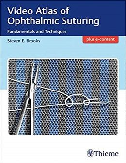
Video Atlas of Ophthalmic Suturing: Fundamentals and Techniques, 1ed + Video
Video Atlas of Ophthalmic Suturing: Fundamentals and Techniques by Steven Brooks, MD, is the first multimedia atlas dedicated to effective suturing and knot tying skills in ophthalmic surgery. The text and videos not only cover essential suturing techniques, but merge these concepts with critical principles of wound healing, tissue biomechanics, ergonomics, and the physical properties of common ophthalmic sutures and surgical needles. This atlas fills a significant knowledge gap in educational ophthalmic surgery literature, and greatly enhances the fundamental knowledge and skills provided in ophthalmic surgical training programs.
The reader is guided step by step starting with history, safety, basic principles, and the role of the suture in wound healing. Subsequent chapters cover all aspects of sutures – from design and proper selection of tools to basic skills and advanced techniques. The instructional videos, accompanying text, and high-quality illustrations provide the resources necessary to develop muscle memory, tactile experience, and conceptual understanding required for eventual mastery. The easy-to-follow didactics enable practicing and replicating various skills in a training lab environment.
Key Features
- Fifty ophthalmic surgery videos utilize non-biological model materials and larger sutures than those used in ophthalmic practice, clearly demonstrating the technical and spatial aspects of suturing maneuvers
- The videos are provided as QR codes directly in the book and are also on the accompanying Media Center site
- A chapter on safe practices, a critical issue rarely discussed in surgical texts, helps prevent the risk of sharps injuries in the operating room
- Discussion of the composition and properties of common surgical threads used in ophthalmic surgery enables proper selection of sutures, needles, holders, forceps, and stitching patterns for specific cases
- Clinical pearls on topics such as tissue stabilization, proper hand position, avoiding common errors, and complications
- A self-assessment quiz ensures a solid understanding of the material presented
Unique in the field, this outstanding reference provides the beginning ophthalmic surgeon practical knowledge of anatomy, pathophysiology, wound healing, biomechanics, and hands-on suturing techniques. Teaching ophthalmologists will discover robust material from which to design didactic and wet lab resident courses.
Contents
۱. Historical Perspective
۲. Safety
۳. Basic Principles
۴. Basics of Wound Healing and the Role of Suture
۵. Ophthalmic Surgical Suture: Propertiesr Design,, and Selection
۶. Suture Needles
۷. Needle Holders and Forceps
۸. Suturing: Basic Skills and Techniques
۹. Common Errors for Beginning Ophthalmic Surgeons
۱۰. Complications
۱۱. Self-Assessment Quiz
Video Contents
Video 2.1: Correct technique to gain control of the needle in order to load a needle holder.
Video 2.2: Proper technique to reduce sharps injury to an assistant in the surgical field.
Video 2.3: Proper techniques to hand a needle back to the scrub nurse, with an instrument or by hand.
Video 8.1: Controlling the needle safely in preparation for loading a needle holder.
Video 8.2: Dangling the needle to reload the needle holder. This technique is useful for loading under an operating microscope.
Video 8.3: A forehand versus backhand needle pass. Note that a needle loaded for a backhand pass with one hand is oriented the same way in needle holder as for a forehand pass with the opposite hand.
Video 8.4: The stability of the needle is affected by the position on the needle body that it is held. The further back on the needle body that it is loaded, the greater the torque experienced at the tip, and the less stable the control.
Video 8.5: Different variations in hand position can be used to suture a wound, offering a range of possibilities to optimize visualization and ergonomics.
Video 8.6: Stabilizing the wound edge to facilitate passage of the needle.
Video 8.7: Forehand versus backhand needle pass.
Video 8.8: Needle holder locking mechanism.
Video 8.9: Needle pass without and with torque.
Video 8.10: Stabilization of exit side to facilitate completion of pass and prevent needle retraction.
Video 8.11: Pulling the needle out of the tissue without damaging the tip.
Video 8.12: Steep versus shallow needle entry.
Video 8.13: Shallow needle pass appropriate for sclera.
Video 8.14: Reducing tissue drag, friction, and shear by pulling the thread in line with the suture pass.
Video 8.15: Guiding the trailing thread with a second instrument to reduce tissue drag, friction, and shear.
Video 8.16: Using the surgical needle to plan entry and exit points across a wound.
Video 8.17: Simple interrupted stitch using 2–۱–۱–۱ surgeon’s knot.
Video 8.18: Figure 8 (cruciate) stitch.
Video 8.19: Simple running stitch.
Video 8.20: Locked running stitch.
Video 8.21: Horizontal mattress stitch.
Video 8.22: Vertical mattress suture.
Video 8.23: Purse string stitch.
Video 8.24: Square knot.
Video 8.25: The grip strength of the first throw is increased by adding additional loops.
Video 8.26: Slip knot.
Video 8.27: Tying a buried knot.
Video 8.28: Rotating the knot to bury.
Video 8.29: Temporary knot.
Video 8.30: Granny knot.
Video 8.31: Surgeon’s knot.
Video 8.32: Effect of asymmetry on knot stability.
Video 8.33: Imbrication stitch.
Video 8.34: Hand tying.
Video 8.35: Instrument tying with tying forceps.
Video 8.36: Instrument tying with needle holders.
Video 8.37: Correct technique to hold leading thread for tying and creating loops on the second instrument.
Video 8.38: Control of the leading thread by keeping the first instrument, or hand, close to the second instrument helps keep the loops in place while the trailing thread is being grasped.
Video 8.39: Moving hands in a coordinated manner helps efficiency and maintains good visualization.
Video 8.40: Holding suture loops back from the second instrument tip to increase efficiency and visualization.
Video 8.41: If the trailing thread is too long, or it is grasped too close to the knot, it is difficult to pull it through the loop to complete each throw, and there is a tendency for redundant loops to get caught in the knot.
Video 8.42: Minimizing disruption of knot to maintain knot tension.
Video 8.43: Maintaining stability of first throw to avoid wound gape.
Video 8.44: Cutting the suture.
لینک کوتاه : https://bookbaz.ir/?p=105560
نویسنده : Steven Brooks
ناشر : Thieme; 1 edition
سال انتشار :
زبان کتاب : انگلیسی
نوع فایل : MP4 + PDF (کیفیت اصلی)
تعداد صفحات : 64
(ISBN) شابک : 1626237166
قیمت کتاب درآمازون : $78.81
حجم فایل : 205 MB
