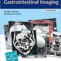
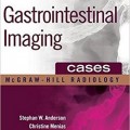
دانلود کتاب تصویربرداری دستگاه گوارش: یک پرونده آموزشی
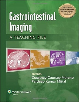
Gastrointestinal Imaging: A Teaching File, 1ed
A brand new resource in The Teaching Files series, Gastrointestinal Imaging: A Teaching File is an exceptional resource for radiology trainees and practicing radiologists who are interested in reviewing the basics of gastrointestinal imaging, including fluoroscopy, CT, and MR. Those studying for board and certifying examinations will also find this volume to be an excellent reference in helping them to review and prepare for these tests.
More than 300 cases, 100 in print and all 300 in an interactive ebook that is included with the print purchase, are portrayed through CT, MR, fluoroscopic, and ultrasound images. This text also covers a wide variety of GI system disorders, including those that affect the solid abdominal organs, intestines, and peritoneal cavity. Each case includes images, along with descriptions of clinical history, findings, differential diagnosis and diagnosis, discussion, relevant questions with appropriate answers, key issues for the report, and important information for the clinician.
Features:
- More than 300 gastrointestinal, including 100 in print and all 300 in the interactive ebook help you make accurate and informed diagnoses.
- Fluoroscopy, CT, and MR images are featured.
- Case information includes images along with management discussion
- Cases are grouped into chapters based on modality to help trainees during modality-based rotations.
Now with the print edition, enjoy the bundled interactive eBook edition, offering tablet, smartphone, or online access to:
- Complete content with enhanced navigation
- A powerful search that pulls results from content in the book, your notes, and even the web
- Cross-linked pages, references, and more for easy navigation
- Highlighting tool for easier reference of key content throughout the text
- Ability to take and share notes with friends and colleagues
- Quick reference tabbing to save your favorite content for future use
- More than 400 Q&A
Contents
CASE 1.1: Achalasia 2
CASE 1.2: Varices 4
CASE 1.3: Sliding hiatal hernia 6
CASE 1.4: Paraesophageal hernia 8
CASE 1.5: Neoplasm (adenocarcinoma) 10
CASE 1.6: Neoplasm (adenocarcinoma) 12
CASE 1.7: Barrett esophagus 14
CASE 1.8: Candida esophagitis 16
CASE 1.9: Boerhaave syndrome 17
CASE 1.10: CMV ulcer 19
CASE 1.11: Extrinsic compression on esophagus due to aberrant left subclavian artery, also right-sided aortic arch 20
CASE 1.12: Fibrovascular polyps 22
CASE 1.13: Submucosal mass (granular cell tumor) 23
CASE 1.14: Long-segment esophageal stricture 25
CASE 1.15: Zenker diverticulum 27
CASE 1.16: Intramural pseudodiverticulosis 29
CASE 1.17: Candidiasis 31
CASE 1.18: Gastric adenocarcinoma 33
CASE 1.19: Adjustable gastric band 35
CASE 1.20: Slipped gastric band 37
CASE 1.21: Eroded gastric band 39
CASE 1.22: Roux-en-y gastric bypass 41
CASE 1.23: Stomal stenosis AND marginal ulcer 43
CASE 1.24: Status post sleeve gastrectomy 44
CASE 1.25: Slipped fundoplication 46
CASE 1.26: Submucosal mass (GIST at biopsy) 48
CASE 1.27: Ulcer 50
CASE 1.28: Gastric adenocarcinoma 51
CASE 1.29: Organoaxial volvulus 52
CASE 1.30: Polyps 53
CASE 1.31: Bezoar 55
CASE 1.32: Linitis plastica appearance of the stomach due to gastric cancer 56
CASE 1.33: Metastatic disease (metastatic melanoma) 57
CASE 1.34: Submucosal mass (lipoma) 58
CASE 1.35: Metastatic disease (melanoma) 59
CASE 1.36: Diverticula 61
CASE 1.37: Scleroderma 63
CASE 1.38: Graft-versus-host disease (GVHD) 65
CASE 1.39: Parasite infection (ascariasis) 67
CASE 1.40: Whipple disease 69
CASE 1.41: Celiac disease 71
CASE 1.42: Delayed diagnosis of Hirschsprung disease 73
CASE 1.43: Rectal cancer 75
CASE 1.44: Adenomatous polyp 76
CASE 1.45: Extrinsic compression due to endometriosis 77
CASE 1.46: Polyp 79
CASE 1.47: Adenocarcinoma 80
CASE 1.48: Sigmoid volvulus 81NDIX
CASE 1.49: Pseudopolyps due to ulcerative colitis 83
CASE 1.50: FAP 85
CASE 1.51: Adenocarcinoma in the setting of ulcerative colitis 86
CASE 2.1: Abscesses 90
CASE 2.2: Hydatid cysts 91
CASE 2.3: Biliary cystadenoma 93
CASE 2.4: SVC occlusion 95
CASE 2.5: Hepatocellular adenoma 97
CASE 2.6: Abscess 100
CASE 2.7: Hepatocellular cancer with tumor thrombus in the right hepatic vein extending into the IVC 102
CASE 2.8: Metastatic carcinoid tumor 104
CASE 2.9: RPC 106
CASE 2.10: Bile duct leak 109
CASE 2.11: Dropped gallstone 111
CASE 2.12: Pancreatic cystosis associated with cystic fibrosis 112
CASE 2.13: Intrapancreatic splenule 114
CASE 2.14: Autoinfarcted spleen in the setting of sickle cell disease 117
CASE 2.15: Delayed splenic rupture 119
CASE 2.16: GIST 120
CASE 2.17: Adenocarcinoma 121
CASE 2.18: Aortoenteric fistula 123
CASE 2.19: Meckel diverticulitis 124
CASE 2.20: Gallstone ileus 125
CASE 2.21: Ischemic enteritis related to systemic lupus erythematosus (SLE) 127
CASE 2.22: Angiotensin-converting enzyme (ACE) inhibitor-induced angioedema of small bowel 129
CASE 2.23: Acute appendicitis 132
CASE 2.24: Active bleeding 133
CASE 2.25: Colonic injury with adjacent mesenteric hematoma 136
CASE 2.26: Complicated diverticulitis with colovesical fistula 137
CASE 2.27: Retained foreign body (blue surgical towel), also known as a gossypiboma 139
CASE 3.1: Hepatocellular carcinoma 142
CASE 3.2: Budd-Chiari syndrome 144
CASE 3.3: FNH 146
CASE 3.4: Hepatocellular carcinoma 148
CASE 3.5: Intrahepatic cholangiocarcinoma 151
CASE 3.6: Lymphoma (secondary, non-Hodgkin) 153
CASE 3.7: Polycystic liver disease associated with autosomal-
dominant polycystic kidney disease (ADPCKD) 155
CASE 3.8: Hemangioma 157
CASE 3.9: Hepatic steatosis with fat sparing 159
CASE 3.10: Confluent hepatic fibrosis 161
CASE 3.11: PTLD 163
CASE 3.12: Gallstones 165
CASE 3.13: Cholangiocarcinoma 167
CASE 3.14: Ischemic cholangiopathy; also intraductal stones and sludge due to stasis 169
CASE 3.15: Adenomyomatosis 171
CASE 3.16: Contained remote prior gallbladder perforation 173
CASE 3.17: SPT of the pancreas 175
CASE 3.18: Autoimmune pancreatitis 177
CASE 3.19: Acute appendicitis 179
CASE 3.20: Appendiceal mucocele due to mucinous cystadenoma 181
CASE 3.21: T4 rectal cancer 183
CASE 3.22: Desmoid tumor 184


